. 2010 Feb 15;21(4):674-85.
doi: 10.1091/mbc.e09-09-0810. Epub 2009 Dec 23.

The cell surface protein gene ecm33+ is a target of the two transcription factors Atf1 and Mbx1 and negatively regulates Pmk1 MAPK cell integrity signaling in fission yeast

Affiliations

The cell surface protein gene ecm33+ is a target of the two transcription factors Atf1 and Mbx1 and negatively regulates Pmk1 MAPK cell integrity signaling in fission yeast

Hirofumi Takada et al. Mol Biol Cell. .

Abstract

The highly conserved fission yeast Pmk1 MAPK pathway plays a key role in cell integrity by regulating Atf1, which belongs to the ATF/cAMP-responsive element-binding (CREB) protein family. We identified and characterized ecm33(+), which encodes a glycosyl-phosphatidylinositol (GPI)-anchored cell surface protein as a transcriptional target of Pmk1 and Atf1. We demonstrated that the gene expression of Ecm33 is regulated by two transcription factors Atf1 and a MADS-box-type transcription factor Mbx1. We identified a putative ATF/CREB-binding site and an RLM1-binding site in the ecm33(+) promoter region and monitored the transcriptional activity of Atf1 or Mbx1 in living cells using a destabilized luciferase reporter gene fused to three tandem repeats of the CRE and six tandem repeats of the Rlm1-binding sequence, respectively. These reporter genes reflect the activation of the Pmk1 pathway by various stimuli, thereby enabling the real-time monitoring of the Pmk1 cell integrity pathway. Notably, the Deltaecm33 cells displayed hyperactivation of the Pmk1 signaling together with hypersensitivity to Ca(2+) and an abnormal morphology, which were almost abolished by simultaneous deletion of the components of the Rho2/Pck2/Pmk1 pathway. Our results suggest that Ecm33 is involved in the negative feedback regulation of Pmk1 cell integrity signaling and is linked to cellular Ca(2+) signaling.


Figures
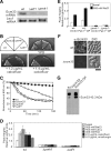
Figure 1.
Figure 1.
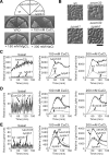
Identification of Ecm33 as a target of Pmk1 and Atf1. (A) Northern blot analysis of total RNA from the wild-type (wt), Δatf1 cells, and Δpmk1 cells. Cells were incubated in YPD medium and collected after culture. Total RNA (20 μg) was subjected to Northern blot analysis using a digoxigenin (DIG)-labeled Ecm33 or Leu1 cRNA. (B) The Δecm33 cells showed hypersensitivity to calcofluor. Wild-type, Δecm33, Δpmk1, and Δatf1 cells were streaked onto the plates as indicated and incubated for 4 d at 27°C. (C) The Δecm33 cells showed hypersensitivity to β-glucanase. Cell lysis was measured at different times during treatment with β-glucanase by determining the OD600. The strains examined were wt, Δecm33, Δpmk1, and Δatf1. (D) Ecm33 (P0.5R2.2) reporter expression is dependent on Pmk1-Atf1 signaling. Ecm33 expression was monitored using the luciferase reporter construct containing the 0.5-kb sequence upstream of ATG of the ecm33+ gene [ecm33 P(0.5)(R2.2)] transformed in wild-type (wt), Δpmk1, and Δatf1 cells. Cells were grown in YPD (basal) or subjected to various stimuli as indicated for 30 min at 27°C, and the assay was performed as described in Deng et al. (2006); data from at least three independent experiments are expressed as mean ± SD. (E) Overexpression of constitutively active Pek1 MAP kinase kinase stimulates Ecm33 expression. Wild-type or Δpmk1 cells harboring [ecm33 P(0.5)(R2.2)] were transformed with either pREP41-Pek1DD (Pek1DD OP) or the control vector (vector) and cultured for 24 h in the absence of thiamine. Cells were either untreated (basal) or treated with 500 mM NaCl. The data were averaged from peak heights of three independent experiments, and each sample was analyzed in triplicate. Error bars, SD. (F) The Ecm33 protein localizes to the cell surface. Immunofluorescence microscopy using anti-Ecm33 antibodies. Exponentially growing wild-type cells were fixed and processed for immunofluorescence microscopy using anti-Ecm33 antibodies (α-Ecm33). Scale bar, 10 μm. (G) Ecm33 monoclonal antibodies specifically recognized the Ecm33 protein. Extracts from wild-type cells containing either the multicopy plasmid carrying ecm33+ or the control vector and Δecm33 were subjected to SDS-PAGE analysis followed by immunoblot analysis with the Ecm33 antibodies.
Figure 2.
Figure 2.
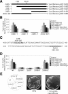
Promoter analysis of ecm33+ gene. (A) Deletion analysis of the ecm33+ promoter. Segment from the ecm33+ upstream region indicated at the left was inserted into the multicopy plasmid containing the luciferase reporter gene. The positions of the CRE (*) and the RLM1 (**) sequences are shown. The numbers refer to the position of the deletion end point relative to the first base of the initiation codon of the gene, which are designated as +1. (B) The upstream region from −500 to −300 of the ecm33+ gene regulates Pmk1-responsive expression of the ecm33+ gene. The luciferase fusion plasmids as indicated were transformed into wild-type cells. Cells were either untreated (basal) or were treated with various stimuli as indicated, and the assay was performed as described in Figure 1E. (C) Identification of CRE and RLM1 in the promoter region of the ecm33+ gene. The sequences of the CRE-like motif (TTACAGTAA) and the RLM1-like motif (GTATATATAG) identified in the ecm33+ promoter are underlined. The numbers refer to the first and last nucleotides of the displayed sequences. (D) The Mbx1 transcription factor is involved in the Ecm33 expression. The luciferase fusion plasmid Ecm33 (P0.5R2.2) was transformed into various strains as indicated. Cells were either untreated (basal) or treated with various stimuli as indicated, and the assay was performed as described in Figure 2B. (E) The Δmbx1, but not the Δmbx2 cells, showed hypersensitivity to calcofluor. The cells as indicated were streaked onto the plates and then incubated for 4 d at 27°C.
Figure 3.
Figure 3.
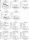
Real-time monitoring of Atf1 activity in living cells. (A) Wild-type cells or Δatf1 cells harboring the multicopy plasmid [3xCREECM33::luc(R2.2) reporter vector] or the mutant version of the reporter vector [3xCREmECM33::luc(R2.2)] were incubated with d-luciferin and treated with 500 mM NaCl. Using a luminometer, light emission levels expressed as relative light units were measured per minute for 2 h. The data shown are representative of multiple experiments. (B) Live-cell monitoring of Atf1 activity in Δpmk1 cells and Δsty1 cells. The cells as indicated were transformed with the multicopy plasmid [3xCREECM33::luc(R2.2) reporter vector] and analyzed as described in A. (C) Overexpression of constitutively active Pek1 MAP kinase kinase stimulates Atf1 activity. Wild-type, Δpmk1, or Δsty1 cells harboring the multicopy plasmid [3xCREECM33::luc(R2.2) reporter vector] were transformed with either pREP2-Pek1DD (Pek1DD OP) or the control vector (vector) and cultured for 24 h in the absence of thiamine. Cells were either untreated (basal) or treated with 500 mM NaCl and analyzed as in Figure 1D.
Figure 4.
Figure 4.
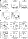
Real-time monitoring of Mbx1 activity in living cells. (A) Wild-type cells or Δmbx1 cells harboring the multicopy plasmid [6xRLMECM33::luc(R2.2) reporter vector] or the mutant version of the reporter vector [6xRLMmECM33::luc(R2.2)) were incubated with d-luciferin and treated with 500 mM NaCl. Using a luminometer, light emission levels expressed as relative light units were measured per min for 2 h. The data shown are representative of multiple experiments. (B) Live-cell monitoring of Mbx1 activity in Δpmk1 cells and Δsty1 cells. The cells as indicated were transformed with the multicopy plasmid [6xRLMECM33::luc(R2.2) reporter vector] and analyzed as described in A. (C) Overexpression of constitutively active Pek1 MAP kinase kinase stimulates Mbx1 activity. Wild-type, Δpmk1, or Δmbx1 cells harboring the multicopy plasmid [6xRLMECM33::luc(R2.2) reporter vector] were transformed with either pREP2-Pek1DD (Pek1DD OP) or the control vector (vector) and cultured for 24 h in the absence of thiamine. Cells were either untreated (basal) or treated with 500 mM NaCl and analyzed as in Figure 1D.
Figure 5.
Figure 5.
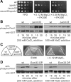
Ecm33 is involved in Pmk1 MAPK-mediated cell integrity signaling. (A) The ecm33+ gene knockout displayed a vic-negative phenotype. The cells as indicated were spotted onto the plates and then incubated for 4 d at 27°C. (B) Knockout of the ecm33+ gene stimulated the phosphorylation of Pmk1 MAPK. Wild-type or Δecm33 cells harboring pREP41-GST-Pmk1 were grown to midlog phase in EMM and then incubated in EMM containing 200 mM CaCl2, or 500 mM NaCl for the indicated time points; cells were collected and lysed at each time point. Immunoblotting using anti-phospho Pmk1 and anti-glutathione S-transferase (GST) antibodies showed that Pmk1 is hyperphosphorylated in the Δecm33 cells. (C) Overexpression of ecm33+ suppressed the Cl hypersensitivity of calcineurin deletion cells (Δppb1). Calcineurin-deleted cells containing either the multicopy plasmid carrying ecm33+ or the control vector, or the wild-type cells carrying the control vector were grown in EMM or EMM containing 0.12 M MgCl2. (D) Overexpression of ecm33+ suppressed the phosphorylation of Pmk1. Wild-type cells transformed with pREP41-GST-Pmk1, containing either the control vector or the multicopy ecm33+ gene, were grown to midlog phase in EMM and analyzed as in B.
Figure 6.
Figure 6.
Altered Ca2+ homeostasis in Δecm33 cells. (A) Δecm33 cells exhibited Ca2+ hypersensitivity. Wild-type cells, Δecm33, Δpmk1, or Δecm33Δpmk1 cells were streaked onto the plates as indicated and then incubated for 4 d at 27°C. (B) Δecm33 cells exhibited abnormal morphology. Morphology of the wild-type cells, Δecm33, Δpmk1, or Δecm33Δpmk1 cells incubated in YPD liquid medium. Scale bar, 10 μm. (C) Δecm33 cells displayed an enhanced calcineurin activity. Wild-type cells, Δecm33, or Δppb1 cells harboring the multicopy plasmid [3xCDRE::luc(R2.2)] reporter vector were incubated with d-luciferin and treated with various concentrations of CaCl2 and then were analyzed as in Figure 1D. (D) Wild-type cells harboring the multicopy plasmid [3xCDRE::luc(R2.2)] reporter vector were transformed with either the control vector (vector) or the ecm33+ gene (Ecm33 OP) and then analyzed as in Figure 6(C). (E) Pmk1 deletion suppressed the enhanced calcineurin activity in Δecm33 cells. Wild-type, Δpmk1, or Δecm33Δpmk1 cells harboring the multicopy plasmid [3xCDRE::luc(R2.2)] reporter vector were analyzed for calcineurin activity as in Figure 6C.
Figure 7.
Figure 7.
Ecm33 regulates Ca2+ influx. The peak response of intracellular Ca2+ monitoring after the addition of CaCl2. Wild-type, Δecm33, and cells overproducing Ecm33 (Ecm33 OP) were transformed with pREP1-AEQ, and their intracellular Ca2+ levels were monitored during the first 10 min. Cells were either untreated or treated with 100 mM CaCl2 or 200 mM CaCl2. The aequorin assay was performed as described in Materials and Methods. The data were averaged from peak heights of three independent experiments, and each sample was done in duplicate. Error bars, SD.
Figure 8.
Figure 8.
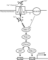
A model for the dual regulation of ecm33+ gene expression and its putative involvement in the negative feedback regulation of the Pmk1 MAPK signaling pathway. Regulatory elements in the promoter are schematically represented. Dotted lines denote hitherto uncharacterized processes. Ecm33 may exert its effect on Ca2+/calcineurin signaling largely via Pmk1 pathway and partly via other Ca2+-influx machineries.

Similar articles

Cited by

References

    1. Agarwal A. K., Rogers P. D., Baerson S. R., Jacob M. R., Barker K. S., Cleary J. D., Walker L. A., Nagle D. G., Clark A. M. Genome-wide expression profiling of the response to polyene, pyrimidine, azole, and echinocandin antifungal agents in Saccharomyces cerevisiae. J. Biol. Chem. 2003;278:34998–35015. - PubMed
    1. Bahler J., Wu J. Q., Longtine M. S., Shah N. G., McKenzie A., III, Steever A. B., Wach A., Philippsen P., Pringle J. R. Heterologous modules for efficient and versatile PCR-based gene targeting in Schizosaccharomyces pombe. Yeast. 1998;14:943–951. - PubMed
    1. Buck V., Ng S. S., Ruiz-Garcia A. B., Papadopoulou K., Bhatti S., Samuel J. M., Anderson M., Millar J. B., McInerny C. J. Fkh2p and Sep1p regulate mitotic gene transcription in fission yeast. J. Cell Sci. 2004;117:5623–5632. - PubMed
    1. Carnero E., Ribas J. C., Garcia B., Duran A., Sanchez Y. Schizosaccharomyces pombe ehs1p is involved in maintaining cell wall integrity and in calcium uptake. Mol. Gen. Genet. 2000;264:173–183. - PubMed
    1. De Groot P. W., Hellingwerf K. J., Klis F. M. Genome-wide identification of fungal GPI proteins. Yeast. 2003;20:781–796. - PubMed

Publication types

MeSH terms