Regulation of tRNA bidirectional nuclear-cytoplasmic trafficking in Saccharomyces cerevisiae
- PMID: 20032305
- PMCID: PMC2820427
- DOI: 10.1091/mbc.e09-07-0551
Regulation of tRNA bidirectional nuclear-cytoplasmic trafficking in Saccharomyces cerevisiae
Abstract
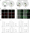
tRNAs in yeast and vertebrate cells move bidirectionally and reversibly between the nucleus and the cytoplasm. We investigated roles of members of the beta-importin family in tRNA subcellular dynamics. Retrograde import of tRNA into the nucleus is dependent, directly or indirectly, upon Mtr10. tRNA nuclear export utilizes at least two members of the beta-importin family. The beta-importins involved in nuclear export have shared and exclusive functions. Los1 functions in both the tRNA primary export and the tRNA reexport processes. Msn5 is unable to export tRNAs in the primary round of export if the tRNAs are encoded by intron-containing genes, and for these tRNAs Msn5 functions primarily in their reexport to the cytoplasm. The data support a model in which tRNA retrograde import to the nucleus is a constitutive process; in contrast, reexport of the imported tRNAs back to the cytoplasm is regulated by the availability of nutrients to cells and by tRNA aminoacylation in the nucleus. Finally, we implicate Tef1, the yeast orthologue of translation elongation factor eEF1A, in the tRNA reexport process and show that its subcellular distribution between the nucleus and cytoplasm is dependent upon Mtr10 and Msn5.
Figures


Similar articles
-
In vivo biochemical analyses reveal distinct roles of β-importins and eEF1A in tRNA subcellular traffic.Genes Dev. 2015 Apr 1;29(7):772-83. doi: 10.1101/gad.258293.115. Genes Dev. 2015. PMID: 25838545 Free PMC article.
-
Utp9p facilitates Msn5p-mediated nuclear reexport of retrograded tRNAs in Saccharomyces cerevisiae.Mol Biol Cell. 2009 Dec;20(23):5007-25. doi: 10.1091/mbc.e09-06-0490. Epub 2009 Oct 7. Mol Biol Cell. 2009. PMID: 19812255 Free PMC article.
-
Genome-wide investigation of the role of the tRNA nuclear-cytoplasmic trafficking pathway in regulation of the yeast Saccharomyces cerevisiae transcriptome and proteome.Mol Cell Biol. 2013 Nov;33(21):4241-54. doi: 10.1128/MCB.00785-13. Epub 2013 Aug 26. Mol Cell Biol. 2013. PMID: 23979602 Free PMC article.
-
tRNA dynamics between the nucleus, cytoplasm and mitochondrial surface: Location, location, location.Biochim Biophys Acta Gene Regul Mech. 2018 Apr;1861(4):373-386. doi: 10.1016/j.bbagrm.2017.11.007. Epub 2017 Nov 28. Biochim Biophys Acta Gene Regul Mech. 2018. PMID: 29191733 Free PMC article. Review.
-
The ins and outs of nuclear re-export of retrogradely transported tRNAs in Saccharomyces cerevisiae.Nucleus. 2010 May-Jun;1(3):224-30. doi: 10.4161/nucl.1.3.11250. Epub 2010 Jan 13. Nucleus. 2010. PMID: 21327067 Free PMC article. Review.
Cited by
-
TNPO3-Mediated Nuclear Entry of the Rous Sarcoma Virus Gag Protein Is Independent of the Cargo-Binding Domain.J Virol. 2020 Aug 17;94(17):e00640-20. doi: 10.1128/JVI.00640-20. Print 2020 Aug 17. J Virol. 2020. PMID: 32581109 Free PMC article.
-
Conservation of structure and mechanism by Trm5 enzymes.RNA. 2013 Sep;19(9):1192-9. doi: 10.1261/rna.039503.113. Epub 2013 Jul 25. RNA. 2013. PMID: 23887145 Free PMC article.
-
Protein kinase A is part of a mechanism that regulates nuclear reimport of the nuclear tRNA export receptors Los1p and Msn5p.Eukaryot Cell. 2014 Feb;13(2):209-30. doi: 10.1128/EC.00214-13. Epub 2013 Dec 2. Eukaryot Cell. 2014. PMID: 24297441 Free PMC article.
-
Separate responses of karyopherins to glucose and amino acid availability regulate nucleocytoplasmic transport.Mol Biol Cell. 2014 Sep 15;25(18):2840-52. doi: 10.1091/mbc.E14-04-0948. Epub 2014 Jul 23. Mol Biol Cell. 2014. PMID: 25057022 Free PMC article.
-
The life and times of a tRNA.RNA. 2023 Jul;29(7):898-957. doi: 10.1261/rna.079620.123. Epub 2023 Apr 13. RNA. 2023. PMID: 37055150 Free PMC article. Review.
References
-
- Arts G. J., Fornerod M., Mattaj I. W. Identification of a nuclear export receptor for tRNA. Curr. Biol. 1998a;8:305–314. - PubMed
Publication types
MeSH terms
Substances
Grants and funding
LinkOut - more resources
Full Text Sources
Molecular Biology Databases
Miscellaneous

