. 2010 Feb 15;21(4):521-37.
doi: 10.1091/mbc.e09-06-0506. Epub 2009 Dec 23.

Plasticity-related gene 5 (PRG5) induces filopodia and neurite growth and impedes lysophosphatidic acid- and nogo-A-mediated axonal retraction

Affiliations

Plasticity-related gene 5 (PRG5) induces filopodia and neurite growth and impedes lysophosphatidic acid- and nogo-A-mediated axonal retraction

Thomas Broggini et al. Mol Biol Cell. .

Abstract

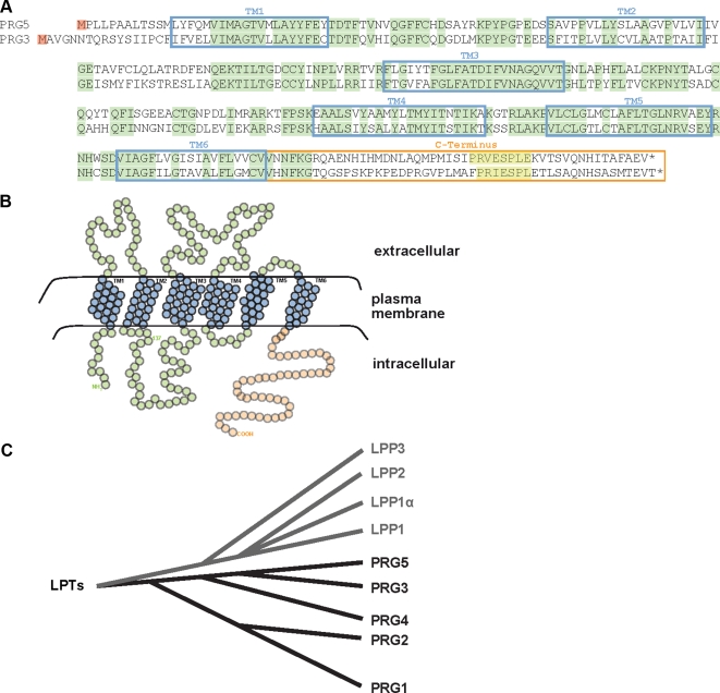
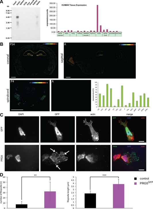
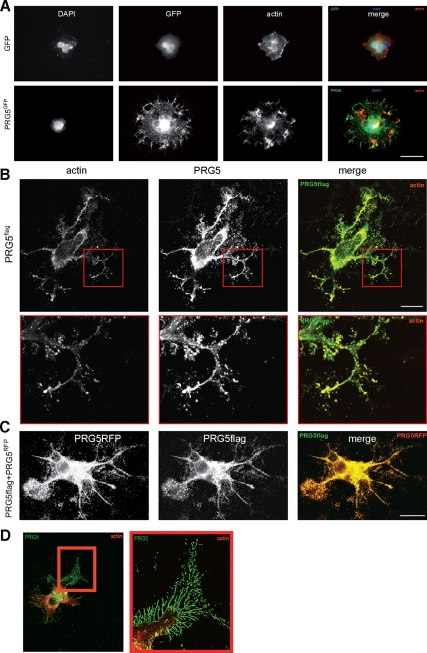
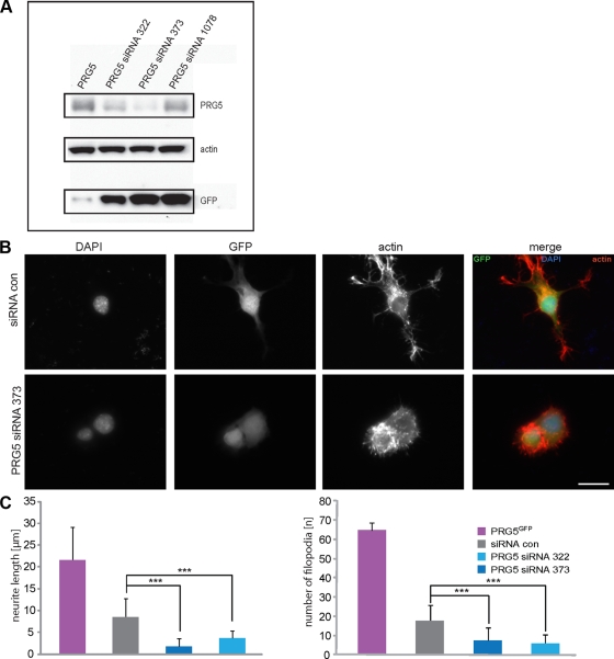
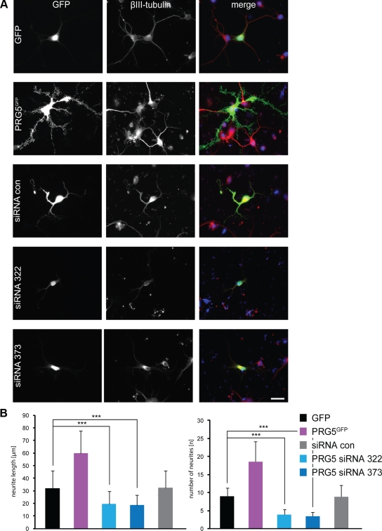
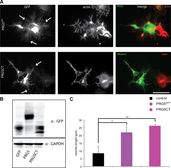
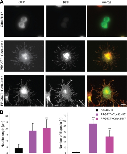
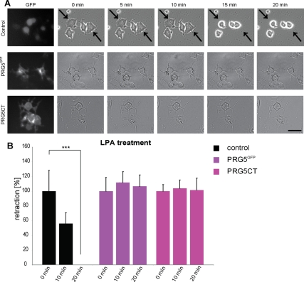
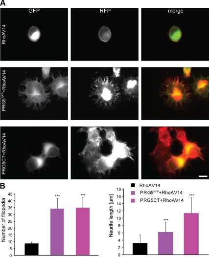
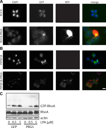
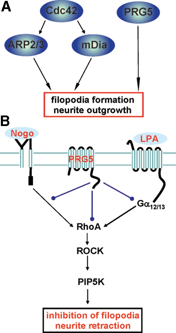
Members of the plasticity-related gene (PRG1-4) family are brain-specific integral membrane proteins and implicated in neuronal plasticity, such as filopodia formation and axon growth after brain lesion. Here we report on the cloning of a novel member of the PRG family, PRG5, with high homologies to PRG3. PRG5 is regulated during brain and spinal cord development and is exclusively allocated within the nervous system. When introduced in neurons, PRG5 is distributed in the plasma membrane and induces filopodia as well as axon elongation and growth. Conversely, siRNA mediated knockdown of PRG5 impedes axon growth and disturbs filopodia formation. Here we show that PRG5 induces filopodia growth independently of Cdc42. Moreover, axon collapse and RhoA activation induced by LPA and myelin-associated neurite inhibitor Nogo-A is attenuated in the presence of PRG5, although direct activation of the RhoA-Rho-PIP5K kinase pathway abolishes PRG5 -formed neurites. Thus, we describe here the identification of a novel member of the PRG family that induces filopodia and axon elongation in a Cdc42-independent manner. In addition, PRG5 impedes brain injury-associated growth inhibitory signals upstream of the RhoA-Rho kinase pathway.


Figures

Figure 1.
Figure 1.
PRG5 is a novel member of the PRG family. PRG5 is a 322-AA-long protein with six transmembrane domains and a 50-AA-long C-terminal tail domain. (A) Amino acid sequence of human PRG5 (GenBank Accession no. FJ472844) aligned to human PRG3 (GenBank Accession no. NM_017753). Putative transmembrane domains (TM) are boxed in cyan, and homologous sequences are indicated in light green. The C-terminus is marked in orange, and the homologous PEST domain at the C-terminus is boxed in yellow. (B) Proposed orientation of PRG5 in the plasma membrane. The six transmembrane domains are shown in blue, the intracellular N-terminal region, three extracellular, and two intracellular loops are shown in green, and the intracellular C-terminal tail (orange). (C) Phylogenetic analysis of the lipid phosphate phosphatase (LPP) superfamily including the PRG subfamily and PRG5. PRG3 is mostly related to PRG5 within the PRG subfamily.
Figure 2.
Figure 2.
PRG5 is expressed during brain development and induces filopodia and membrane extension growth. (A) Multitissue Northern blot analysis of PRG5 mRNA shows a strong band at 4.4 kb in brain tissue and a slight band in testis. Right, GeneNote analysis (NCBI profile graph) of PRG5 gene expression in various human tissues. Samples are composed from a pool of 10–25 individuals. (B) Representative in situ hybridization images of PRG5 mRNA in the adult brain (left, coronal; right, sagittal). Bottom, representative images of PRG5 mRNA in situ expression in spinal cord at postnatal stage (P4) and in adult (A). Color coded bars illustrates the relative expression level of PRG5 in various adult mouse brain regions. Scales bars in all images, 800 μm. Right, quantitative analysis of PRG5 mRNA expression in various brain regions. CB, cerebellum; CTX, cerebral cortex; ISO, isocortex; OLF, olfactory area; HPF, hippocampal formation; HIP, hippocampal region; STR, striatum; STRd, striatum dorsal region; STRv, striatum ventral region; RHP, retrohippocampal region; PAL, pallidum; sAMY, striatum-like amygdala; HY, hypothalamus; P, pons; LSX, lateral septal complex. (C) PRG5-GFP fusion construct expression (PRG5GFP) in P19 cells compared with GFP-transfected controls. PRG5 induces strong filopodia growth and is localized mainly in the plasma membrane and end tips of filopodia. Solely expressed GFP is predominantly found in the cytosol and nucleus. Actin is given in red; nuclei are shown in blue. Scale bar in top right image, 11.1 μm, bottom image, 20 μm. (D) Quantification of filopodia formation in P19 cells. The values were averaged for at least three independent experiments. Statistical analysis was performed with Student's t test (n = 8); **p ≤ 0.01, ***p ≤ 0.001; mean values are shown; error bars, ±SD.
Figure 3.
Figure 3.
PRG5 induces filopodia and is located at the plasma membrane and tips of filopodia and neurites. (A) PRG5 expression in neuronal cells induces dramatic neurite and filopodia growth. PRG5GFP (green) is localized at the tips of filopodia and neurites, whereas sole GFP (green) is mainly found in the cytosol. Actin is given in red; nuclei are shown in blue. Scale bar, 15 μm. (B) PRG5 expression in neuronal cells induces massive neurite and filopodia growth independent of the tag. For PRG5 expression a flag-tagged PRG5 (green) construct has been utilized and N1E-115 cells transfected. Immunostained flag-tagged PRG5 is localized at the tips of filopodia, neurites, and internal membrane structures. Actin is given in red. Bottom, higher magnification of framed region in top images. Scale bar, 20 μm. (C) Comparison of localization of RFP-PRG5 fusion protein (red) with PRG5-flag tagged protein (green). Both PRG5 constructs show equal subcellular distribution and phenotype. Scale bar, 20 μm. (D) High-resolution image of PRG5-expressing neuron. Higher magnification of the red boxed area (right) indicates the distribution of PRG5 in single actin-rich filopodia. Scale bar, 50 μm.
Figure 4.
Figure 4.
siRNA-mediated PRG5 knockdown inhibits filopodia formation and neurite growth. (A) Validation of PRG5 expression in neuronal cells and RNAi-mediated PRG5 knockdown. PRG5-GFP fusion protein runs at ∼60 kDa. RNAi-mediated knockdown of PRG5 with two functional siRNAs (siRNA322 and siRNA373) reduces PRG5 expression significantly, as revealed by immunoblotting. Notably, siRNA1078 construct does not affect PRG5 expression as scramble siRNA and therefore serves as a control siRNA. Actin serves as a control for equal loading. Because GFP and siRNA expression is driven by a bicistronic expression cassette, GFP serves as control for efficient siRNA expression levels independently of RNAi efficacy. (B) siRNA-mediated PRG5 knockdown blocks filopodia formation and neurite growth. Representative images of scramble siRNA (siRNAcon) serving as a control and PRG5 siRNA373 knockdown in neuronal cells. Scale bar, 20 μm. (C) Quantification of neurite length and number of filopodia. Statistical analysis contains values averaged for three independent experiments and was performed with Student's t test; ***p < 0.001; error bars, ±SD of each group.
Figure 5.
Figure 5.
PRG5 promotes neurite formation in primary cortical neurons and PRG5 silencing attenuates neurite formation and growth. (A) PRG5GFP expression in E18 wild-type rat cortical neurons promotes neurite formation and neurite growth. GFP alone (green) is mainly found in the cytosol of cell body, whereas PRG5 (green) is mainly localized at the end tips of neurites. The neuronal marker ßIII tubulin is shown in red. Scale bar, 20 μm. (B) Quantification of neurite length and number in cortical neurons. Measurements of number of neurites are given per neuron. Values are averaged for three independent experiments; statistical analysis was performed with Student's t test; ***p < 0.001; error bars, ±SD of each group.
Figure 6.
Figure 6.
Membrane targeting and localization of PRG5 is essential for axon growth. (A) Neuronal cells were transfected with PRG5 wild-type construct (PRG5GFP, green) and with the PRG5CT consistent of PRG5 C-terminus fused to the myristylation consensus sequence of the YES-kinase for membrane targeting (green). PRG5 and PRG5CT are both localized in neurites and promote similar phenotypes. Arrows indicate branching points. Actin, red. Scale bar, 50 μm. (B) Total cell lysates from neuronal cells transfected with GFP or GFP fusion constructs, PRG5, or PRG5CT were examined by immunoblotting with a GFP-specific antibody. GAPDH serves as a control for equal loading. (C) Quantification of neurite length and number of filopodia in controls, PRG5 and PRG5CT neurons. Statistical analysis contains values averaged for three independent experiments and was performed with Student's t test; *p < 0.01, **p < 0.005; error bars, ±SD of each group.
Figure 7.
Figure 7.
PRG5 promotes filopodia formation and neurite growth independently of Cdc42. (A) Neuronal cells were transfected with dominant negative Cdc42 (pIRES-GFP-Cdc42N17), and neurite length and filopodia were monitored. Representative images of Cdc42N17-expressing neurons and cotransfected neurons are shown. PRG5-expressing neurons cotransfected with Cdc42N17 show normal neurite length and filopodia number (mean neurite length 18 ± 5.9 μm; 54.6 ± 5.9 filopodia/cell) comparable to solely PRG5RFP-expressing cells (mean neurite length 21.6 ± 7.5 μm; 64.7 ± 8 filopodia/cell). Bottom images, neurons coexpressing Cdc42N17 (green) and C-terminal tail of PRG5 (PRG5CT, red). Scale bar, 20 μm. (B) Quantification of neurite length and number of filopodia in transfected neurons compared with Cdc42N17-expressing neurons. Measurements of number of filopodia are given per neuron. Statistical analysis was performed with Student's t test; ***p < 0.001; error bars, ±SD.
Figure 8.
Figure 8.
PRG5 inhibits lysophosphatidic acid (LPA)-induced rapid neurite retraction. (A) Time-lapse measurements of neurons transfected with GFP, PRG5, and PRG5CT. PRG5-positive cells and PRG5CT do not show changes in neurite length after LPA treatment, whereas controls display neurite collapse. Arrows indicate retracting neurites in representative examples. Neurons were monitored for 20 min after treatment. Scale bar, 50 μm. (B) Quantification of neurite length in LPA-treated neurons. Statistical analysis was performed with Student's t test (n = 3); values are mean ± SD; ***p < 0.001.
Figure 9.
Figure 9.
PRG5 attenuates Nogo-A–induced axon collapse. (A) Time-lapse measurements of neurons after Nogo-A treatment. Representative examples of time-lapse images are shown. Note, that PRG5GFP positive cells and PRG5CT remain their neurite length after Nogo-A treatment, whereas neurites from controls (GFP-transfected cells) collapsed rapidly. Arrows indicate retracting neurites in controls. Time scale of treatment was 40 min. Scale bar, 50 μm. (B) Quantification of neurite length after Nogo-A treatment of neurons. Statistical analysis was performed with Student's t test (n = 3), values are mean ± SD; *p < 0.05. (C) Quantification of neurite length after control IgG-antibody treatment in neuronal cells. Statistical analysis was performed with Student's t test (n = 3); *p < 0.01, error bar, ±SD.
Figure 10.
Figure 10.
PRG5-dependent effects on neurites are impeded by RhoA signaling. (A) Representative images of neuronal cells expressing constitutive active RhoA (RhoAV14, green). RhoAV14-expressing neurons show massive reduction in neurite length and retraction of filopodia. RhoV14 (green)- and PRG5 (red)-coexpressing neurons show dramatically reduced neurite length (6.2 ± 2.7 μm) and filopodia number (34.2 ± 7.7 filopodia per cell) compared with solely PRG5-expressing neurons (neurite length 21.6 ± 7.5 μm, 64.7 ± 10.9 filopodia per cell). Bottom images, neurons coexpressing RhoAV14 (green) and C-terminal tail of PRG5 (PRG5CT, red). Scale bar, 20 μm. (B) Quantification of number of filopodia and neurite length in transfected neurons compared with RhoAV14-expressing cells. Measurements of number of filopodia are given per neuron. Statistical analysis was performed with Student's t test; ***p < 0.001; error bars, ±SD.
Figure 11.
Figure 11.
PRG5 acts upstream from RhoA-ROCK-PIP5K pathway and interferes with LPA-induced RhoA activation. (A) Neuronal cells were transfected with ROCK and PRG5, and neurite growth and filopodia formation was monitored. Solely ROCK-expressing neurons show dramatic cell rounding. Note, that neurons expressing both PRG5 (red) and ROCK (green) also display round cell shape. (B) Neurons transfected with PRG5 and PIP5Kβ (type 1 phosphatidylinositol 4-phosphate 5-kinase β) show a phenotype comparable to solely PIP5K1β (green)-expressing neurons. Nuclei in A and B are shown in blue. Scale bar, 20 μm. (C) Neurons were transfected with either GFP or PRG5 as indicated and stimulated with various concentrations of LPA and levels of activated GTP-RhoA were determined by Rhotekin-binding assay (top panel). Total RhoA and actin served as controls for equal loading (bottom panels). Details on the Rhotekin-binding assay are described in Materials and Methods.
Figure 12.
Figure 12.
PRG5 signaling in axon growth and neurite retraction. PRG5 induces filopodia and axon growth independent of Cdc42 and impedes RhoA-dependent neurite retraction. (A) Proposed model of PRG5 action on filopodia and axon growth. (B) Summary and proposed model of PRG5 signaling in the context of neurite growth inhibitors. The common neurite growth inhibitors Nogo-A and LPA (underscored light blue) act on their receptors, and their signaling pathway converge at the RhoA-Rho kinase pathway. PRG5 is localized in the plasma membrane and attenuates LPA and Nogo-A—induced neurite retraction and alleviates basal RhoA activation, whereas direct RhoA, ROCK or PIP5Kβ activation overcomes PRG5 induced neurites. Thus, PRG5 may act upstream of RhoA interfering with Nogo-A and LPA receptor signaling or alternatively PRG5 directly balances or impacts RhoA activation.

References

    1. Abe T., Kato M., Miki H., Takenawa T., Endo T. Small GTPase Tc10 and its homologue RhoT induce N-WASP-mediated long process formation and neurite outgrowth. J. Cell Sci. 2003;116:155–168. - PubMed
    1. Arakawa Y., Bito H., Furuyashiki T., Tsuji T., Takemoto-Kimura S., Kimura K., Nozaki K., Hashimoto N., Narumiya S. Control of axon elongation via an SDF-1alpha/Rho/mDia pathway in cultured cerebellar granule neurons. J. Cell Biol. 2003;161:381–391. - PMC - PubMed
    1. Atwal J. K., Pinkston-Gosse J., Syken J., Stawicki S., Wu Y., Shatz C., Tessier-Lavigne M. PirB is a functional receptor for myelin inhibitors of axonal regeneration. Science. 2008;322:967–970. - PubMed
    1. Benfey M., Aguayo A. J. Extensive elongation of axons from rat brain into peripheral nerve graft. Nature. 1982;296:150–152. - PubMed
    1. Bräuer A. U., Savaskan N. E., Kühn H., Prehn S., Ninnemann O., Nitsch R. A new phospholipid phosphatase, PRG-1, is involved in axon growth and regenerative sprouting. Nat. Neurosci. 2003;6:572–578. - PubMed

Publication types

MeSH terms