. 2010 Feb 15;21(4):547-61.
doi: 10.1091/mbc.e09-06-0505. Epub 2009 Dec 23.

Autophosphorylation of polo-like kinase 4 and its role in centriole duplication

Affiliations

Autophosphorylation of polo-like kinase 4 and its role in centriole duplication

James E Sillibourne et al. Mol Biol Cell. .

Abstract

Centrosome duplication occurs once every cell cycle in a strictly controlled manner. Polo-like kinase 4 (PLK4) is a key regulator of this process whose kinase activity is essential for centriole duplication. Here, we show that PLK4 autophosphorylation of serine S305 is a consequence of kinase activation and enables the active fraction to be identified in the cell. Active PLK4 is detectable on the replicating mother centriole in G1/S, with the proportion of active kinase increasing through interphase to reach a maximum in mitosis. Activation of PLK4 at the replicating daughter centriole is delayed until G2, but a level equivalent to the replicating mother centriole is achieved in M phase. Active PLK4 is regulated by the proteasome, because either proteasome inhibition or mutation of the degron motif of PLK4 results in the accumulation of S305-phosphorylated PLK4. Autophosphorylation probably plays a role in the process of centriole duplication, because mimicking S305 phosphorylation enhances the ability of overexpressed PLK4 to induce centriole amplification. Importantly, we show that S305-phosphorylated PLK4 is specifically sequestered at the centrosome contrary to the nonphosphorylated form. These data suggest that PLK4 activity is restricted to the centrosome to prevent aberrant centriole assembly and sustained kinase activity is required for centriole duplication.


Figures
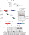
Figure 1.
Figure 1.
PLK4 autophosphorylates. (A) Results of an in vitro kinase assay using full-length human PLK4 purified from bacteria showing that it autophosphorylates. It should be noted that PLK4 is readily degraded, explaining the presence of multiple bands. (B) An N-terminal fragment of PLK4 consisting of residues 1–367 that possessed a mutation within the catalytic domain rendering the kinase inactive (PLK41-367 K41M) was used as a substrate in an in vitro kinase assay using the kinase domain of PLK4 (PLK41-285). Autoradiography showed that PLK41-367 k41M was phosphorylated when mixed with the kinase domain of PLK4, indicating that intermolecular phosphorylation had occurred as well as autophosphorylation of the kinase domain. (C) Schematic showing the location of the three PEST sequences (numbered light blue boxes), kinase (red box), crypto Polo-box (dark blue box), and Polo-box (green box) domains in PLK4. The fragments of PLK4 used as substrates in in vitro phosphorylation assays with the kinase domain of PLK4 are also shown. (D) Results of in vitro kinase assays using as substrates the different fragments of PLK4 depicted in C. The kinase and central and crypto Polo-box domains of PLK4 were all found to be phosphorylated. (E) Diagram showing the location of predicted autophosphorylation sites in PLK4. (F) Results of in vitro phosphorylation assays using peptides spanning predicted autophosphorylation sites showed that S305 was a major site within PLK4. Peptide numbers are noted on top of the gels and sequences are given in the boxes with potential phosphorylation sites marked in red with S/T to A mutations marked in blue. Serine residue 305 seemed to be the major autophosphorylation site within PLK4. The plus sign indicates a positive control carried out using a 12 amino acid peptide from RAF containing serine 338 closely matching the consensus phosphorylation sequence of PLK4.
Figure 2.
Figure 2.
PLK4 localizes asymmetrically to centrioles. (A) HeLa cells stably expressing GFP-centrin-1 (green) were fixed and stained with DAPI (blue) to label DNA, anti-PLK4 KD (red), and anti-ninein (blue insets) antibodies. PLK4 was found to localize to two sites on the mother centriole, with one site at the proximal end and the other site close to the subdistal appendages between two ninein-positive staining foci. In contrast, the daughter centriole possessed a single focus of PLK4 staining at the proximal end of the centriole, which often colocalized with ninein staining. A cartoon depicting the arrangement of mother (M), daughter (D), and procentrioles (PC) is superimposed on the enlargements of the merged centriole images presented. (B) RPE1 cells transiently transfected with EGFP-PLK4 (green) and stained with centrin (red) and ninein (blue) antibodies. Mother centrioles consistently possessed more EGFP-PLK4 than daughter centrioles. Bar, 10 μm; inset, 1 μm.
Figure 3.
Figure 3.
Serine 305 of PLK4 is autophosphorylated in vitro and in cells. (A) PLK41-367 and PLK41-367 K41M were subjected to in vitro phosphorylation and then treated with or without lambda phosphatase. Sypro Ruby staining was used to detect the PLK4 fragments, and autoradiography was used to determine whether they had been phosphorylated. Only the untreated PLK41-367 fragment was found to have autophosphorylated, and this form was specifically recognized by the PLK4 pS305 antibody. HRP-conjugated molecular weight markers were loaded onto the PLK4 pS305 blot (left lane). (B) Lysates were prepared from HCT116 cells that had been transiently transfected with EGFP-PLK4 or -PLK4 K41M in conjunction with control or PLK4 siRNA and Western blotted with anti-PLK4 CPB and anti-PLK4 pS305 antibodies. The anti-PLK4 pS305 antibody was found to recognize specifically the active form of the kinase in cells. (C) Western blotting of lysates from HeLa cells simultaneously transfected with siRNA and FLAG-tagged PLK4 expression constructs. The results show that both PLK4 siRNA duplexes are efficacious to the wild-type PLK4 mRNA, whereas PLK4 siRNA 2 is no longer effective against the siRNA-resistant form of PLK4 (siRes PLK4). α-Tubulin blotting was carried out as a loading control. (D) Images of HeLa cells transfected with siRNA-resistant EGFP-PLK4, -PLK4 K41M, or -PLK4 S305A (all green) and PLK4 siRNA 2, stained with DAPI (blue) to label DNA and PLK4 pS305 antibody (red). The PLK4 pS305 antibody recognizes wild-type siRes EGFP-PLK4, but neither siRes EGFP-PLK4 K41M nor siRes EGFP-PLK4 S305A in the absence of endogenous PLK4. Bar, 10 μm; inset, 1 μm.
Figure 4.
Figure 4.
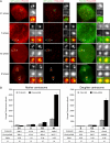
PLK4 autophosphorylation peaks in mitosis. (A) HeLa cells stably expressing GFP-centrin (green) were stained with anti-PLK4 KD (red) and anti-PLK4 pS305 (red) antibodies. GFP-tagged centrin served as a marker for centrioles. Staining with the anti-PLK4 KD antibody enabled detection of all the PLK4 present within the cell, whereas the anti-PLK4 pS305 antibody detected only the phosphorylated form of the kinase. In G1, the abundance of S305 phosphorylated PLK4 at centrioles is extremely low, being barely detectable, whereas in S phase levels increase to produce a clearly discernible signal that rises to reach a maximum in mitosis. Strikingly, more active PLK4 seemed to be present at mother centriole (marked as D, daughter; and M, mother). Bar, 10 μm; inset, 1 μm. (B) Results of fluorescence intensity measurements of PLK4 and PLK4 pS305 at the mother and daughter centriole pairs. The table below the graphs lists the average fluorescence intensity at each centriole pair at G1, S, G2, and M phases of the cell cycle. The ratio of PLK4 pS305 to PLK4 is also given to enable comparison of the level of PLK4 autophosphorylation between the cell cycle phases. The acquisition settings for mitotic cells were not the same as those for interphasic cells, because the exposure times were reduced to avoid saturating and exceeding the dynamic range of the digital camera. Fluorescence intensity measurements were multiplied by a factor corresponding to the reduction in exposure time to produce the final results for the mitotic cells.
Figure 5.
Figure 5.
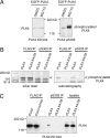
Proteasome inhibition causes the accumulation of S305 autophosphorylated PLK4. (A) RPE1 cells were transfected with EGFP-PLK4 and treated with either DMSO, as a control, or 1 μM MG115 for 3 h before preparing total cell lysates. Western blotting was carried out using the anti-PLK4 kinase domain and PLK4 pS305 antibodies to determine the relative quantities of PLK4- and S305-autophosphorylated PLK4. This demonstrated that proteasome inhibition specifically causes the accumulation of autophosphorylated PLK4. (B) PC3 cells were transiently transfected with either PLK4 or PLK4 K41M, tagged at their C termini with a 3xFLAG motif and treated with 1 μM MG115 for 3 h. Immunoprecipitations were carried out using either anti-FLAG or anti-PLK4 pS305 antibodies. Approximately 70% of the immunoprecipitated material was subjected to an in vitro phosphorylation assay, migrated on a gradient gel, stained with silver, and exposed to a phosphorimager screen. The open arrowhead points to immunoprecipitated PLK4 and PLK4 K41M. Autoradiography demonstrated that phosphorylated PLK4 was present in the anti-FLAG and anti-PLK4 pS305 immunoprecipitates from PC3 cells transfected with wild-type PLK4 but not the kinase inactive mutant PLK4 K41M. (C) Western blotting was carried out on the remaining immunoprecipitated material with the PLK4 KD antibody. This showed that approximately equal amounts of PLK4 and PLK4 K41M had been immunoprecipitated with the anti-FLAG antibody, whereas the PLK4 pS305 antibody only immunoprecipitated wild-type PLK4.
Figure 6.
Figure 6.
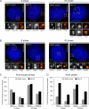
PLK4 S305 autophosphorylation increases at centrioles upon proteasome inhibition. (A and B) HeLa GFP-centrin-1 cells (green) were treated with either DMSO, as a control, or 1 μM MG115 for 3 h; fixed; and stained with DAPI (blue) to label DNA and either anti-PLK4 kinase domain (A) or anti-PLK4 pS305 antibody (B) (both red). Inhibition of the proteasome in S phase cells caused an increase in the abundance of S305-phosphorylated PLK4 at both the mother and daughter centriole pairs. In G2 cells, an increase was observed only at the daughter centriole pair. Bar, 10 μm; inset, 1 μm. (C and D) Fluorescence intensity measurements showing PLK4 and S305 phosphorylated PLK4 levels at mother and daughter centriole pairs in S and G2 phase with and without proteasome inhibitor treatment. At least 19 centrosomes were quantified in each group.
Figure 7.
Figure 7.
Autophosphorylation enhances centriole amplification. (A) RPE1 cells were transiently transfected with pEGFP-PLK4 constructs and Western blotting was carried out using anti-PLK4 KD and anti-PLK4 pS305 antibodies to determine total and S305 phosphorylated PLK4 levels. An anti-α-tubulin blot was carried out as a loading control. The results show that mutation of S305, either to mimic or prevent phosphorylation, has no impact upon the stability of the kinase. Wild-type PLK4 ND containing two mutations in the degron motif, S285A and T289A, to prevent phosphorylation was expressed to high levels, whereas PLK4 K41M and PLK4 K41M ND were both expressed at a lower level, indicating that kinase activity may influence protein stability. Western blotting with the PLK4 pS305 antibody shows PLK4 ND is heavily S305 phosphorylated and suggests that stabilization allows active kinase to accumulate. (B) HeLa cells were transiently transfected with EGFP-PLK4 (green) expression constructs, fixed, and stained with DAPI (blue) to label DNA and anti-centrin antibody (red). Bar, 10 μm; inset, 1 μm. (C) Results of three independent experiments quantifying the number of transfected cells exhibiting centriole amplification (n = 200). The results showed that wild-type PLK4 and PLK4 S305A triggered centriole amplification to a similar extent, whereas PLK4 S305E triggered centriole amplification more frequently, indicating that mimicking S305 autophosphorylation enhances centriole amplification. In agreement with published results in Drosophila S2 cells, PLK4 ND expression resulted in a higher incidence of centriole amplification compared with wild-type PLK4.
Figure 8.
Figure 8.
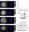
S305-autophosphorylated PLK4 is important for centriole duplication and is sequestered at the centrosome. (A) HeLa cells were transiently transfected with EGFP-PLK4 (green), plated onto 37-μm-diameter circular micropatterned coverslips, fixed, and stained DAPI (blue) to label DNA, anti-centrin (red), and anti-γ-tubulin antibodies (black and white). (B) EGFP-PLK4 fluorescence intensity was quantified in transfected cells exhibiting centriole amplification by drawing a region around the centrosomal area and measuring the fluorescence intensity (n = 28–33 cells). No significant difference was found in EGFP-PLK4 levels at the centrosome between the mutant forms and wild-type PLK4. (C) RPE1 cells were transiently transfected with EGFP, EGFP-PLK4, or varying amounts of EGFP-PLK4 ND, and Western blotting was carried out with PLK4 KD, PLK4 pS305 and α-tubulin antibodies. The results show more of the nondegradable form of PLK4 is phosphorylated compared with the wild-type kinase, indicating that the fraction of active kinase in PLK4 ND transfected cells is greater than in those transfected with wild-type PLK4. (D) Results of Western blotting using anti-PLK4 pS305, anti-FLAG, and anti-α-tubulin antibodies to probe soluble and insoluble extracts that had been prepared from RPE1 cells transiently transfected with EGFP-PLK4–3xFLAG expression constructs. Probing with the anti-FLAG antibody showed that overexpressed PLK4 was present in both the soluble and insoluble extracts, whereas S305-phosphorylated PLK4 partitioned in the insoluble fraction in extracts prepared from cells transfected with wild-type and nondegradable wild-type PLK4 (lanes 4 and 6). These data suggest that the active form of PLK4 is restricted to the centrosome because it is present in the insoluble, and not the soluble, extract.

Similar articles

Cited by

References

    1. Azimzadeh J., Hergert P., Delouvee A., Euteneuer U., Formstecher E., Khodjakov A., Bornens M. hPOC5 is a centrin-binding protein required for assembly of full-length centrioles. J. Cell Biol. 2009;185:101–114. - PMC - PubMed
    1. Azioune A., Storch M., Bornens M., Thery M., Piel M. Simple and rapid process for single cell micro-patterning. Lab Chip. 2009;9:1640–1642. - PubMed
    1. Bettencourt-Dias M., Rodrigues-Martins A., Carpenter L., Riparbelli M., Lehmann L., Gatt M. K., Carmo N., Balloux F., Callaini G., Glover D. M. SAK/PLK4 is required for centriole duplication and flagella development. Curr. Biol. 2005;15:2199–2207. - PubMed
    1. Bornens M. Centrosome composition and microtubule anchoring mechanisms. Curr. Opin. Cell Biol. 2002;14:25–34. - PubMed
    1. Chen Z., Indjeian V. B., McManus M., Wang L., Dynlacht B. D. CP110, a cell cycle-dependent CDK substrate, regulates centrosome duplication in human cells. Dev. Cell. 2002;3:339–350. - PubMed

MeSH terms