. 2010 Feb 26;285(9):6327-36.
doi: 10.1074/jbc.M109.074633. Epub 2009 Dec 23.

Interactions between copper-binding sites determine the redox status and conformation of the regulatory N-terminal domain of ATP7B

Affiliations

Interactions between copper-binding sites determine the redox status and conformation of the regulatory N-terminal domain of ATP7B

Erik S LeShane et al. J Biol Chem. .

Abstract

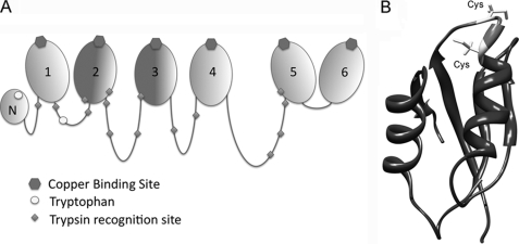
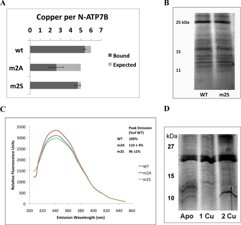
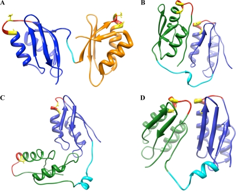
Copper-transporting ATPase ATP7B is essential for human copper homeostasis and normal liver function. ATP7B has six N-terminal metal-binding domains (MBDs) that sense cytosolic copper levels and regulate ATP7B. The mechanism of copper sensing and signal integration from multiple MBDs is poorly understood. We show that MBDs communicate and that this communication determines the oxidation state and conformation of the entire N-terminal domain of ATP7B (N-ATP7B). Mutations of copper-coordinating Cys to Ala in any MBD (2, 3, 4, or 6) change the N-ATP7B conformation and have distinct functional consequences. Mutating MBD2 or MBD3 causes Cys oxidation in other MBDs and loss of copper binding. In contrast, mutation of MBD4 and MBD6 does not alter the redox status and function of other sites. Our results suggest that MBD2 and MBD3 work together to regulate access to other metal-binding sites, whereas MBD4 and MBD6 receive copper independently, downstream of MBD2 and MBD3. Unlike Ala substitutions, the Cys-to-Ser mutation in MBD2 preserves the conformation and reduced state of N-ATP7B, suggesting that hydrogen bonds contribute to interdomain communications. Tight coupling between MBDs suggests a mechanism by which small changes in individual sites (induced by copper binding or mutation) result in stabilization of distinct conformations of the entire N-ATP7B and altered exposure of sites for interactions with regulatory proteins.


Figures

FIGURE 1.
FIGURE 1.
Organization of N-ATP7B. A, schematic illustrating the relative length of the loops connecting the N-terminal MBDs; copper-binding sites, tryptophan residues, and trypsin recognition sites are as indicated. B, structure of a representative MBD (MBD5; adapted from Ref. 8) (Protein Data Bank code 2ew9) The GMxCxxC metal-binding loop is shown in light gray; Cys residues are indicated.
FIGURE 2.
FIGURE 2.
Atox1-mediated copper transfer is impaired in the MBD2 mutant. A, diagram of the N-ATP7B mutants used in this work. B, copper transfer from Atox1 to N-ATP7B. Points represent the amount of copper that remains bound to N-ATP7B after incubation with the indicated amounts of Cu-Atox1 and removal of Cu-Atox1 from the mixture. Error bars indicate S.D. across three independent experiments. wt, wild-type N-ATP7B.
FIGURE 3.
FIGURE 3.
Mutation m2A induces structural changes in N-ATP7B. The Coomassie Blue-stained gel compares limited proteolytic patterns of wild-type (WT) and m2A N-ATP7B.
FIGURE 4.
FIGURE 4.
Mutations in MBDs have distinct effects on the copper-binding stoichiometry of N-ATP7B mutants. Wild-type (wt) and mutant N-ATP7B were loaded with copper in E. coli prior to purification. Light gray bars indicate the number of CxxC motifs left intact. Dark gray bars represent experimental values as determined by atomic absorption (copper) and Lowry assay (protein). Error bars indicate S.D. across three experimental repeats.
FIGURE 5.
FIGURE 5.
Comparison of Cys-directed fluorescent labeling for wild-type and mutant N-ATP7B. A, fluorescent (upper) and Coomassie Blue-stained gel (lower) images of wild-type (WT) and m2A N-ATP7B labeled with CPM. The tables indicate intensity of fluorescent labeling per protein compared with wild-type apo-N-ATP7B. The indicated samples were treated with TCEP prior to labeling. The lane on the right shows CPM labeling of copper-loaded wild-type N-ATP7B used as a background control. N/A, not applicable. B, fluorescent (upper) and Coomassie Blue-stained gel (lower) images of additional mutants shown in Fig. 2A, with label/protein ratios indicated in the table. Average percentages and S.D. are indicated for three experimental repeats.
FIGURE 6.
FIGURE 6.
MBDs in N-ATP7B are tightly packed. A, wild-type N-ATP7B was subjected to limited proteolysis as described in the legend to Fig. 3 and separated on a native Tris/glycine gel. Under these conditions, most of the fragments stayed together. The gel was stained with Coomassie Blue. B, the gel lane containing fragments run on a native gel was then cut and placed on top of a Laemmli gel and separated in the second dimension under reducing and denaturing conditions. The fragments no longer migrated together (silver staining).
FIGURE 7.
FIGURE 7.
Molecular dynamics simulation of MBD2 of ATP7A illustrates the mobility and exposure of cysteines. A, relevant distances calculated for each frame of a 10,000-ps simulation. Black dots (RMSD) indicate overall backbone r.m.s.d. for the protein. Green dots (Cα–Cα) indicate the distance between α-carbons of metal-binding cysteines. Blue dots (S–S) indicate the distance between sulfur atoms of metal-binding cysteines. B, different orientation of the metal-binding loop from molecular dynamics simulation. Upper, loop with metal-binding cysteines facing away from each other; lower, loop with metal-binding cysteines facing toward each other.
FIGURE 8.
FIGURE 8.
Mutations m2A and m2S have different effects on the conformation and oxidation state of N-ATP7B. A, copper-binding stoichiometries of wild-type N-ATP7B (wt) and the m2A and m2S mutants. B, Coomassie Blue-stained gel comparing limited proteolysis of wild-type and m2S N-ATP7B. C, fluorescence emission spectra of apo-N-ATP7B when excited with 280 nm UV light. Representative curves are shown for wild-type (blue), m2A (red), and m2S (green) N-ATP7B, with average peak emissions (342 nm) for three experiments shown in the inset. D, proteolytic patterns of wild-type apo-N-ATP7B and N-ATP7B with one or two Cu+ atoms transferred from Atox1.
FIGURE 9.
FIGURE 9.
Structures of pairs of neighboring MBDs. A, structure of MBD5 and MBD6 of ATP7B (adapted from Achila et al. (8); Protein Data Bank code 2ew9). B–D, ab initio structures for MBD1 and MBD2. MBD1 is shown in green, the inter-MBD loop in cyan, and MBD2 in blue. The GMxCxxC loop is shown in red, and copper-binding Cys residues are shown in yellow.

Similar articles

Cited by

References

    1. Bertini I., Rosato A. (2008) Cell. Mol. Life Sci. 65, 89–91 - PMC - PubMed
    1. Lutsenko S., LeShane E. S., Shinde U. (2007) Arch. Biochem. Biophys. 463, 134–148 - PMC - PubMed
    1. Bartee M. Y., Ralle M., Lutsenko S. (2009) Biochemistry 48, 5573–5581 - PMC - PubMed
    1. Guo Y., Nyasae L., Braiterman L. T., Hubbard A. L. (2005) Am. J. Physiol. Gastrointest. Liver Physiol. 289, G904–G916 - PubMed
    1. Lim C. M., Cater M. A., Mercer J. F., La Fontaine S. (2006) J. Biol. Chem. 281, 14006–14014 - PubMed

Publication types

LinkOut - more resources