. 2010 Mar 5;285(10):7619-32.
doi: 10.1074/jbc.M109.057182. Epub 2009 Dec 23.

Inhibition of calcineurin-mediated endocytosis and alpha-amino-3-hydroxy-5-methyl-4-isoxazolepropionic acid (AMPA) receptors prevents amyloid beta oligomer-induced synaptic disruption

Affiliations

Inhibition of calcineurin-mediated endocytosis and alpha-amino-3-hydroxy-5-methyl-4-isoxazolepropionic acid (AMPA) receptors prevents amyloid beta oligomer-induced synaptic disruption

Wei-Qin Zhao et al. J Biol Chem. .

Abstract

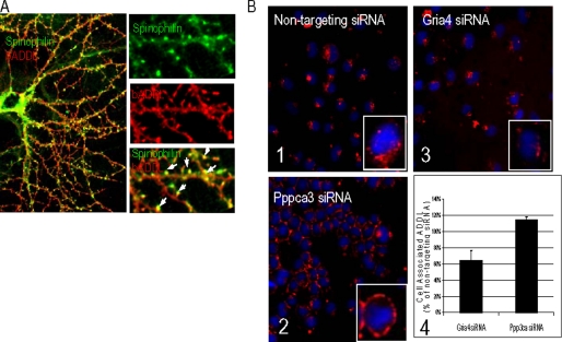
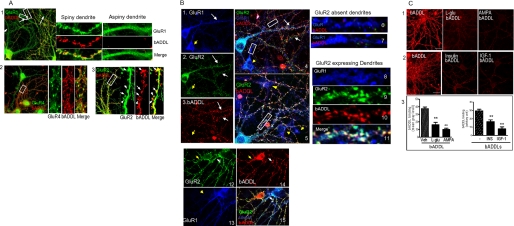
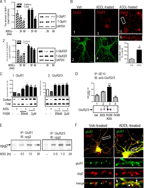
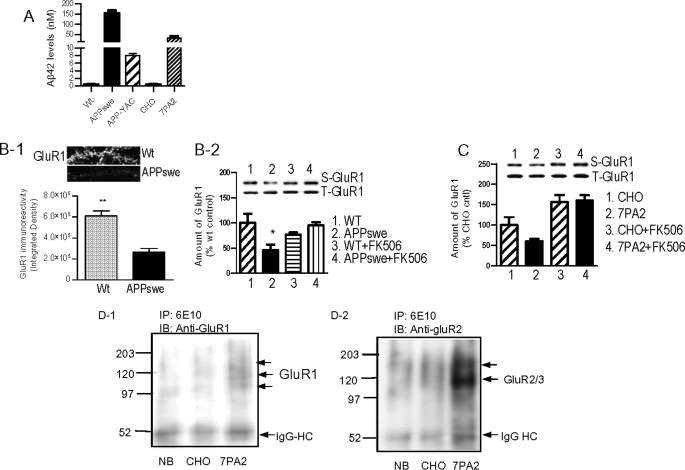
Synaptic degeneration, including impairment of synaptic plasticity and loss of synapses, is an important feature of Alzheimer disease pathogenesis. Increasing evidence suggests that these degenerative synaptic changes are associated with an accumulation of soluble oligomeric assemblies of amyloid beta (Abeta) known as ADDLs. In primary hippocampal cultures ADDLs bind to a subpopulation of neurons. However the molecular basis of this cell type-selective interaction is not understood. Here, using siRNA screening technology, we identified alpha-amino-3-hydroxy-5-methyl-4-isoxazolepropionic acid (AMPA) receptor subunits and calcineurin as candidate genes potentially involved in ADDL-neuron interactions. Immunocolocalization experiments confirmed that ADDL binding occurs in dendritic spines that express surface AMPA receptors, particularly the calcium-impermeable type II AMPA receptor subunit (GluR2). Pharmacological removal of the surface AMPA receptors or inhibition of AMPA receptors with antagonists reduces ADDL binding. Furthermore, using co-immunoprecipitation and photoreactive amino acid cross-linking, we found that ADDLs interact preferentially with GluR2-containing complexes. We demonstrate that calcineurin mediates an endocytotic process that is responsible for the rapid internalization of bound ADDLs along with surface AMPA receptor subunits, which then both colocalize with cpg2, a molecule localized specifically at the postsynaptic endocytic zone of excitatory synapses that plays an important role in activity-dependent glutamate receptor endocytosis. Both AMPA receptor and calcineurin inhibitors prevent oligomer-induced surface AMPAR and spine loss. These results support a model of disease pathogenesis in which Abeta oligomers interact selectively with neurotransmission pathways at excitatory synapses, resulting in synaptic loss via facilitated endocytosis. Validation of this model in human disease would identify therapeutic targets for Alzheimer disease.


Figures

FIGURE 1.
FIGURE 1.
Cell selectivity of bADDL binding. A, cultured hippocampal neurons (21 days in vitro) were treated with 500 nm bADDLs at 37 °C for 10 min. Cells were immunostained with an anti-spinophilin antibody followed by a secondary antibody-Alexa 488 and streptavidin-Alexa 555. bADDL staining shows a high degree of colocalization with spinophilin. B, bADDL biding in siRNA-transfected N2A cells: Panels 1–3, representative images of cells transfected with siRNA silencing Gria4 and Ppp3ca and control stained to visualize cell-associated ADDL (red) and nuclei (blue; stained by 4,6-diamidino-2-phenylindole). Gria4 and Ppp3ca were the two top hits from a screen aimed at identifying key proteins involved in the binding and processing of ADDL to neuronal cells. Images were acquired using an automated confocal imager (IN Cell 3000, GE Healthcare) equipped with a ×40 air objective. Image analysis and quantitation of the ADDL-associated fluorescent signal was performed using the imager proprietary software (Raven, GE Healthcare) and its “translocation” algorithm. Approximately 400 cells/well were imaged and analyzed. Images are reproduced using the same intensity scale. Panel 1, bADDL binding under non-target siRNA condition; panel 2, Pppca3 siRNA reduced ADDL internalization; panel 3, inhibited ADDL binding by Gria4 siRNA; panel 4, bar graph showing quantitation of cell-associated ADDL in Gria4 and Ppp3ca siRNA-treated samples (n = 3) obtained during the confirmation screen. Data are expressed as the percentage of cell-associated ADDL measured in samples treated with non-targeting siRNA. Both Gria4 and Ppp3ca siRNAs were the highest ranking genes among those that inhibited binding and blocked internalization of ADDL, respectively.
FIGURE 2.
FIGURE 2.
AMPAR-dependent bADDL binding. A, bADDL bindings are observed in AMPAR-containing dendritic spines. Panel 1, bADDL binding was observed only in those dendrites with spines. The bADDL binding shows partial colocalization with GluR1 immunoreactivity. Panel 2, bADDL binding shows a low degree of colocalization with GluR4. Panel 3, bADDL binding colocalizes well with GluR2. B, bADDLs bind to GluR2-expressing neurons. Triple immunostaining for GluR1, GluR2, and bADDL binding was applied to bADDL-treated neurons. Panels 1–3 show representative GluR1 (blue) and GluR2 (green) immunostaining and bADDL binding (red), respectively, acquired from the same microscopy plane under different filters. bADDL binding was observed largely in GluR2-expressing neurons (white arrows), whereas neurons expressing GluR1 but not GluR2 show negative bADDL binding (yellow arrows). Panel 4 is a merged image of panels 1–3. Panels 6 and 7 show enlarged images of dendritic fragments (rectangular boxes in panel 4) in which only GluR1 but not GluR2 is expressed. These dendrites display negative bADDL binding. Panel 5 is another merged image showing similar results. Panels 8–11 show an enlarged image of a dendritic fragment taken from panel 5 (rectangular box) with abundant GluR2 and GluR1. These dendrites show strong bADDL binding. Panels 12–15 show that although neurons expressing both GluR2 and GluR1 bind bADDLs (white arrows), neurons expressing only GluR2 (panels 12, yellow arrow) but not GluR1 (panels 13, yellow arrow) still bind bADDLs (panels 14 and 15, red color, yellow and white arrows). Together the results suggest that GluR2, but not GluR1, is required for bADDL binding. C, treating hippocampal neurons with different reagents that internalize AMPAR cause marked reductions in bADDL binding. l-Glu (50 μm), AMPA (100 μm), insulin (1 μm), and IGF-1 (1 μm) were preincubated with hippocampal neurons for 30 min at 37 °C followed by incubation with bADDL for 10 min at 37 °C. One-way analysis of variance showed significant differences in bADDL binding between neurons with and without pharmacological pretreatment. **, p < 0.01.
FIGURE 3.
FIGURE 3.
Synaptic uptake of bADDLs. After being treated with 500 nm bADDLs for various lengths of time, neurons were subjected to high salt acid stripping to remove the membrane surface bADDLs. Cells were then fixed and permeabilized before stained with Alexa Fluor 555-labeled-streptavidin. A, panels 1–4, bADDL staining from pre- and post-stripping ADDL binding; panel 5, summary of bADDL binding quantification. *, p < 0.05. Bar scale: 10 μm. B, bADDL trafficking revealed by double staining with CTb-Alexa 488, a lipid raft marker, and streptavidin-Alexa 555. Representative dendritic segments show that at 1 min following bADDL treatment, CTb (green) binding on dendritic spines is highly colocalizated with bADDL (red) staining, displayed as yellow-colored (panel 1). After stripping off of the surface bADDLs, the internalized bADDLs (red) is seen in the internal area of the dendrites (panel 2). At 30 min following bADDL treatment (panels 3 and 4), bADDL stays colocalized with CTb after stripping (panel 4), indicating that the majority of bADDLs has been transported to intraspine compartments. Bar scale: 3 μm. C, bound and internalized bADDLs were highly colocalized with cpg2. D, FK506-blocked bADDL internalization correlated with the length of bADDL treatment time. Scale bar: 10 μm. **, p < 0.001.
FIGURE 4.
FIGURE 4.
ADDL-induced AMPAR loss. A, panel 1, rat hippocampal neurons were treated with 500 nm ADDLs for 30 and 60 min, respectively. Changes in the surface and the total amount of GluR1 and GluR2/3 were examined by surface biotinylation. The results indicate ADDL-induced reductions of the surface and total amounts of GluR1 (panel 1) and reduction of surface GluR2/3 (panel 2). S-GuR1, surface GluR1; T-GluR1, total GluR1; S-GluR2/3, surface GluR2/3; T-GluR2/3, total GluR2/3; GAPDH, glyceraldehyde-3-phosphate dehydrogenase. B, immunocytochemistry detecting ADDL-induced GluR2 endocytosis (see “Experimental Procedures” for details). The internalized GluR2 was detected with an anti-mouse IgG-Alexa Fluor 555 (1:500). CTb-Alexa Fluor 488 (1:10,000) was used to label the neuronal membrane. Thirty neurons from 14 vehicle (Veh)-treated image planes and 35 neurons from 16 ADDL-treated image planes were quantified. C, effects of FK506 on AMPAR internalization. Hippocampal neurons were pretreated with 200 nm and 2 μm FK506 followed by treatment with 500 nm ADDLs for 1 h. The amounts of surface AMPAR subunits were assessed by surface biotinylation. The results showed that FK506 increased the surface GluR and prevented ADDL-induced surface GluR loss. D, detection of ADDL-GluR2/3 complex by co-IP. The ADDL-treated hippocampal neuronal lysates in the presence and absence of FK506 were precipitated by 6E10 followed by detection of AMPAR on Western blots (IB) by anti-GluR antibodies. The results showed that GluR2/3 was co-precipitated with ADDLs, the amount of which was increased under inhibition of endocytosis by FK506. A–D, **, p < 0.01. Scale bar, 8 μm. E, ADDL-induced association of GluRs with cpg2. Hippocampal neurons were treated with 500 nm ADDLs for various lengths of time. The treated neuronal lysates were precipitated by anti-GluR1 and anti-GluR2/3 antibodies, respectively. The complexes were then resolved on SDS-PAGE, and co-IP of cpg2 was detected on Western blots with an anti-cpg2 antibody. The results showed that the 500 nm ADDL treatment induced associations of cpg2 with GluR1 and Glur2/3 that lasted as long as 24 h after treatment. F, ADDL-induced colocalization of GluR1 with cpg2 detected with immunocytochemistry. The results showed that cpg2 and GluR1 immunostaining signals were separated localized in untreated hippocampal neurons, but they became partially colocalized following ADDL treatment.
FIGURE 5.
FIGURE 5.
Surface AMPAR removal by cell-derived Aβ oligomers. A, ELISA show high levels of Aβ42 from APPswe neurons and 7PA2 cells. Wt, wild type. B, panel 1, immunocytochemical results showing APPswe medium cause a striking loss of dendritic GluR1 in rat hippocampal neurons after 6-h incubation. Twelve dendrites from five neurons in total were quantified and compared with a t test. **, p < 0.001. Panel 2, measured by surface biotinylation, APPswe medium caused significant reduction in the surface GluR1 in rat hippocampal neurons after 1 h of incubation. The APPswe-meduated surface GluR1 loss was prevented by FK506. *, p < 0.05. C, reduction of surface GluR1 caused by treatment with condition medium from 7PA2 cells for 6 h. FK506 prevented the effect and increased surface GluR1. D, photoreactive cross-link experiments show that cell-derived Aβ and/or Aβ oligomers preferentially interact with GluR2. IB, immunoblotting; IgG-HC, IgG heavy chain; NB, neurobasal culture medium.
FIGURE 6.
FIGURE 6.
Effect of AMPA receptor antagonists on bADDL synaptic binding. Rat hippocampal neurons (21 days in vitro) were incubated with 500 nm bADDLs for 10 and 60 min in the presence of GYKI52466 (50 μm or 100 μm), IEM1460 (100 μm), or CNQX (50 μm). A, representative neuronal images showing changes in bADDL binding in the absence (panel 1) or presence (panel 2) of GYKI51466. B-1, bar graph summarizing the quantification of bADDL binding intensities from each treatment (n ≥ 30 neurons). B-2, dose-dependent changes in bADDL binding by GYKI52466 (B-1 and B-2, *, p < 0.05; **, p < 0.001). Veh, vehicle.
FIGURE 7.
FIGURE 7.
AMPA receptor antagonists prevent ADDL-induced surface AMPA receptor and spine loss. A, rat primary hippocampal cultures were treated with ADDLs (500 nm) for 1 h in the presence or absence of GYKI52466 (100 μm), IEM1460 (100 μm), and CNQX (100 μm). Changes in surface AMPA receptors were examined by surface biotinylation. Both GYKI52466 and CNQX were shown to reduce ADDL-induced GluR1 and GluR2/3 loss. B, GYKI52466 alone has no effect on surface AMPA receptor expression. C, GYKI52466 prevented ADDL-induced spine loss. Veh, vehicle. A–C, *, p < 0.05; **, p < 0.01.
FIGURE 8.
FIGURE 8.
A schematic diagram showing hypothetic cascades through which Aβ oligomers induce surface AMPAR loss leading to inhibition of synaptic efficacy. Binding of Aβ oligomers to receptors on dendritic spines triggers activation of calcineurin (PP2B), probably via Ca2+/calmodulin-dependent kinase II (CaMKII) activity. The calcineurin activity activates the clathrin-dependent endocytosis of AMPARs and/or NMDARs via the involvement of dynamin. Activation of the group I metabotropic glutamate receptors (mGluR1/5) is involved in this cascade, which in turn facilitates the internalization of AMPARs. On the other hand, the interaction of Aβ oligomers with GluR2 may potentially interfere with the Ca2+ impermeability of the AMPAR channel leading to increased voltage-dependent Ca2+ influx, thus generating aberrant Ca2+ toxicity. The abnormal Ca2+ signals could enhance the internalization of dendritic AMPAR. Removal of the surface AMPAR and NMDA lead to LTD and reduced spine numbers. If LTD and loss of spine structure persist, the resulting synaptic transmission failure would lead to memory impairment.

Similar articles

Cited by

References

    1. Selkoe D. J. (2002) Science 298, 789–791 - PubMed
    1. Coleman P. D., Yao P. J. (2003) Neurobiol. Aging 24, 1023–1027 - PubMed
    1. Lassmann H., Fischer P., Jellinger K. (1993) Ann. N.Y. Acad. Sci. 695, 59–64 - PubMed
    1. Mucke L., Masliah E., Yu G. Q., Mallory M., Rockenstein E. M., Tatsuno G., Hu K., Kholodenko D., Johnson-Wood K., McConlogue L. (2000) J. Neurosci. 20, 4050–4058 - PMC - PubMed
    1. Terry R. D., Masliah E., Salmon D. P., Butters N., DeTeresa R., Hill R., Hansen L. A., Katzman R. (1991) Ann. Neurol. 30, 572–580 - PubMed

MeSH terms

Substances