. 2010 Feb 26;285(9):6612-22.
doi: 10.1074/jbc.M109.065854. Epub 2009 Dec 23.

Carbohydrate recognition properties of human ficolins: glycan array screening reveals the sialic acid binding specificity of M-ficolin

Affiliations

Carbohydrate recognition properties of human ficolins: glycan array screening reveals the sialic acid binding specificity of M-ficolin

Evelyne Gout et al. J Biol Chem. .

Abstract

Ficolins are oligomeric innate immune recognition proteins consisting of a collagen-like region and a fibrinogen-like recognition domain that bind to pathogen- and apoptotic cell-associated molecular patterns. To investigate their carbohydrate binding specificities, serum-derived L-ficolin and recombinant H- and M-ficolins were fluorescently labeled, and their carbohydrate binding ability was analyzed by glycan array screening. L-ficolin preferentially recognized disulfated N-acetyllactosamine and tri- and tetrasaccharides containing terminal galactose or N-acetylglucosamine. Binding was sensitive to the position and orientation of the bond between N-acetyllactosamine and the adjacent carbohydrate. No significant binding of H-ficolin to any of the 377 glycans probed could be detected, providing further evidence for its poor lectin activity. M-ficolin bound preferentially to 9-O-acetylated 2-6-linked sialic acid derivatives and to various glycans containing sialic acid engaged in a 2-3 linkage. To further investigate the structural basis of sialic acid recognition by M-ficolin, point mutants were produced in which three residues of the fibrinogen domain were replaced by their counterparts in L-ficolin. Mutations G221F and A256V inhibited binding to the 9-O-acetylated sialic acid derivatives, whereas Y271F abolished interaction with all sialic acid-containing glycans. The crystal structure of the Y271F mutant fibrinogen domain was solved, showing that the mutation does not alter the structure of the ligand binding pocket. These analyses reveal novel ficolin ligands such as sulfated N-acetyllactosamine (L-ficolin) and gangliosides (M-ficolin) and provide precise insights into the sialic acid binding specificity of M-ficolin, emphasizing the essential role of Tyr(271) in this respect.


Figures

FIGURE 1.
FIGURE 1.
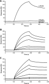
Analysis by SPR spectroscopy of the interaction of the three human ficolins with various immobilized BSA glycoconjugates. L-ficolin (A) and H-ficolin (B) at a concentration of 4.2 μg/ml were injected over immobilized GlcNAc-, Gal-, and GalNAc-BSA (3800–4200 RU) in 150 mm NaCl, 20 mm Hepes, pH 7.4, containing 0.005% surfactant P20 at a flow rate of 20 μl/min in the presence of 1 mm CaCl2 (solid lines) or 1 mm EDTA (dashed lines). M-ficolin (C) at a concentration of 1.1 μg/ml was injected over immobilized GlcNAc-, Gal-, GalNAc-, LacNAc-, and Neu5Acα2-3LacNAc-BSA (3800–4200 RU) under the same conditions in the presence of 1 mm CaCl2. The specific signal shown was obtained by subtracting the background signal recorded over a surface with immobilized BSA.
FIGURE 2.
FIGURE 2.
Glycan array screening of human ficolins. Version 3.0 of the printed array of the Consortium for Functional Glycomics was probed with L-ficolin (200 μg/ml) (A), H-ficolin (200 μg/ml) (B), M-ficolin (100 μg/ml) (C), and M-ficolin (1 μg/ml) (D). Error bars represent the means ± S.D. of four measurements. Glycans yielding significant binding are shown with their numbers and structures, and the glycan groups defined in the text are indicated. One representative experiment of three is shown. The asterisk indicates non-reproducible binding from one experiment to the other.
FIGURE 3.
FIGURE 3.
SPR analysis of the interaction of ficolins with immobilized heparin. A, L-, H- and M-ficolins were injected at a concentration of 1.7 μg/ml over immobilized 6-kDa biotinylated heparin (50 RU) as described under “Experimental Procedures.” B, L-ficolin was injected at 6 concentrations ranging from 0.25 to 4 nm (from bottom to top) over immobilized heparin. C, the FBG domain of L-ficolin was injected at 6 concentrations ranging from 15 to 240 nm (from bottom to top) over immobilized heparin. Results are representative of two independent experiments.
FIGURE 4.
FIGURE 4.
A, shown is SDS-PAGE analysis of the M-ficolin variants; Coomassie Blue staining of unreduced wild-type M-ficolin (lane 1) and mutants G221F (lane 2), A256V (lane 3), G221F/A256V (lane 4), and Y271F (lane 5). B, shown is analysis by SPR spectroscopy of the interaction of the M-ficolin variants with immobilized Neu5Acα2-3LacNAc-BSA. Immobilization of the BSA glycoconjugate and injection of 12 nm M-ficolin (protomer concentration) were performed as described under “Experimental Procedures.” WT, wild type. C, analysis of the ability of the M-ficolin variants to activate the lectin complement pathway. Increasing concentrations of each M-ficolin variant were added to ficolin-depleted human serum in microtiter wells coated with 50 μg/ml acetylated BSA, and the resulting mannan-binding lectin-associated serine protease-2 C4-cleaving activity was measured by a C4b deposition assay as described under “Experimental Procedures.” Results are expressed as the means ± S.D. of three independent experiments.
FIGURE 5.
FIGURE 5.
Effect of M-ficolin mutations on its ability to bind sialylated glycans. Binding of wild-type (WT) M-ficolin and its G221F, A256V, and Y271F mutants at a concentration of 5 μg/ml is shown for selected sialylated glycans of the array version 3.2. Error bars represent the means ± S.D. of four measurements. The glycan numbers of array version 3.0 are indicated in parentheses.
FIGURE 6.
FIGURE 6.
Comparative views are shown of the external S1 ligand binding site in wild-type M-ficolin (A) and in the Y271F mutant (B). A bound isopropanol molecule is shown in purple sticks. The red dashed lines represent hydrogen bonds. The electron density map (2mFo-DFc) is shown in blue.

Similar articles

Cited by

References

    1. Medzhitov R. (2007) Nature 449, 819–826 - PubMed
    1. Fearon D. T., Locksley R. M. (1996) Science 272, 50–53 - PubMed
    1. Fraser D. A., Tenner A. J. (2008) Curr. Drug Targets 9, 113–122 - PubMed
    1. Matsushita M., Fujita T. (2002) Immunobiology 205, 490–497 - PubMed
    1. Ohashi T., Erickson H. P. (2004) J. Biol. Chem. 279, 6534–6539 - PubMed

Publication types

Associated data

LinkOut - more resources