. 2010 Feb 26;285(9):6170-8.
doi: 10.1074/jbc.M109.054338. Epub 2009 Dec 23.

Role of WNT-5a in the determination of human mesenchymal stem cells into preadipocytes

Affiliations

Role of WNT-5a in the determination of human mesenchymal stem cells into preadipocytes

Roman Bilkovski et al. J Biol Chem. .

Abstract

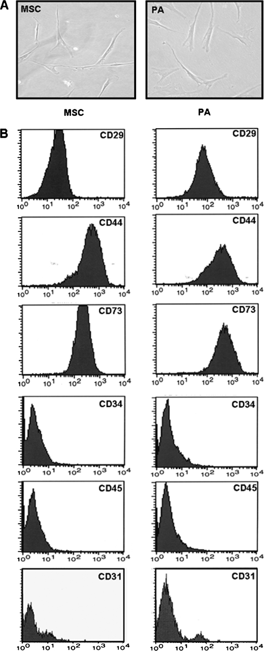
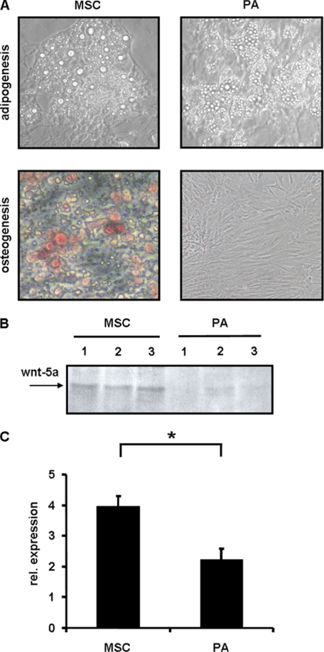
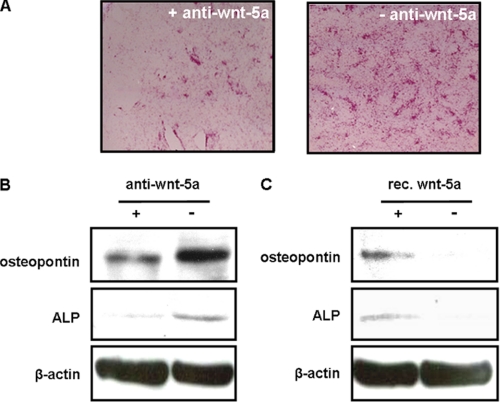
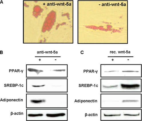
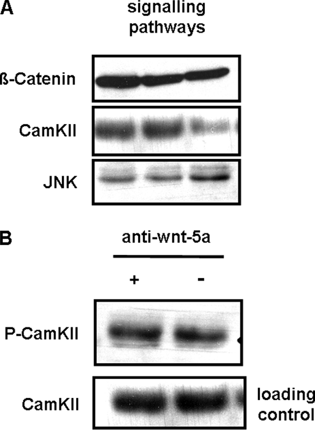
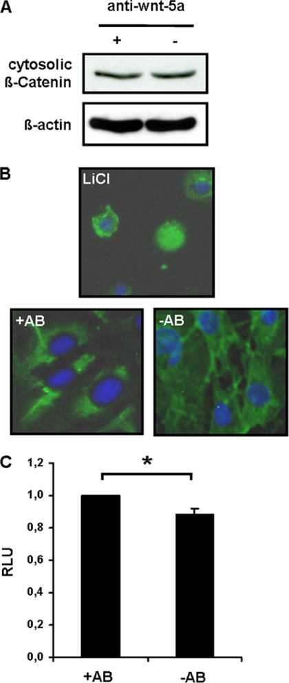
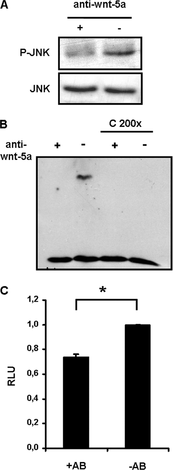
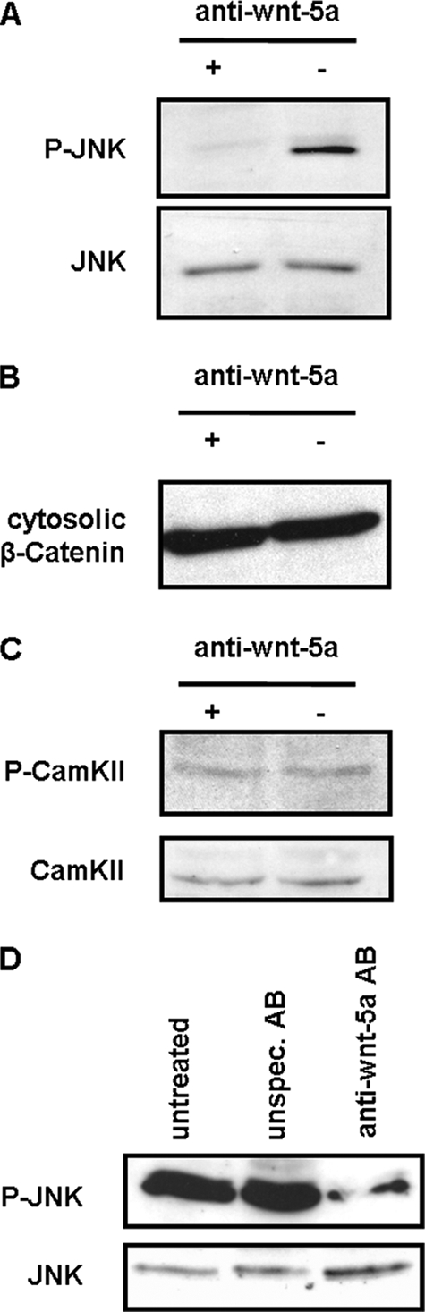
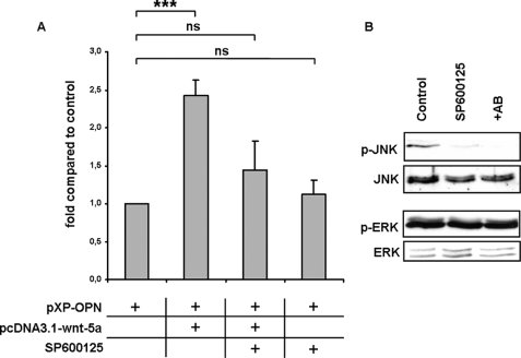
Increasing adipocyte size as well as numbers is important in the development of obesity and type 2 diabetes, with adipocytes being generated from mesenchymal precursor cells. This process includes the determination of mesenchymal stem cells (MSC) into preadipocytes (PA) and the differentiation of PA into mature fat cells. Although the process of differentiation has been highly investigated, the determination in humans is poorly understood. In this study, we compared human MSC and human committed PA on a cellular and molecular level to gain further insights into the regulatory mechanisms in the determination process. Both cell types showed similar morphology and expression patterns of common mesenchymal and hematopoietic surface markers. However, although MSC were able to differentiate into adipocytes and osteocytes, PA were only able to undergo adipogenesis, indicating that PA lost their multipotency during determination. WNT-5a expression showed significantly higher levels in MSC compared with PA suggesting that WNT-5a down-regulation might be important in the determination process. Indeed, incubation of human MSC in medium containing neutralizing WNT-5a antibodies abolished their ability to undergo osteogenesis, although adipogenesis was still possible. An opposite effect was achieved using recombinant WNT-5a protein. On a molecular level, WNT-5a was found to promote c-Jun N-terminal kinase-dependent intracellular signaling in MSC. Activation of this noncanonical pathway resulted in the induction of osteopontin expression further indicating pro-osteogenic effects of WNT-5a. Our data suggest that WNT-5a is necessary to maintain osteogenic potential of MSC and that inhibition of WNT-5a signaling therefore plays a role in their determination into PA in humans.


Figures

FIGURE 1.
FIGURE 1.
Comparison of human mesenchymal stem cells and preadipocytes with respect to morphology and expression of common cell surface markers. A, morphology of hMSC and PA, light microscopy, ×200. B, expression of surface markers. Preconfluent human MSC and PA were analyzed by FACS analysis for expression of the mesenchymal markers CD29, CD44, CD73, the hematopoietic markers CD34, CD45, and the endothelial marker CD31.
FIGURE 2.
FIGURE 2.
A, induction of adipogenesis and osteogenesis in human mesenchymal stem cells and preadipocytes. MSC and PA were incubated in induction medium for adipogenesis or osteogenesis for 7–10 days. Upper panel, adipogenesis in MSC and PA, light microscopy (×100); lower panel, osteogenesis in MSC and PA, Alizarin staining and light microscopy (×100). B and C, comparison of WNT-5a expression in human mesenchymal stem cells and preadipocytes. Preconfluent human MSC and PA were lysed by RIPA buffer, and Western blotting was performed for WNT-5a. B, representative Western blot of MSC and PA from three independent human individuals each. C, statistical analysis of Western blotting experiments of MSC or PA from n = 5 independent individuals each. Wnt-5a expression was normalized to the amount of total protein. Mean ± S.E. Student's t test; *, p < 0.05. rel., relative.
FIGURE 3.
FIGURE 3.
Wnt-5a effect on osteogenesis of human mesenchymal stem cells. Human MSC were induced to undergo osteogenesis in the presence or absence of neutralizing anti-WNT-5a antibodies or recombinant WNT-5a protein within the tissue culture medium. A, alizarin staining. B and C, Western blotting for osteopontin and alkaline phosphatase (ALP), two molecular markers for osteogenesis. β-Actin was used to normalize for total protein content. rec. = recombinant. Shown is one example of n = 3 independent experiments using different cells from independent human individuals.
FIGURE 4.
FIGURE 4.
Wnt-5a effect on adipogenesis of human mesenchymal stem cells. Human MSC were induced to undergo adipogenesis in the presence or absence of neutralizing anti-WNT-5a antibodies or recombinant WNT-5a protein within the tissue culture medium. A, Oil Red O lipid staining, microscopically imaged (×200). B and C, Western blotting of molecular markers for adipogenesis (PPAR-γ and SREBP-1c) and the adipokine adiponectin. β-Actin was used to normalize for total protein content. rec. = recombinant. Shown is one example of n = 3 independent experiments using different cells from independent human individuals.
FIGURE 5.
FIGURE 5.
A, expression of intracellular signaling molecules of the canonical and noncanonical pathways in human mesenchymal stem cells. Western blotting of whole cell lysates of MSC from three independent human individuals is shown. B, Wnt-5a does not activate the CamKII-dependent noncanonical pathway in C3H10T1/2 mesenchymal stem cells. C3H10 cells were incubated in medium containing neutralizing anti-WNT-5a antibodies or control medium for 24 h. Afterward, cells were lysed using RIPA buffer containing phosphatase inhibitors, and Western blotting was performed for phospho-CamKII (upper panel) and whole CamKII (loading control, lower panel). Shown is one example of n = 3 independent experiments.
FIGURE 6.
FIGURE 6.
Wnt-5a does not activate the canonical signaling pathway in C3H10T1/2 mesenchymal stem cells. A, comparison of the amount of cytosolic β-catenin in stem cells treated with neutralizing anti-WNT-5a antibodies for 24 h or control cells. In this experiment, cells were lysed using a hypotonic lysis buffer to separate cytosolic from membrane-bound β-catenin. β-Actin was used to normalize for total protein content. Shown is one example of n = 3 independent experiments. B, immunofluorescence for β-catenin in stem cells treated with neutralizing anti-WNT-5a antibodies (+AB) or control cells (−AB). Incubation of cells in LiCl was used as a positive control for translocation of β-catenin into the nucleus. Shown is one example of n = 3 independent experiments. C, promoter-reporter gene analysis using the TOPFLASH plasmid in cells treated with neutralizing anti-WNT-5a antibodies (+AB) and controls (−AB). pRL-CMV was used for normalization of transfection efficiency and unspecific promoter effects in this experiment. Mean ± S.E. of n = 5 independent experiments. Student's t test; *, p < 0.05. RLU, relative luciferase units.
FIGURE 7.
FIGURE 7.
Wnt-5a activates the JNK-dependent noncanonical signaling pathway in C3H10T1/2 mesenchymal stem cells. A, comparison of the amount of phosphorylated JNK in control cells and C3H10T1/2 cells incubated in medium containing neutralizing anti-WNT-5a antibodies (upper panel). Amount of whole JNK (phosphorylated and nonphosphorylated) was used as a loading control (lower panel). Shown is one example of n = 3 independent experiments. B, electromobility gel shift assay for c-Jun binding to an oligonucleotide containing a c-Jun-binding site. + = nuclear proteins from cells treated with neutralizing anti-WNT-5a antibodies; − = nuclear proteins from control cells. Competitive reaction was performed under identical conditions adding 200-fold molar ratio of unlabeled oligonucleotides. Samples were separated by 5% PAGE, and final analysis was performed according to the LightShift® chemiluminescent electromobility gel shift assay kit (Pierce). Shown is one example of n = 3 independent experiments. C, promoter-reporter gene analysis using a pGL plasmid containing the AP1 promoter in C3H10T1/2 cells treated with neutralizing anti-WNT-5a antibodies (+AB) and control cells (−AB). The AP1 promoter is a known target of c-Jun. pRL-CMV was used for normalization of transfection efficiency and unspecific promoter effects. Mean ± S.E. of n = 5 independent experiments. Student's t test; *, p < 0.05. RLU, relative luciferase units.
FIGURE 8.
FIGURE 8.
Wnt-5a specifically activates the JNK-dependent noncanonical signaling pathway in human mesenchymal stem cells. A–C, comparison of the amount of phospho-JNK, β-catenin, and phospho-CamKII in control cells and human mesenchymal stem cells incubated in medium containing neutralizing anti-WNT-5a antibodies. Shown is one example of n = 3 independent experiments. D, to exclude unspecific effects due to the neutralizing antibody, human mesenchymal stem cells were either untreated or treated with an unspecific control antibody (unspec. AB) of the same subclass or a specific antibody for WNT-5a (anti-WNT-5a AB) for 24 h. Afterward, cells were lysed using RIPA buffer containing phosphatase inhibitors, and Western blotting was performed for phospho-JNK (upper panel) as well as total JNK (lower panel). Shown is one example of n = 4 independent experiments.
FIGURE 9.
FIGURE 9.
Wnt-5a induces osteopontin promoter activity in C3H10T1/2 mesenchymal stem cells via JNK signaling pathway. A, C3H10T1/2 cells were transiently co-transfected with 500 ng of an expression plasmid containing the coding sequence of human WNT-5a (pcDNA3.1-WNT-5a) and 200 ng of a luciferase construct (pXP2-OPN) with −88 to + 79 bp of the proximal osteopontin promoter containing a c-Jun-binding site (TGAGCCA) kindly provided by David T. Denhardt (36). 10 ng of pRL-CMV were co-transfected for normalization of transfection efficiency and unspecific promoter effects. Cells were lysed after 24–48 h, and luciferase activity was measured by Dual-Luciferase reporter gene assay (Promega). To test that the effect of WNT-5a on osteopontin promoter activity is mediated via the JNK signaling pathway, luciferase experiments were performed in the presence of the pharmacological JNK inhibitor SP600125 as indicated in the figure. ***, p < 0.001 by Student's t test of n = 8 independent experiments. Mean ± S.E. ns is nonsignificant. B, Western blot for phospho-JNK and total JNK as well as phospho-ERK and total ERK in pre-confluent mesenchymal stem cells treated with the JNK inhibitor SP600125 and anti-WNT-5a (AB) antibodies for 24 h to demonstrate that SP600125 is specific for the JNK signaling pathway in these cells. Shown is one example of n = 3 independent experiments.

References

    1. Mokdad A. H., Ford E. S., Bowman B. A., Dietz W. H., Vinicor F., Bales V. S., Marks J. S. (2003) JAMA 289, 76–79 - PubMed
    1. Kahn B. B., Flier J. S. (2000) J. Clin. Invest. 106, 473–481 - PMC - PubMed
    1. Bowers R. R., Lane M. D. (2007) Cell Cycle 6, 385–389 - PubMed
    1. Tchoukalova Y. D., Sarr M. G., Jensen M. D. (2004) Am. J. Physiol. Regul. Integr. Comp. Physiol. 287, R1132–R1140 - PubMed
    1. Tang W., Zeve D., Suh J. M., Bosnakovski D., Kyba M., Hammer R. E., Tallquist M. D., Graff J. M. (2008) Science 322, 583–586 - PMC - PubMed

Publication types