

PGC-1alpha plays a functional role in exercise-induced mitochondrial biogenesis and angiogenesis but not fiber-type transformation in mouse skeletal muscle
- PMID: 20032509
- PMCID: PMC3353735
- DOI: 10.1152/ajpcell.00481.2009
PGC-1alpha plays a functional role in exercise-induced mitochondrial biogenesis and angiogenesis but not fiber-type transformation in mouse skeletal muscle
Abstract
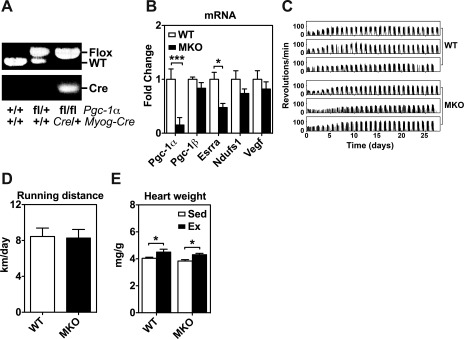
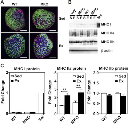
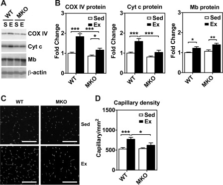
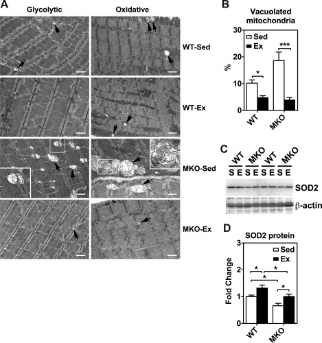
Endurance exercise stimulates peroxisome proliferator-activated receptor gamma coactivator-1alpha (PGC-1alpha) expression in skeletal muscle, and forced expression of PGC-1alpha changes muscle metabolism and exercise capacity in mice. However, it is unclear if PGC-1alpha is indispensible for endurance exercise-induced metabolic and contractile adaptations in skeletal muscle. In this study, we showed that endurance exercise-induced expression of mitochondrial enzymes (cytochrome oxidase IV and cytochrome c) and increases of platelet endothelial cell adhesion molecule-1 (PECAM-1, CD31)-positive endothelial cells in skeletal muscle, but not IIb-to-IIa fiber-type transformation, were significantly attenuated in muscle-specific Pgc-1alpha knockout mice. Interestingly, voluntary running effectively restored the compromised mitochondrial integrity and superoxide dismutase 2 (SOD2) protein expression in skeletal muscle in Pgc-1alpha knockout mice. Thus, PGC-1alpha plays a functional role in endurance exercise-induced mitochondrial biogenesis and angiogenesis, but not IIb-to-IIa fiber-type transformation in mouse skeletal muscle, and the improvement of mitochondrial morphology and antioxidant defense in response to endurance exercise may occur independently of PGC-1alpha function. We conclude that PGC-1alpha is required for complete skeletal muscle adaptations induced by endurance exercise in mice.
Figures
Similar articles
-
MicroRNA-23a has minimal effect on endurance exercise-induced adaptation of mouse skeletal muscle.Pflugers Arch. 2015 Feb;467(2):389-98. doi: 10.1007/s00424-014-1517-z. Epub 2014 Apr 23. Pflugers Arch. 2015. PMID: 24756198
-
Sirtuin 1 (SIRT1) deacetylase activity is not required for mitochondrial biogenesis or peroxisome proliferator-activated receptor-gamma coactivator-1alpha (PGC-1alpha) deacetylation following endurance exercise.J Biol Chem. 2011 Sep 2;286(35):30561-30570. doi: 10.1074/jbc.M111.261685. Epub 2011 Jul 11. J Biol Chem. 2011. PMID: 21757760 Free PMC article.
-
The role of PGC-1alpha on mitochondrial function and apoptotic susceptibility in muscle.Am J Physiol Cell Physiol. 2009 Jul;297(1):C217-25. doi: 10.1152/ajpcell.00070.2009. Epub 2009 May 13. Am J Physiol Cell Physiol. 2009. PMID: 19439529
-
PGC-1alpha regulation by exercise training and its influences on muscle function and insulin sensitivity.Am J Physiol Endocrinol Metab. 2010 Aug;299(2):E145-61. doi: 10.1152/ajpendo.00755.2009. Epub 2010 Apr 6. Am J Physiol Endocrinol Metab. 2010. PMID: 20371735 Free PMC article. Review.
-
PGC-1alpha-mediated adaptations in skeletal muscle.Pflugers Arch. 2010 Jun;460(1):153-62. doi: 10.1007/s00424-010-0834-0. Epub 2010 Apr 19. Pflugers Arch. 2010. PMID: 20401754 Review.
Cited by
-
The Drosophila PGC-1α homolog spargel modulates the physiological effects of endurance exercise.PLoS One. 2012;7(2):e31633. doi: 10.1371/journal.pone.0031633. Epub 2012 Feb 13. PLoS One. 2012. PMID: 22348115 Free PMC article.
-
Combined whole-body vibration, resistance exercise, and sustained vascular occlusion increases PGC-1α and VEGF mRNA abundances.Eur J Appl Physiol. 2013 Apr;113(4):1081-90. doi: 10.1007/s00421-012-2524-4. Epub 2012 Oct 20. Eur J Appl Physiol. 2013. PMID: 23086295 Clinical Trial.
-
Effect of Normobaric Hypoxia on Alterations in Redox Homeostasis, Nitrosative Stress, Inflammation, and Lysosomal Function following Acute Physical Exercise.Oxid Med Cell Longev. 2022 Feb 25;2022:4048543. doi: 10.1155/2022/4048543. eCollection 2022. Oxid Med Cell Longev. 2022. PMID: 35251471 Free PMC article.
-
Regulation of exercise-induced fiber type transformation, mitochondrial biogenesis, and angiogenesis in skeletal muscle.J Appl Physiol (1985). 2011 Jan;110(1):264-74. doi: 10.1152/japplphysiol.00993.2010. Epub 2010 Oct 28. J Appl Physiol (1985). 2011. PMID: 21030673 Free PMC article. Review.
-
Perm1 regulates CaMKII activation and shapes skeletal muscle responses to endurance exercise training.Mol Metab. 2019 May;23:88-97. doi: 10.1016/j.molmet.2019.02.009. Epub 2019 Feb 27. Mol Metab. 2019. PMID: 30862473 Free PMC article.
References
-
- Adhihetty PJ, Uguccioni G, Leick L, Hidalgo J, Pilegaard H, Hood DA. The role of PGC-1α on mitochondrial function and apoptotic susceptibility in muscle. Am J Physiol Cell Physiol 297: C217–C225, 2009 - PubMed
-
- Akimoto T, Pohnert SC, Li P, Zhang M, Gumbs C, Rosenberg PB, Williams RS, Yan Z. Exercise stimulates Pgc-1alpha transcription in skeletal muscle through activation of the p38 MAPK pathway. J Biol Chem 280: 19587–19593, 2005 - PubMed
-
- Akimoto T, Ribar TJ, Williams RS, Yan Z. Skeletal muscle adaptation in response to voluntary running in Ca2+/calmodulin-dependent protein kinase IV-deficient mice. Am J Physiol Cell Physiol 287: C1311–C1319, 2004 - PubMed
-
- Akimoto T, Sorg BS, Yan Z. Real-time imaging of peroxisome proliferator-activated receptor-gamma coactivator-1α promoter activity in skeletal muscles of living mice. Am J Physiol Cell Physiol 287: C790–C796, 2004 - PubMed
MeSH terms
Substances
Grants and funding
LinkOut - more resources
Full Text Sources
Molecular Biology Databases
Miscellaneous