. 2010 Mar;298(3):C656-64.
doi: 10.1152/ajpcell.00456.2009. Epub 2009 Dec 23.

Ca2+ influx via TRPC channels induces NF-kappaB-dependent A20 expression to prevent thrombin-induced apoptosis in endothelial cells

Affiliations

Ca2+ influx via TRPC channels induces NF-kappaB-dependent A20 expression to prevent thrombin-induced apoptosis in endothelial cells

Prabhakar B Thippegowda et al. Am J Physiol Cell Physiol. 2010 Mar.

Abstract

NF-kappaB signaling is known to induce the expression of antiapoptotic and proinflammatory genes in endothelial cells (ECs). We have shown recently that Ca(2+) influx through canonical transient receptor potential (TRPC) channels activates NF-kappaB in ECs. Here we show that Ca(2+) influx signal prevents thrombin-induced apoptosis by inducing NF-kappaB-dependent A20 expression in ECs. Knockdown of TRPC1 expressed in human umbilical vein ECs with small interfering RNA (siRNA) suppressed thrombin-induced Ca(2+) influx and NF-kappaB activation in ECs. Interestingly, we observed that thrombin induced >25% of cell death (apoptosis) in TRPC1-knockdown ECs whereas thrombin had no effect on control or control siRNA-transfected ECs. To understand the basis of EC survival, we performed gene microarray analysis using ECs. Thrombin stimulation increased only a set of NF-kappaB-regulated genes 3- to 14-fold over basal levels in ECs. Expression of the antiapoptotic gene A20 was the highest among these upregulated genes. Like TRPC1 knockdown, thrombin induced apoptosis in A20-knockdown ECs. To address the importance of Ca(2+) influx signal, we measured thrombin-induced A20 expression in control and TRPC1-knockdown ECs. Thrombin-induced p65/RelA binding to A20 promoter-specific NF-kappaB sequence and A20 protein expression were suppressed in TRPC1-knockdown ECs compared with control ECs. Furthermore, in TRPC1-knockdown ECs, thrombin induced the expression of proapoptotic proteins caspase-3 and BAX. Importantly, thrombin-induced apoptosis in TRPC1-knockdown ECs was prevented by adenovirus-mediated expression of A20. These results suggest that Ca(2+) influx via TRPC channels plays a critical role in the mechanism of cell survival signaling through A20 expression in ECs.


Figures

Fig. 1.
Fig. 1.
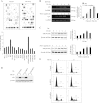
Canonical transient receptor potential channel 1 (TRPC1) knockdown prevents thrombin-induced Ca2+ influx and nuclear factor-κB (NF-κB) activation and induces apoptosis in endothelial cells (ECs). A: ECs were transfected with either 100 nM scrambled small interfering RNA (Sc-siRNA) or TRPC1 siRNA. At 48 h after transfection, cells were lysed and immunoblotted (IB) with anti-TRPC1 polyclonal Ab (top). The membrane was stripped and blotted with anti-β-actin (bottom). B: control ECs and ECs transfected with 100 nM Sc-siRNA or TRPC1-siRNA were used to measure thrombin (Thr)-induced store Ca2+ release and Ca2+ release-activated Ca2+ influx. The results were compared with control cells (not transfected). The experiment was repeated 3 times with similar results. C: control ECs and ECs transfected with TRPC1-siRNA (100 nM) were exposed to thrombin (50 nM) or TNF-α (100 U) for the indicated time periods, and then electrophoretic mobility shift assay (EMSA) was performed. The experiment was repeated 3 times with similar results. D: ECs transfected with either 100 nM Sc-siRNA or TRPC1-siRNA were treated with or without thrombin (50 nM) for various time intervals. The whole cell lysate was immunoblotted with anti-phospho-ERK1/2 and anti-ERK1/2 antibody. E: ECs were transfected with either 100 nM Sc-siRNA or TRPC1-siRNA. At 48 h after transfection, cells were treated with thrombin for 0, 6, and 12 h. After thrombin treatment, cells were collected and stained with 5 μg of Hoechst 33342 and subjected to fluorescence-activated cell sorting (FACS) analysis to determine apoptosis in ECs. The experiment was repeated 3 times. Results from representative experiments are shown.
Fig. 2.
Fig. 2.
Protease-activated receptor (PAR)-1 signaling-induced A20 expression prevents apoptosis in ECs. A: ECs grown to confluence were challenged with thrombin (50 nM) for 4 h. After thrombin treatment, total RNA isolated was used for microarray analysis. The experiment was repeated twice with duplicates. Results from representative experiments are shown (top). The gene induction fold increase was calculated over basal with housekeeping gene GAPDH expression levels (bottom). B: ECs were treated with or without thrombin (50 nM) or PAR-1 agonist peptide (40 μM) for various time intervals. Total RNA was isolated and RT-PCR was performed to determine A20 and GAPDH expressions. The experiment was repeated 4 times, and results from a representative experiment are shown (left). RT-PCR gel images were quantified, and A20 mRNA induction fold increase over basal was calculated by measuring the ratio of A20 to GAPDH (right). *P < 0.05, significantly different from control. C: after thrombin (50 nM) or PAR-1 agonist peptide (40 μM) treatment, cell lysates were immunoblotted with anti-A20 MAb. The membrane was stripped and reprobed with anti-β-actin Ab. The experiment was repeated 4 times, and results from representative experiment are shown (left). Immunoblots were quantified by densitometry, and A20 protein induction fold increase over basal was calculated by measuring the ratio of A20 to β-actin (right). *P < 0.05, significantly different from control. D: ECs were transfected with either 100 nM Sc-siRNA or A20-siRNA. At 48 h after transfection cells were immunoblotted with anti-A20 MAb. E: ECs were transfected with either 100 nM Sc-siRNA or A20-siRNA as described above. At 48 h after transfection, cells were treated with or without thrombin (50 nM) for 0 and 12 h. After thrombin treatment, cells were used for FACS analysis to determine apoptosis. The experiment was repeated 3 times, and results shown are from a representative experiment.
Fig. 3.
Fig. 3.
Pharmacological inhibition of Ca2+ influx or knockdown of p65/RelA prevents thrombin-induced A20 expression in ECs. A: human (h)A20 promoter-specific NF-κB oligonucleotide probe used for EMSA. B: ECs grown to confluence were incubated in serum-free medium for 2 h. After this incubation period, cells were exposed to 2-aminoethoxydiphenyl borate (2-APB; 75 μM) for 30 min and then treated with thrombin (50 nM) for up to 60 min. After thrombin treatment, the nuclear extracts prepared were used for EMSA utilizing hA20 promoter-specific NF-κB probe (left). Nuclear extracts prepared from ECs exposed to thrombin for 60 min were incubated with either anti-p65 or anti-p50 antibodies for 30 min before addition of labeled probe (right). The experiment was repeated 3 times, and results shown are from a representative experiment. C: ECs were pretreated with 2-APB (75 μM) for 30 min and then challenged with thrombin (50 nM) up to 4 h. After thrombin treatment, total RNA was isolated and RT-PCR was performed to determine A20 and GAPDH transcript expression levels. The experiment was repeated 4 times, and results from a representative experiment are shown (top). RT-PCR gel images were quantified, and the A20 mRNA induction fold increase over basal was calculated by measuring the ratio of A20 to GAPDH (bottom). *P < 0.05, significantly different from control. D: ECs pretreated with 2-APB as described in C were challenged with thrombin (50 nM) for up to 4 h. After thrombin treatment, cell lysates were immunoblotted with anti-A20 MAb. The membrane was stripped and reprobed with anti-β-actin Ab. A representative blot is shown at left. The blots were quantified as described in Fig. 2C. Results are means ± SE from 4 separate experiments shown (right). *P < 0.05, significantly different from control. E: ECs were transfected with either 50 nM Sc-siRNA or p65-siRNA. At 48 h after transfection, cell lysates were immunoblotted with anti-p65 Ab (top). The membrane was stripped and reprobed with anti-β-actin Ab (bottom). F: ECs transfected with either 50 nM Sc-siRNA or p65-siRNA were challenged with thrombin (50 nM) for up to 4 h, and then cell lysates were immunoblotted with anti-A20 MAb or anti-β-actin Ab. A representative blot is shown at left. The results from 4 separate experiments were quantified by densitometry, and the A20 protein induction fold increase over basal was calculated by measuring the ratio of A20 to β-actin (right). *P < 0.05, significantly different from control.
Fig. 4.
Fig. 4.
TRPC1 expression is required for thrombin-induced A20 expression in ECs. A: control ECs and ECs transfected with 100 nM Sc-siRNA or TRPC1-siRNA were exposed to thrombin (50 nM) or TNF-α (100 U) for the indicated time periods. After this treatment, EMSA was performed with A20 promoter-specific NF-κB probe as shown in Fig. 3B. The experiment was repeated 3 times with similar results, and results from representative experiments are shown. B: ECs were transfected with either 100 nM Sc-siRNA or TRPC1-siRNA. At 48 h after transfection, cells were exposed to thrombin (50 nM) for the indicated time periods. Total RNA was isolated, and RT-PCR was performed to determine A20 and GAPDH transcript expression levels. The experiment was repeated 3 times, and results from representative experiments are shown. C: control ECs and ECs transfected with 100 nM Sc-siRNA or TRPC1 siRNA were exposed to thrombin as described above. After thrombin exposure, cells were lysed and immunoblotted with anti-A20 MAb (top). The membrane was stripped and probed with β-actin Ab (bottom). D: control ECs and ECs transfected with 100 nM Sc-siRNA or TRPC1-siRNA were exposed to TNF-α (100 U) for 0 and 4 h. After TNF-α exposure, cells were used to measure A20 mRNA expression by RT-PCR. The experiment was repeated 3 times, and results from representative experiments are shown. E: control ECs and ECs transfected with TRPC1 siRNA as described above were treated with thrombin (50 nM) for 0, 6, and 12 h. After thrombin treatment, cells were lysed and immunoblotted with anti-BAX and anti-caspase-3 antibodies. The experiment was repeated 3 times, and results from representative experiments are shown.
Fig. 5.
Fig. 5.
Adenovirus (Ad)-mediated A20 expression rescues TRPC1 knockdown phenotype in ECs. A: ECs were transfected with or without 100 nM TRPC1 siRNA as described in Fig. 4. At 48 h after transfection, ECs were infected with either Ad-null or Ad-A20 virus (100 pfu/cell) for 12 h, and then ECs were challenged with thrombin (50 nM) for 0 and 4 h. After thrombin treatment, cell lysates were immunoblotted with anti-A20 MAb. The experiment was repeated 3 times, and results from representative experiments are shown. B: ECs were transfected with or without 100 nM TRPC1-siRNA. At 48 h after transfection, ECs were infected with either Ad-null or Ad-A20 virus (100 pfu/cell) for 24 h, and then ECs were challenged with thrombin (50 nM) for 0 and 12 h. After thrombin treatment, cells were used for FACS analysis. Percentages of apoptotic cells are indicated on the histograms. The experiment was repeated 3 times, and results from representative experiments are shown.

Similar articles

Cited by

References

    1. Bair AM, Thippegowda PB, Freichel M, Cheng N, Ye RD, Vogel SM, Yu Y, Flockerzi V, Malik AB, Tiruppathi C. Ca2+ entry via TRPC channels is necessary for thrombin-induced NF-kappaB activation in endothelial cells through AMP-activated protein kinase and protein kinase Cdelta. J Biol Chem 284: 563–574, 2009 - PMC - PubMed
    1. Boone DL, Turer EE, Lee EG, Ahmad RC, Wheeler MT, Sui C, Hurley P, Chien M, Chai S, Hitotsumatsu O, McNally E, Pickart C, Ma A. The ubiquitin-modifying enzyme A20 is required for termination of Toll-like receptor responses. Nat Immunol 10: 1052–1060, 2004 - PubMed
    1. Chalmers CJ, Balmanno K, Hadfield K, Ley R, Cook SJ. Thrombin inhibits Bim (Bcl-2-interacting mediator of cell death) expression and prevents serum-withdrawal-induced apoptosis via protease-activated receptor 1. Biochem J 375: 99–109, 2003 - PMC - PubMed
    1. Cheng KT, Liu X, Ong HL, Ambudkar IS. Functional requirement for Orai1 in store-operated TRPC1-STIM1 channels. J Biol Chem 283: 12935–129340, 2008 - PMC - PubMed
    1. Cooper JT, Stroka DM, Brostjan C, Palmetshofer A, Bach FH, Ferran C. A20 blocks endothelial cell activation through a NF-kappaB-dependent mechanism. J Biol Chem 271: 18068–18073, 1996 - PubMed

Publication types

MeSH terms