. 2010 Mar;298(3):C665-78.
doi: 10.1152/ajpcell.00153.2009. Epub 2009 Dec 23.

CYP1B1 and endothelial nitric oxide synthase combine to sustain proangiogenic functions of endothelial cells under hyperoxic stress

Affiliations

CYP1B1 and endothelial nitric oxide synthase combine to sustain proangiogenic functions of endothelial cells under hyperoxic stress

Yixin Tang et al. Am J Physiol Cell Physiol. 2010 Mar.

Abstract

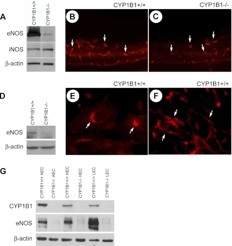
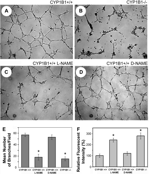
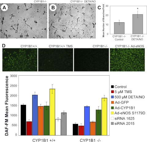
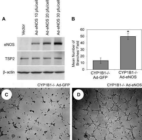
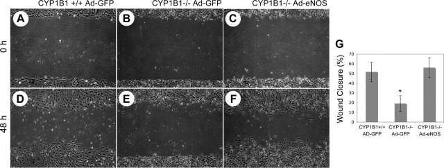
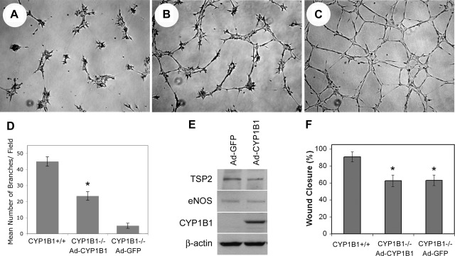
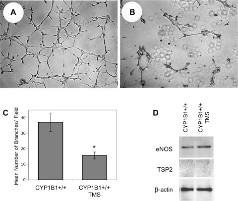
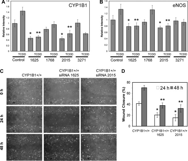
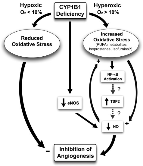
We have recently shown that deletion of constitutively expressed CYP1B1 is associated with attenuation of retinal endothelial cell (EC) capillary morphogenesis (CM) in vitro and angiogenesis in vivo. This was largely caused by increased intracellular oxidative stress and increased production of thrombospondin-2, an endogenous inhibitor of angiogenesis. Here, we demonstrate that endothelium nitric oxide synthase (eNOS) expression is dramatically decreased in the ECs prepared from retina, lung, heart, and aorta of CYP1B1-deficient (CYP1B1(-/-)) mice compared with wild-type (CYP1B1(+/+)) mice. The eNOS expression was also decreased in retinal vasculature of CYP1B1(-/-) mice. Inhibition of eNOS activity in cultured CYP1B1(+/+) retinal ECs blocked CM and was concomitant with increased oxidative stress, like in CYP1B1(-/-) retinal ECs. In addition, expression of eNOS in CYP1B1(-/-) retinal ECs or their incubation with a nitric oxide (NO) donor enhanced NO levels, lowered oxidative stress, and improved cell migration and CM. Inhibition of CYP1B1 activity in the CYP1B1(+/+) retinal ECs resulted in reduced NO levels and attenuation of CM. In contrast, expression of CYP1B1 increased NO levels and enhanced CM of CYP1B1(-/-) retinal ECs. Furthermore, attenuation of CYP1B1 expression with small interfering RNA proportionally lowered eNOS expression and NO levels in wild-type cells. Together, our results link CYP1B1 metabolism in retinal ECs with sustained eNOS activity and NO synthesis and/or bioavailability and low oxidative stress and thrombospondin-2 expression. Thus CYP1B1 and eNOS cooperate in different ways to lower oxidative stress and thereby to promote CM in vitro and angiogenesis in vivo.


Figures

Fig. 1.
Fig. 1.
Decreased eNOS expression in the absence of CYP1B1. A: lysates prepared from CYP1B1+/+ and CYP1B1−/− retinal endothelial cells (ECs) were analyzed for eNOS and iNOS expression by Western blotting. Antibody to β-catenin was used as loading control. Please note a significant decrease in eNOS levels in retinal ECs from CYP1B1−/− mice. Frozen eye sections prepared from p17 CYP1B1+/+ (B) and CYP1B1−/− mice (C) during oxygen-induced ischemic retinopathy (OIR) were also stained with antibodies to eNOS. Eye sections were stained, and images were obtained under identical conditions. Please note the marked decrease in fluorescence staining of eNOS in CYP1B1−/− retinal vasculature compared with CYP1B1+/+ retinal vasculature. D: Western blot analysis of whole retinal extracts prepared from p21 CYP1B1+/+ and CYP1B1−/− mice. Antibody to β-actin was used as loading control. Please note a significant decrease in eNOS level in retina extracts from CYP1B1−/− mice compared with CYP1B1+/+ mice. These experiments were repeated twice with eyes from 4 different mice with similar results. E and F: eNOS localization in CYP1B1+/+ retinal ECs at subconfluent (E) and confluent (F) states. Cells were grown on glass coverslips and stained for eNOS as described in materials and methods. Please note cellular localization of eNOS (arrows), which changes with degree of confluence. Comparisons of CYP1B1 and eNOS expression in CYP1B1+/+ and CYP1B1−/− ECs from aorta, heart, and lung were made by Western blot analysis of cell lysates (G). Antibody to β-actin was used as loading control. Please note a significant decrease in eNOS level in cell extracts from CYP1B1−/− ECs compared with CYP1B1+/+ ECs.
Fig. 2.
Fig. 2.
eNOS activity is essential for capillary morphogenesis of CYP1B1+/+ retinal ECs in Matrigel. CYP1B1+/+ (A) and CYP1B1−/− (B) retinal ECs were plated in Matrigel without (A and B) or with (C) nitro-l-arginine methyl ester (l-NAME; 3 mM), an inhibitor of eNOS activity. After 17 h of incubation, CYP1B1+/+ retinal ECs formed a well-organized capillary-like network (A), whereas the ability of these cells to undergo capillary morphogenesis was attenuated upon incubation with l-NAME (C). These results are similar to what we observed in CYP1B1−/− retinal ECs, which express little or no eNOS (B). The inactive analog of eNOS inhibitor, nitro-d-arginine methyl ester (d-NAME; 3 mM), had no effect on CYP1B1+/+ retinal EC capillary morphogenesis (D). E: quantitative assessment of the data. Data in each bar are mean number of branches per 5 high-power fields (×100). Error bars indicate SD (*P < 0.05; n = 5). Reactive oxygen species (ROS) levels were determined in CYP1B1+/+ retinal ECs incubated with or without l-NAME or d-NAME as described in materials and methods (F). Please note a significant increase in oxidative stress of CYP1B1+/+ cells incubated with l-NAME compared with d-NAME, which was comparable with levels in CYP1B1−/− retinal ECs (*P < 0.05; n = 15).
Fig. 3.
Fig. 3.
Restoration of capillary morphogenesis in CYP1B1−/− retinal ECs upon incubation with a nitric oxide (NO) donor, diethylenetriamine-NO (DETA/NO). CYP1B1−/− retinal ECs were preincubated with solvent control (A) or DETA/NO (20 μM; B) for 24 h, plated in Matrigel with freshly added DETA/NO (20 μM), and photographed after 17 h of incubation at 37°C. The capillary morphogenesis of CYP1B1−/− retinal ECs was significantly improved in the presence of the NO donor. C: quantitative assessment of the data. Data in each bar are mean number of branches per 5 high-power fields (×100). Error bars indicate SD (*P < 0.05; n = 5). D: NO levels were assessed in CYP1B1+/+ and CYP1B1−/− retinal ECs under various conditions as described in materials and methods. D, top: representative fluorescent images of retinal ECs incubated with DAF-FM. Please note reduced NO level in CYP1B1+/+ retinal ECs incubated with tetramethoxystilbene (TMS) and CYP1B1−/− retinal ECs. Expression of eNOS in CYP1B1−/− retinal ECs restored NO levels to near normal levels. NO levels were also assessed in retinal ECs incubated with TMS or DETA/NO or infected with viruses expressing GFP (control), CYP1B1, eNOS, or CYP1B1-specific small interfering RNAs (siRNAs). D, bottom: quantitative assessment of data. Data in each bar are mean fluorescence intensities of 3 separate experiments. Error bars indicate SD (P < 0.05 for wild-type cells expressing CYP1B1 siRNA or incubated with TMS compared with control or null cells expressing CYP1B1, eNOS, or incubated with DETA/NO compared with control).
Fig. 4.
Fig. 4.
Reexpression of eNOS restored capillary morphogenesis of CYP1B1−/− retinal ECs. A: Western blot analysis of whole cell lysates prepared from CYP1B1−/− retinal ECs infected with the adenoviruses expressing a constitutively active form of eNOS (CA-eNOS) or empty vector at different multiplicity of infection. Cells were lysed 2 days after infection, and eNOS levels were determined by Western blot. Antibody to β-actin was used to control for loading. Cells infected with control (C) or CA-eNOS (D) adenoviruses (20 pfu/cell) were plated in Matrigel. Images were obtained 17 h after incubation at 37°C. Expression of the CA-eNOS in CYP1B1−/− retinal ECs restored their ability to undergo capillary morphogenesis. B: quantitative assessment of the data. Data in each bar are mean number of branches per 5 high-power fields (×100). Error bars indicate SD (*P < 0.05; n = 5).
Fig. 5.
Fig. 5.
Reexpression of eNOS restored the migration of CYP1B1−/− retinal ECs. Confluent cultures of CYP1B1+/+ retinal ECs infected with control (A and D), CYP1B1−/− retinal ECs infected with control (B and E), and CYP1B1−/− retinal ECs infected with CA-eNOS (C and F) adenoviruses were wounded with a 1-ml micropipette tip 1 day after infection. Pictures were taken at 0 h (immediately after wounding) or 48 h after wounding. G: quantitative assessment of the data. Data in each bar are mean percentages of wound closed. Error bars indicate SD (*P < 0.05, n = 3). Please note that the CYP1B1−/− retinal ECs with restored eNOS expression/activity, similar to CYP1B1+/+ vector control cells, migrated significantly faster than CYP1B1−/− vector control cells.
Fig. 6.
Fig. 6.
Reexpression of CYP1B1 in CYP1B1−/− retinal ECs restored capillary morphogenesis and lowered thrombospondin-2 (TSP2) level but did not affect eNOS level. Semiconfluent cultures of CYP1B1−/− retinal ECs were infected with control (A) or CYP1B1 expressing adenoviruses (B), and their ability to undergo capillary morphogenesis was assessed in Matrigel. C: capillary morphogenesis of CYP1B1+/+ cells. D: quantitative assessment of the date. Please note a significant enhancement of capillary morphogenesis in null cells expressing CYP1B1 (*P < 0.05; n = 5). E: Western blot of cell lysates prepared from CYP1B1 or vector control-infected cell. Please note decreased expression of TSP2 in null cells expressing CYP1B1 compared with vector control cells. No differences in the levels of eNOS were observed. β-Actin staining was used to control for loading. The effect of CYP1B1 expression on migration of null cells was assessed by scratch wound assays. F: quantitative assessment of the data. CYP1B1 expression had minimal effect on the migration of null cells. *P < 0.05; n = 5.
Fig. 7.
Fig. 7.
Inhibition of CYP1B1 activity in CYP1B1+/+ retinal ECs blocked capillary morphogenesis without a significant effect on TSP2 and eNOS levels. Semiconfluent cultures of CYP1B1+/+ retinal ECs were incubated with DMSO (solvent control; A) or TMS (5 μM; B) overnight. The next day, cells were removed, and their ability to undergo capillary morphogenesis was evaluated in Matrigel as described in materials and methods (A and B). C: quantitative assessment of the data. Please note a significant decrease in capillary morphogenesis of cells incubated with TMS compared with control (*P < 0.05; n = 5). D: levels of eNOS and TSP2 were determined by Western blot analysis of cell lysates prepared from CYP1B1+/+ retinal ECs incubated with solvent control or TMS as described above. Please note TMS treatment had minimal effect on the level of eNOS or TSP2.
Fig. 8.
Fig. 8.
Knockdown of CYP1B1 in CYP1B1+/+ retinal ECs resulted in decreased expression of eNOS and migration. Semiconfluent cultures of CYP1B1+/+ retinal ECs were infected with retroviruses encoding specific or control CYP1B1 siRNA and stable lines were established as described in materials and methods. A quantitative comparisons of Western blot of lysates prepared from CYP1B1+/+ cells expressing a specific CYP1B1 or control siRNA probed for CYP1B1 (A) or eNOS (B) are shown. Antibody to β-actin was used to control for loading. Please note significant knockdown of CYP1B1 relative to β-actin in cells expressing CYP1B1 siRNAs 1625 and 2015 (Table 1 and A) in the presence or absence of 2,3,7,8-tetrachlorodibenzo-p-dioxin (TCDD; 10−7 M for 24 h) compared with control. A similar trend was observed when levels of eNOS were compared (B). Please note decreased levels of eNOS in cells expressing lower level of CYP1B1 (*P and **P < 0.05; n = 3). C: effect of CYP1B1 knockdown on migration of CYP1B1+/+ cells. D: quantitative assessment of data. Please note a significant decrease in migration of cells expressing siRNAs 1625 or 2015 compared with control (*P and **P < 0.05; n = 3).
Fig. 9.
Fig. 9.
CYP1B1 and modulation of angiogenesis. In the absence of CYP1B1, especially under ambient/hyperoxic (>10%) oxygen conditions, there is an increase in oxidative stress. The identity of the CYP1B1 metabolites/substrates, whose accumulation in the absence of CYP1B1 is responsible for increased oxidative stress, remains elusive. However, there is a significant decline in eNOS expression, resulting in decreased EC migration, and inhibition of capillary morphogenesis in vitro and angiogenesis in vivo. The increase in TSP2 expression in the absence of CYP1B1 is mediated, at least in part, through increased oxidative stress. Increased oxidative stress may activate NF-κB transcription factor, which drives TSP2 expression. CYP1B1−/− retinal ECs exhibit enhanced NF-κB activity in transient transfection with NF-κB reporter plasmids compared with wild-type cells (our unpublished date). Whether increased activity of NF-κB drives expression of TSP2 is currently under investigation. Our data indicated that downregulation of TSP2 in CYP1B1−/− retinal ECs was sufficient to improve capillary morphogenesis of CYP1B1−/− retinal ECs independent of changes in eNOS expression. Although TSP1, a TSP2 closely related family member with antiangiogenic activity, is shown to inhibit NO-mediated angiogenesis, the role of TSP2 in this process needs further investigation. The lack of CYP1B1 minimally affects angiogenesis under low-oxygen (<10%; hypoxic) conditions.

Similar articles

Cited by

References

    1. Ando A, Yang A, Mori K, Yamada H, Yamada E, Takahashi K, Saikia J, Kim M, Melia M, Fishman M, Huang P, Campochiaro PA. Nitric oxide is proangiogenic in the retina and choroid. J Cell Physiol 191: 116–124, 2002 - PubMed
    1. Beauchamp MH, Sennlaub F, Speranza G, Gobeil JF, Checchin D, Kermorvant-Duchemin E, Abran D, Hardy P, Lachapelle P. Redox-dependent effects of nitric oxide on microvascular integrity in oxygen-induced retinopathy. Free Radic Biol Med 37: 1885–1894, 2004 - PubMed
    1. Bejjani BA, Stockton DW, Lewis RA, Tomey KF, Dueker DK, Jabak M, Astle WF, Lupski JR. Multiple CYP1B1 mutations and incomplete penetrance in an inbred population segregating primary congenital glaucoma suggest frequent de novo events and a dominant modifier locus. Hum Mol Genet 9: 367–374, 2000 - PubMed
    1. Bhattacharyya KK, Brake PB, Eltom SE, Otto SA, Jefcoate CR. Identification of a rat adrenal cytochrome P450 active in polycyclic hydrocarbon metabolism as rat CYP1B1. Demonstration of a unique tissue-specific pattern of hormonal and aryl hydrocarbon receptor-linked regulation. J Biol Chem 270: 11595–11602, 1995 - PubMed
    1. Capdevila JH, Falck JR, Imig JD. Roles of the cytochrome P450 arachidonic acid monooxygenases in the control of systemic blood pressure and experimental hypertension. Kidney Int 72: 683–689, 2007 - PubMed

Publication types

MeSH terms