. 2010 Mar;298(3):C725-39.
doi: 10.1152/ajpcell.00455.2009. Epub 2009 Dec 23.

Tumor protein D52 expression and Ca2+-dependent phosphorylation modulates lysosomal membrane protein trafficking to the plasma membrane

Affiliations

Tumor protein D52 expression and Ca2+-dependent phosphorylation modulates lysosomal membrane protein trafficking to the plasma membrane

Diana D H Thomas et al. Am J Physiol Cell Physiol. 2010 Mar.

Abstract

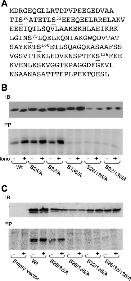
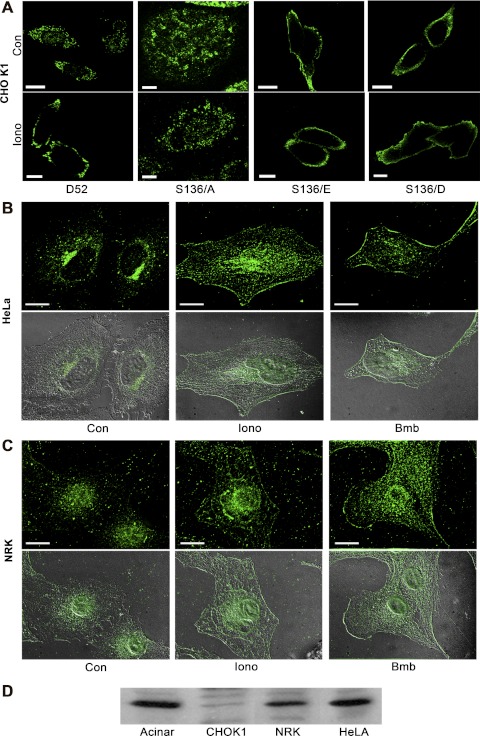
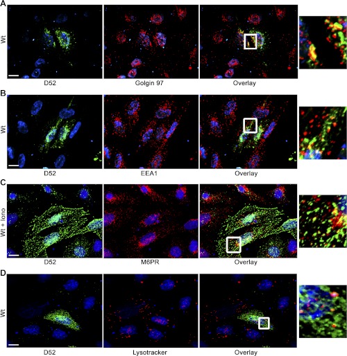
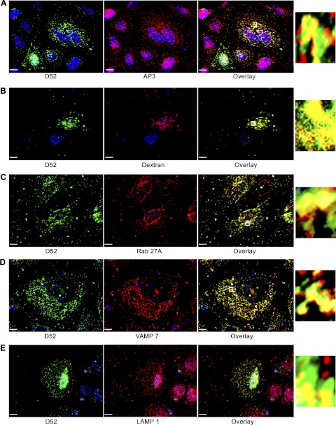
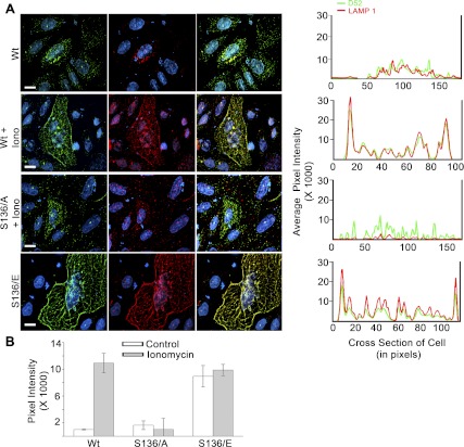
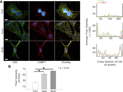
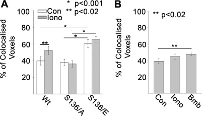
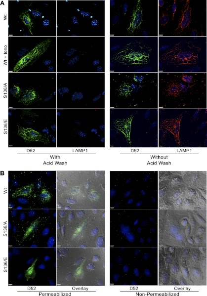
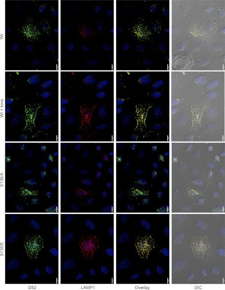
Tumor protein D52 (also known as CRHSP-28) is highly expressed in multiple cancers and tumor-derived cell lines; however, it is normally abundant in secretory epithelia throughout the digestive system, where it has been implicated in Ca(2+)-dependent digestive enzyme secretion (41). Here we demonstrate, using site-specific mutations, that Ca(2+)-sensitive phosphorylation at serine 136 modulates the accumulation of D52 at the plasma membrane within 2 min of cell stimulation. When expressed in Chinese hamster ovary CHO-K1 cells, D52 colocalized with adaptor protein AP-3, Rab27A, vesicle-associated membrane protein VAMP7, and lysosomal-associated membrane protein LAMP1, all of which are present in lysosome-like secretory organelles. Overexpression of D52 resulted in a marked accumulation of LAMP1 on the plasma membrane that was further enhanced following elevation of cellular Ca(2+). Strikingly, mutation of serine 136 to alanine abolished the Ca(2+)-stimulated accumulation of LAMP1 at the plasma membrane whereas phosphomimetic mutants constitutively induced LAMP1 plasma membrane accumulation independent of elevated Ca(2+). Identical results were obtained for endogenous D52 in normal rat kidney and HeLA cells, where both LAMP1 and D52 rapidly accumulated on the plasma membrane in response to elevated cellular Ca(2+). Finally, D52 induced the uptake of LAMP1 antibodies from the cell surface in accordance with both the level of D52 expression and phosphorylation at serine 136 demonstrating that D52 altered the plasma membrane recycling of LAMP1-associated secretory vesicles. These findings implicate both D52 expression and Ca(2+)-dependent phosphorylation at serine 136 in lysosomal membrane trafficking to and from the plasma membrane providing a novel Ca(2+)-sensitive pathway modulating the lysosome-like secretory pathway.


Figures

Fig. 1.
Fig. 1.
D52 phosphorylation occurs on serine 136. A: human D52 amino acid sequence. Serines 26, 32, 75, and 136 are predicted casein kinase II (CKII) sites. Serine 100 is a predicted CaM kinase II site. B: Chinese hamster ovary (CHO-K1) cells were transiently transfected with indicated constructs for 18–24 h and then metabolically labeled with [32P]orthophosphate for 2 h. Cells were treated as control (Con) or with 2 μM ionomycin (Iono) for 5 min. D52 was immunoprecipitated from lysates and analyzed by SDS-PAGE and autoradiography. Upper gels are immunoblots (IB; 20 μg/lane) of D52 demonstrating transfection levels for each construct. Note the lack of incorporation of phosphate in all mutants containing the S136/A mutation. Wt, wild-type.
Fig. 2.
Fig. 2.
Phosphorylation at serine 136 mediates D52 accumulation on the plasma membrane. A: a single confocal optical section of CHO-K1 cells transfected with Wt-D52 or the indicated S136 mutants that were treated as control or with 2 μM ionomycin for 5 min prior to fixation in 2% formaldehyde. D52 immunoreactivity was detected in A by using anti-hemagglutinin (HA) tag (1:100) with Alexa Fluor 546-conjugated anti-mouse IgG (1:500). Note the pronounced accumulation of Wt-D52 to the plasma membrane in response to elevated cellular Ca2+, which was abolished in the S136/A mutants. Likewise, note the pronounced accumulation of phosphomimetic mutants along the plasma membrane independent of elevated Ca2+. B and C: endogenous D52 (1:100) was analyzed in HeLa and normal rat kidney (NRK) cells treated as control or stimulated with 2 μM ionomycin or 100 nM of the bombesin analog Lys3-bombesin (Bmb) for 5 min and detected by use of Alexa Fluor 488-conjugated anti-rabbit IgG (1:500). Images are a reconstructed z-series acquired by brightfield microscopy. Corresponding differential interference microscopy (DIC) images are shown below each immunofluorescence image. All images are representative of multiple determinations performed on at least 3 separate tissue preparations. Bars, 20 μm. D: immunoblot of D52 (1:1,000) in lysates (100 μg) from pancreatic acinar cells, CHO-K1, NRK, and HeLa cells. Note that acinar, NRK, and HeLa cells express significant levels of endogenous D52, whereas CHO-K1 cells exhibit much less immunoreactivity.
Fig. 3.
Fig. 3.
Partial colocalization of D52 with Golgi. D52 was analyzed in CHO-K1 cells transfected with Wt-D52. Following 2% formaldehyde fixation, D52 (1:100) immunoreactivity was detected by use of Alexa Fluor 546-conjugated anti-rabbit IgG (1:500) (A and B). Golgin 97 (1:50) (A) and early endosomal antigen 1 (EEA1) (1:100) (B) immunoreactivities were detected by use of Alexa Fluor 488-conjugated anti-mouse IgG (1:250). C: D52 immunoreactivity was detected with anti-HA tag (1:100) by using Alexa Fluor 546-conjugated anti-mouse IgG (1:250), and mannose-6-phosphate receptor (M-6-P) (1:100) immunoreactivity was detected with Alexa Fluor 488-conjugated anti-rabbit IgG (1:500). Note that cells costained for M-6-P had been treated with ionomycin for 5 min prior to fixation. D: cells were labeled with LysoTracker (5 μM, 1 h at 37°C) prior to fixation and staining for D52. Nuclei were labeled with 4,6-diamidino-2-phenylindole (DAPI) except in LysoTracker-labeled cells where TOPO-3 iodide was used. Postcollection, D52 and costained antigens were applied green and red pseudo-colors, respectively. Note the moderate colocalization of D52 with Golgin 97 but more modest colocalization with EEA1, M-6-P, and LysoTracker. Each image is a reconstructed z-series obtained by brightfield microscopy. All images are representative of multiple determinations performed on at least 3 separate tissue preparations. Bars, 13 μm.
Fig. 4.
Fig. 4.
D52 is present on a lysosome-like secretory compartment. CHO-K1 cells transfected with Wt-D52 were fixed in 2% formaldehyde and labeled with antibodies for D52, AP-3, Rab27A, and vesicle-associated membrane protein 7 (VAMP7) (all at 1:100). Immunoreactivities were detected by using Alexa Fluor 546-conjugated anti-rabbit IgG (1:500) for D52, and Alexa Fluor 488-conjugated anti-mouse IgG (1:250) (A, B, D, and E) or Alexa 546-conjugated anti-goat (1:100) (C) for the costained antigen. For dextran labeling of lysosomes, cells were incubated with FITC-dextran (10,0000 MW) for 3 h at 37°C followed by a 2-h chase period prior to fixation and staining for D52. The FITC-dextran signal was amplified by use of anti-FITC goat-conjugated Alexa Fluor 488 antibody (1:100). Note the extensive colocalization of D52 with each marker (see Table 1 for quantification). Insets are enlargements of regions denoted by the small boxed region in each overlay. Each image is a reconstructed z-series obtained by brightfield microscopy. All images are representative of multiple determinations performed on at least 3 separate tissue preparations. Bars, 13 μm.
Fig. 5.
Fig. 5.
Lysosome-associated membrane protein 1 (LAMP1) exocytosis is dependent on D52 expression levels and its Ca2+-regulated phosphorylation at serine 136. A, left: intracellular D52 and externalized LAMP1 were analyzed in CHO-K1 cells transfected with Wt-D52, S136/A, or S136/E. Cells were treated with 0.01% DMSO as control or with 2 μM ionomycin for 5 min and then incubated at 4°C with anti-LAMP1 (1:100) to label externalized antigen. Cells were then fixed in 2% formaldehyde, permeabilized, and further labeled with anti-D52 (1:100). D52 and LAMP1 immunoreactivities were detected postfixation, by use of Alexa Fluor 546-conjugated anti-rabbit IgG (1:500) and Alexa Fluor 488-conjugated anti-mouse IgG (1:250), respectively. Nuclei were labeled with DAPI. Postcollection, D52 and LAMP1 images were applied green and red pseudo-colors, respectively. Each image is a reconstructed z-series obtained by brightfield microscopy. All images are representative of multiple determinations performed on at least 3 separate tissue preparations. Bars, 13 μm. A, right: line density analysis of D52 (green) and LAMP1 (red) staining across a single cell. B: quantification of LAMP1 localization at the plasma membrane designated as the first 20 pixels of signal acquired at the cell periphery from multiple line density plots. Data are means ± SE (n = 10 for each experimental condition) performed in at least 3 separate tissue preparations.
Fig. 6.
Fig. 6.
Endogenous D52 translocates to plasma membrane with LAMP1 during Ca2+-stimulated exocytosis. Intracellular D52 and externalized LAMP1 were analyzed in NRK cells treated with 0.01% DMSO as control, 2 μM ionomycin, or 100 nM Lys3-bombesin for 5 min. Cells were incubated at 4°C with anti-LAMP1 to label externalized LAMP1 as described in Fig. 5. Each image is a reconstructed z-series obtained by brightfield microscopy. All images are a single representative experiment performed in at least 3 separate tissue preparations. Bars, 13 μm. A, right: line density analysis of D52 (green) and LAMP1 (red) staining across a single cell. B: quantitative analysis of LAMP1 localization at the plasma membrane designated as the first 20 pixels of signal acquired at the cell periphery from multiple line density plots. Data are means ± SE (n = 10 for each experimental condition) performed in at least 3 separate tissue preparations.
Fig. 7.
Fig. 7.
Colocalization of externalized LAMP1 and intracellular D52. The percent colocalized voxels of surfaced labeled LAMP1 with intracellular D52 was analyzed in multiple reconstructed z-series images in CHO-K1 cells transfected with D52 or D52 phospho-mutants (A) or NRK cells expressing endogenous D52 (B) (n ≥ 10 for each determination).
Fig. 8.
Fig. 8.
Conformation of LAMP1 surface labeling and intracellular labeling of D52. A: CHO-K1 cells were transfected with Wt-D52, S136/A, or S136/E and treated as control or with 2 μM ionomycin for 5 min. Cells were immunolabeled for external LAMP1 as described in Figs. 5 and 6. Following LAMP1 labeling, coverslips were rinsed 3 times with ice-cold PBS followed by fixation or washed an additional 5 times in acid wash buffer containing (in mM) 100 glycine, 20 mg acetate, 50 KCl, pH 2.2, all at 4°C. After being washed with PBS, cells were fixed in 2% formaldehyde, blocked, permeabilized, and immunolabeled for D52 (1:100) by using Alexa Fluor 546-conjugated anti-rabbit IgG (1:500). Note the complete loss of LAMP1 surface label with acid washing. B: cells were surfaced labeled for LAMP1 as described in Figs. 5 and 6. Following 2% formaldehyde fixation, cells were permeabilized or not with Triton X-100 during labeling with D52 antibodies. Note that cell permeabilization is essential to detect D52 immunoreactivity.
Fig. 9.
Fig. 9.
Overexpression of D52 and D52 phosphorylation at serine 136 stimulates LAMP1 trafficking and retrieval from the plasma membrane. CHO-K1 cells were transfected with Wt-D52 or D52 phospho-mutants for 18 h prior to a 2-h incubation at 37°C with Alexa 488-conjugated LAMP1 antibodies (1:20). Where indicated, 2 μM ionomycin was added for the last 5 min of incubation. Cells were then acid washed as described in Fig. 8, fixed in 2% formaldehyde, permeabilized, and labeled for D52 (1:100) with Alexa Fluor 546-conjugated anti-rabbit IgG (1:500). Note that compared with adjacent cells expressing low levels of D52, transfected cells show a marked accumulation of LAMP1 that is concentrated in perinuclear regions. Treatment of cells expressing Wt-D52 with ionomycin or expression of S136/E mutants under basal conditions significantly increased LAMP1 and D52 labeling just underneath peripheral plasma membrane regions. Also note that cells expressing S136/A mutants accumulate significant LAMP1 label compared with surrounding nontransfected cells but fail to show the enhanced peripheral label in response to ionomycin. Each image is a reconstructed z-series obtained by brightfield microscopy. All images are a single representative experiment performed in at least 3 separate tissue preparations. Bars, 13 μm.

Similar articles

Cited by

References

    1. Arantes RM, Andrews NW. A role for synaptotagmin VII-regulated exocytosis of lysosomes in neurite outgrowth from primary sympathetic neurons. J Neurosci 26: 4630– 4637, 2006 - PMC - PubMed
    1. Ashrafi K, Chang FY, Watts JL, Faser AG, Kamath RS, Ahringer J, Ruvkun G. Genome-wide RNAi analysis of Caenorhabditis elegans fat regulatory genes. Nature 421: 268–272, 2003 - PubMed
    1. Bard F, Casano L, Mallabiabarrena A, Wallace E, Saito K, Kitayama H, Guizzunti G, Hu Y, Wendler F, DasGupta R, Perrimon N, Malhotra V. Functional genomics reveals genes involved in protein secretion and Golgi organization. Nature 439: 604–607, 2006 - PubMed
    1. Borner GHH, Harbour M, Hester S, Lilley KS, Robinson MS. Comparative proteomics of clathrin-coated vesicles. J Cell Biol 175: 571–578, 2006 - PMC - PubMed
    1. Boutros R, Bailey AM, Wilson SHD, Byrne JA. Alternative splicing as a mechanism for regulating 14-3-3 binding: interactions between hD53 (TPD52L1) and 14-3-3 proteins. J Mol Biol 332: 675–687, 2003 - PubMed

Publication types

MeSH terms