

Review
doi: 10.1038/ki.2009.489.
Epub 2009 Dec 23.
T cells and dendritic cells in glomerular disease: the new glomerulotubular feedback loop
Affiliations
- PMID: 20032960
- PMCID: PMC3039448
- DOI: 10.1038/ki.2009.489
Item in Clipboard
Review
T cells and dendritic cells in glomerular disease: the new glomerulotubular feedback loop
Kidney Int.
2010 Mar.
Abstract
A newly described glomerulotubular feedback loop may explain the relationship between glomerular damage, epitope spreading, tubulointerstitial nephritis, proteinuria as a progression factor, and the importance of the local milieu in kidney damage. It also opens the horizons for exciting innovative approaches to therapy of both acute and chronic kidney diseases.
Conflict of interest statement
The authors declared no competing interests.
Figures
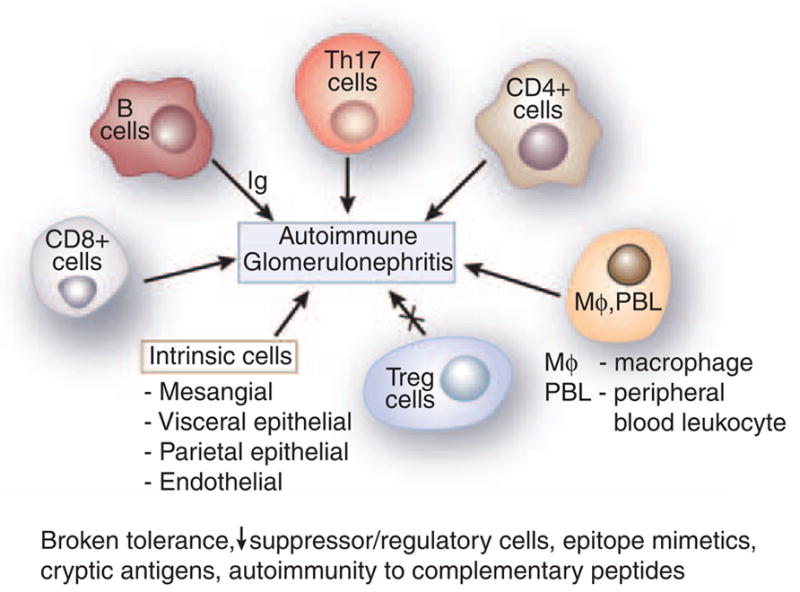
Figure 1 represents an aggregate of data derived from animal models. B cells
were classically considered to be involved in the pathogenesis of GN by
elaboration of immunoglobulin (Ig). Th-17, CD4 +, and CD8
+ cells have a significant role as shown by
abrogation of activity leading to amelioration of GN. Macrophages and
peripheral blood leukocyte (PBL) are essential in the histological changes
of GN. All result in a variable increase in the mesangial matrix, and
involvement of the visceral and parietal epithelial cells. T regulatory
cells downregulate disease. Experimental autoimmune glomerulonephritis
presumably arises secondary to various etiologies, including broken
tolerance, a decrease in suppressor/regulatory cells, epitope mimetics,
exposure of cryptic antigens, and possibly autoimmunity to complementary
peptides.
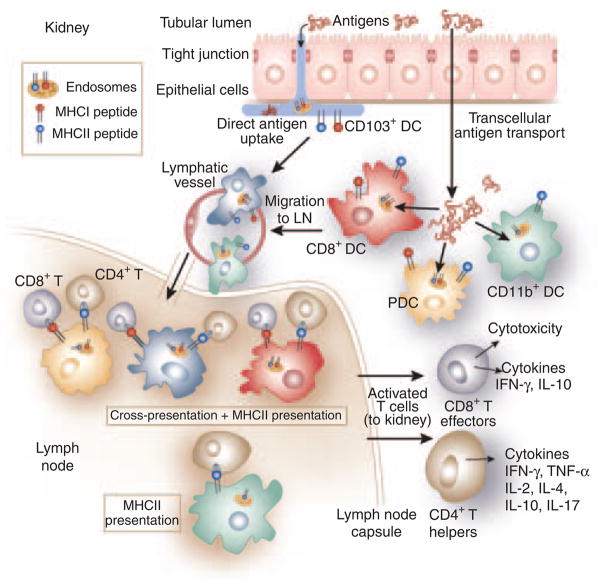
Uptake and presentation of tubular proteins by renal DCs. The figure shows
the potential pathways by which tubular antigens are internalized,
processed, and presented to CD8 + and CD4
+ T-cells in the kidney draining lymph nodes. DC
subsets in the interstitium internalize antigens in the tubular lumen either
directly by inserting pseudopods into the lumen (CD103 +
DCs) or indirectly by internalizing antigen transported and/or processed by
transcellular antigen transport through tubular epithelial cells (TECs). The
presence of all DC types shown has been published, except for CD103
+ DCs, which have been identified in preliminary
studies (Sung SJ). For cross-presentation, antigens can be directly
processed in the endosomes and the MHC-I molecules are loaded by the
released peptides and presented to CD8 + T cells. Other
possible pathways of exogenous antigen loading—by transit through
the cytosol with processing by proteosomes and MHC-I loading in the
endoplasmic reticulum, and processing and MHC-I loading in the
phagosomes—are not shown. In cross-presentation in the KDLNs, CD4
+ T-cell help is shown. Cross-presentation by CD11b
+ DCs has been shown to be much inferior to other DC
types and is not shown. The main functions of CD8 + T
cells and CD4 + helper cells, but not those of Treg
cells, are shown.
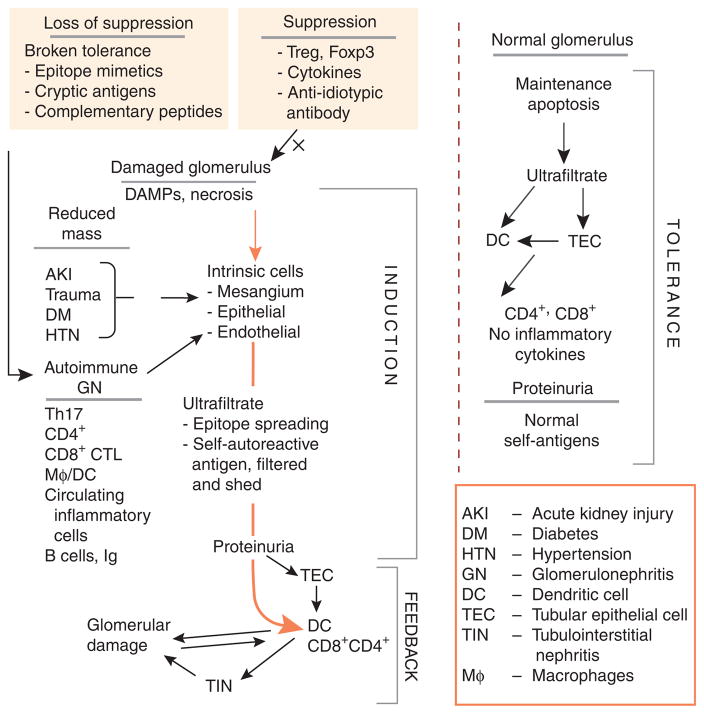
The figure depicts the glomerulotubular feedback loop involving new paradigms
as described by Macconi and Heymann. Under normal circumstances the
glomerular filtrate contains many peptides, proteins, and other substances
that are filtered or shed into the urinary space. These are taken up by
dendritic cells directly from the tubular lumen or after processing by
tubular epithelial cells (TECs). Normally, cell death is affected by
apoptosis with the presentation of these antigens to T cells without
inflammatory cytokines, resulting in tolerance and suppression. Even
proteinuria in this circumstance, consisting of a large amount of
self-antigens, is not associated with damage sans an inflammatory process.
On the other hand, damaged kidney, whether by reduced mass or by autoimmune
injury, induces cell necrosis rather than apoptosis, with the release of
damage-associated molecular patterns (DAMPs), recruitment of exogenous
cells, and stimulation of endogenous cells to proliferate and elaborate the
matrix and inflammatory cytokines. The ultrafiltrate contains normal
constituents, but also now contains constituents released by renal injury.
This inductive phase may progress, depending on regulatory feedback.
However, when DCs process and present antigen to T cells in this
inflammatory milieu, the result is tubulointerstitial nephritis (TIN) and
feedback to the glomerulus, either directly or through a periglomerular
infiltrate, which somehow communicates with the glomerulus across
Bowman’s capsule. Thus, a glomerulotubular feedback loop is
established in the setting of inflammation. This leads to further glomerular
damage with CD8 + CTL cross-presentation of previously
irrelevant or self-antigens and progression of TIN and
glomerulosclerosis.
References
-
- Rocklin R, Lewis E, David J. In-vitro evidence for cellular hypersensitivity to glomerular-basement membrane antigens in human glomerulonephritis. N Engl J Med. 1970;283:497–501. - PubMed
-
- Dixon FJ. What are sensitized cells doing in glomerulonephritis? N Engl J Med. 1970;283:536–537. - PubMed
-
- Nikolic-Paterson DJ, Lan HY, Atkins R. Macrophages in immune renal injury. In: Neilson EG, Couser WG, editors. Immunologic Renal Diseases. Lippincott-Raven; Philadelphia: 1997. pp. 575–592.
Publication types
MeSH terms
Grants and funding
LinkOut - more resources
Full Text Sources
Medical
Molecular Biology Databases
