. 2010 Apr;130(4):985-94.
doi: 10.1038/jid.2009.384. Epub 2009 Dec 24.

Sebum free fatty acids enhance the innate immune defense of human sebocytes by upregulating beta-defensin-2 expression

Affiliations

Sebum free fatty acids enhance the innate immune defense of human sebocytes by upregulating beta-defensin-2 expression

Teruaki Nakatsuji et al. J Invest Dermatol. 2010 Apr.

Abstract

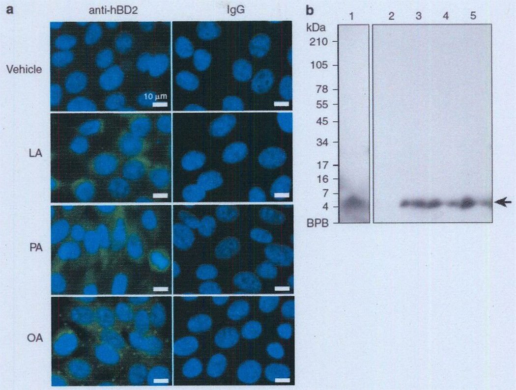
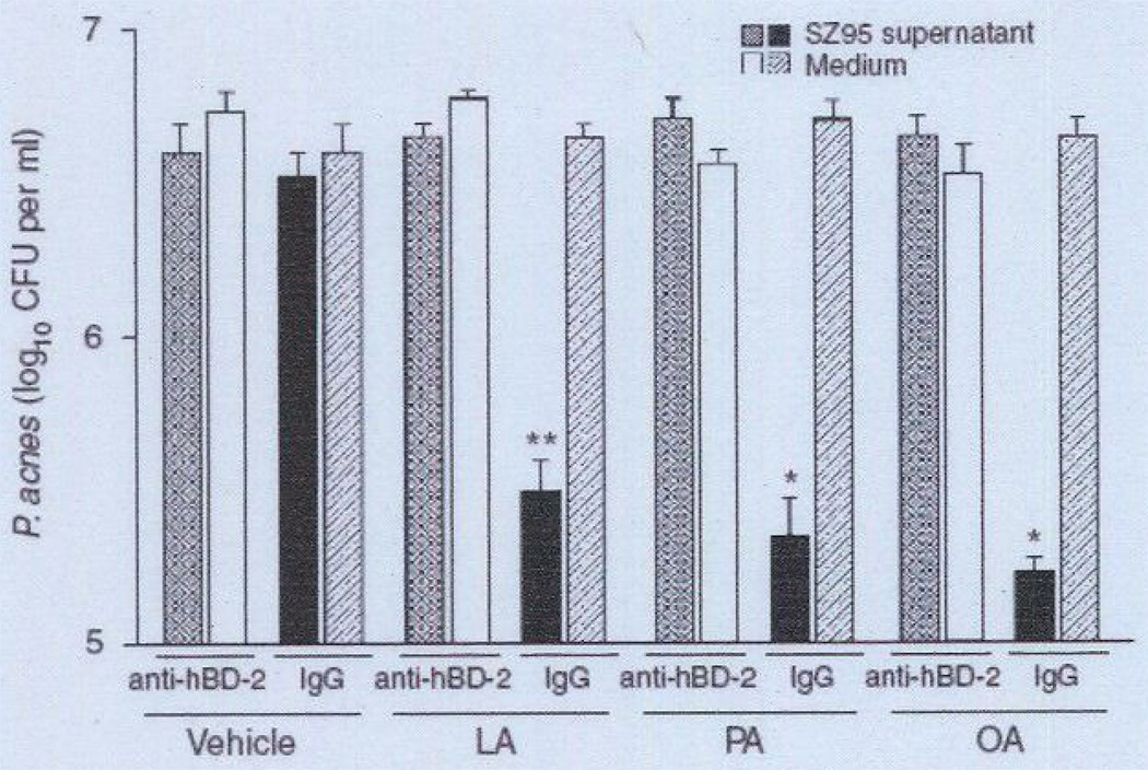
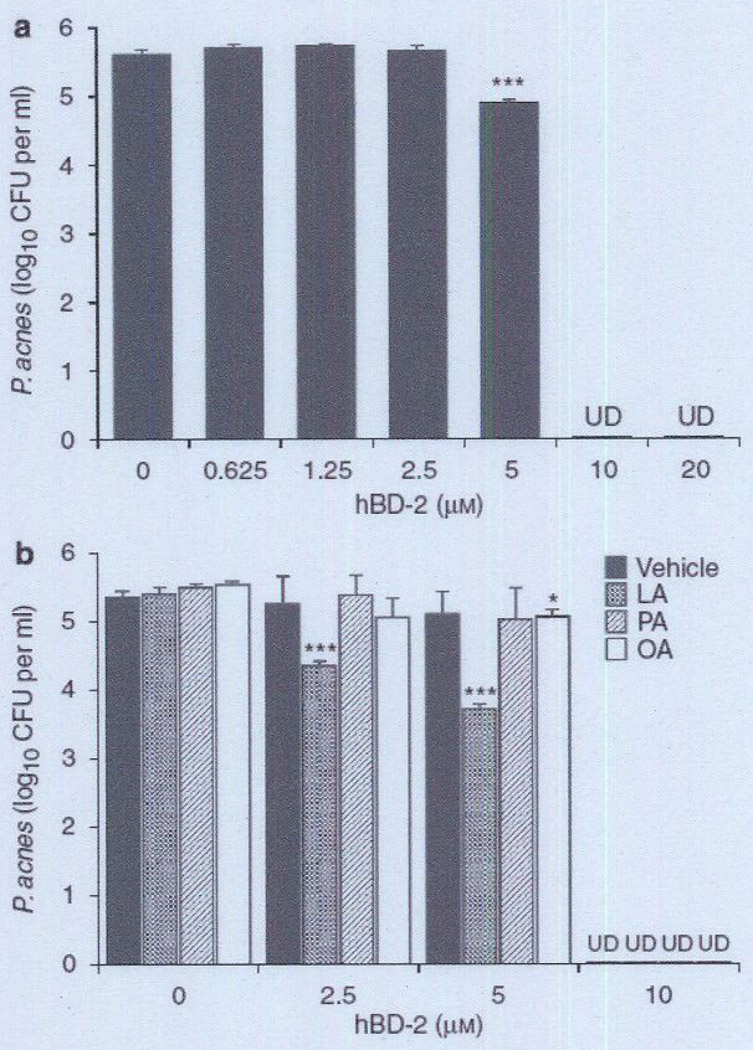
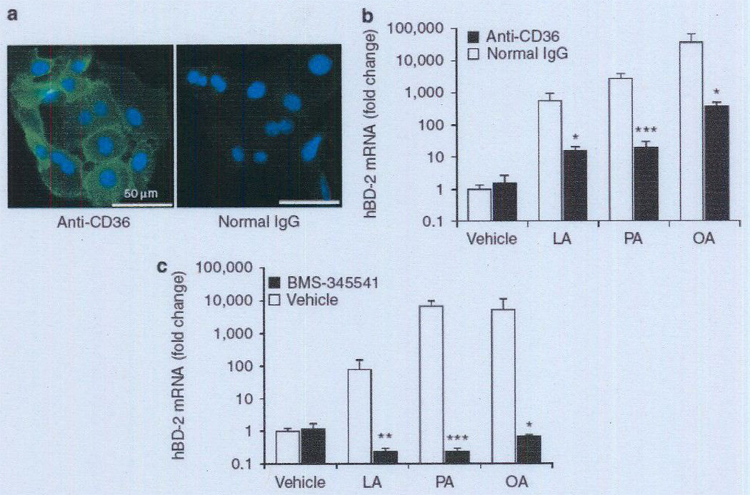
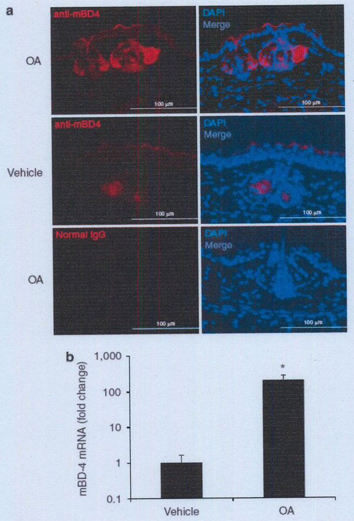
Various sebum free fatty acids (FFAs) have shown antibacterial activity against a broad range of gram-positive bacteria, resulting in the suggestion that they are accountable, at least partially, for the direct antimicrobial activity of the skin surface. In this study, we examined the effects of sebum FFAs on the antimicrobial peptide (AMP)-mediated innate immune defense of human sebocytes. Incubation of lauric acid, palmitic acid, or oleic acid (OA) with human sebocytes dramatically enhanced their expression of human beta-defensin (hBD)-2, one of the predominant AMPs found in the skin, whereas remarkable increases in hBD-1, hBD-3, and human cathelicidin LL-37 were not observed. Secreted hBD-2 was detectable by western blotting in the supernatant of sebocyte culture incubated with each FFA, but not with a vehicle control. The supernatant of FFA-incubated sebocyte culture showed antimicrobial activity against Propionibacterium acnes, whereas the enhanced antimicrobial activity of human sebocytes was neutralized by anti-hBD-2 IgG. In addition, the FFA-induced hBD-2 expression was suppressed by blocking the cluster of differentiation (CD)36 fatty acid translocase on the surface of sebocytes with anti-human CD36 IgG or blocking the NF-kappaB signaling pathway with BMS-345541, a highly selective inhibitor of inhibitory kappaB kinase. These data suggest that sebum FFAs upregulate the expression of hBD-2 in human sebocytes, which may enhance the disinfecting activity of the human sebaceous gland. The FFA-induced upregulation of hBD-2 is facilitated by CD36-mediated FFA uptake and NF-kappaB-mediated transactivation. The upregulation of mouse beta-defensin 4, a mouse ortholog for hBD-2, was also observed in the hair follicle sebaceous glands of mouse ear skin after an epicutaneous application of OA, the most hBD-2-inducible FFA tested. This report highlights the potential of using FFAs as a multifunctional antimicrobial therapy agent for acne vulgaris treatment; FFAs may provide direct antibacterial activities against P. acnes and enhance the skin's innate antibacterial defense by inducing the expression of hBD-2 in sebocytes as well.


Conflict of interest statement

CONFLICT OF INTEREST

The authors state no conflict of interest.

Figures

Figure 1
Figure 1. Effect of FFAs on the antimicrobial peptide expression in human sebocytes
Immortalized human sebocytes, SZ95 (1 × l06 per well), were incubated with LA, PA, or OA (25 µg ml−1) in 1% FBS-Sebomed containing 0.5% (w/v) DMSO for 24 hours at 37 °C. The control received an equal amount of DMSO. (a–d) mRNA expression of hBD-1 (a), hBD-2 (b). hBD-3 (c), and human cathelicidin LL-37 (d) was evaluated by real-time qPCR, normalized to that of GAPDH. and then plotted as relative expression compared with that of vehicle-treated cells. Data represent means ± SE of four individual experiments (*P<0.05, **P <0.01, ***P<0.005 by Student’s t-test vs. vehicle control). FBS, fetal bovine serum; FFA, free fatty acid; GAPDH, glyceraldehyde-3-phosphate dehydrogenase; hBD, human β-defensin; LA, lauric acid; OA, oleic acid; PA, palmitic acid; qPCR. quantitative PCR.
Figure 2
Figure 2. FFA-enhanced hBD-2 peptide level in human sebocytes
(a) Immortalized human sebocytes, SZ95 (2 × 10⁶ per well), were incubated with LA, PA, or OA (25 µg ml−1) in 1% FBS-Sebomed containing 0.5% (w/v) DMSO for 24 hours at 37 °C. The control received an equal amount of DMSO. After incubation, the cells were stained with rabbit anti-hBD-2, followed by FITC-labeled anti-rabbit IgCs. The nuclei were counterstained with DAPI. Bar = 10 µm. (b) To detect secreted hBD-2 from SZ95 sebocytes by western blotting, the supernatants of the cell culture media were concentrated using a Sep-Pak cartridge as described in the Materials and Methods section. The reconstituted samples (400 µl equivalent of medium) from cell culture media, incubated with vehicle, LA, PA, and OA (lanes 2–5. respectively), were separated by SDS-PACE (16% acrylamide). Synthetic hBD-2 (60 nmol) was used as a positive control (lane 1) for detection of hBD-2. hBD-2 was detected by rabbit anti-hBD-2 IgCs. Data are representative of three separate experiments with similar results. Lane 1 and lanes 2–5 are from the same original gel. Because of different film exposure times, two images were obtained and later merged again. DAPI, 40′-6-diamidino-2-phenylindole; FBS, fetal bovine serum; FFA, free fatty acid; hBD-2, human (β-defensin-2; LA, lauric acid; OA, oleic acid; PA, palmitic acid.
Figure 3
Figure 3. Neutralization of FFA-induced antimicrobial activity of human sebocytes with anti-hBD-2 IgG
Immortalized human sebocytes, SZ95 (2 × 10⁶ per well), were incubated with LA, PA, or OA (25 µg ml−1) in 1% FBS-Sebomed containing 0.5% (w/v) DMSO for 24 hours at 37 °C. An equal amount of DMSO was used as vehicle control. To examine the antimicrobial activity of each FFA contained in the medium, the FFA-conditioned medium was incubated without sebocytes. After incubation, the supermatant of the cell culture was filtrated to remove cell debris and then preincubated with rabbit anti-hBD-2 IgG or normal rabbit IgG for 2 hours at 37 °C. The mixture was incubated with Propionibacterium acnes (1 × 10⁶ CFU per ml) for 5 hours at 37 °C under anaerobic conditions. After incubation, the P. acnes suspension was diluted 1:1–1:104 with PBS. A volume of 5 µl of the diluted solution was spotted on a Brucella broth agar plate supplemented with 5% defibrinated sheep blood and hemin and with vitamin K. After the liquid in the P. acnes suspension was absorbed into the agar, the plate was incubated under anaerobic conditions to quantify the CFUs of P. acnes Data represent means ± SE of three individual experiments (*P<0.05, **P <0.01 by Student0’s t-test vs. medium control). CFU, colony-forming unit: FBS, fetal bovine serum: FFA, free fatty acid; hBD-2, human β-defensin-2; LA, lauric acid; OA, oleic acid; PA, palmitic acid; PBS, phosphate-buffered saline.
Figure 4
Figure 4. Bactericidal effect of synthetic hBD-2 on Propionibacterium acnes
(a) P. acnes (1 × 10⁶ CFU per ml) was incubated with 0–20 µm synthetic hBD-2 peptide in 20 mm phosphate buffer, pH 6.5, containing 100 mm NaCI for 5 hours under anaerobic conditions, (b) To enable examination of the synergistic antimicrobial activity of hBD-2 and FFAs, P. acnes (1 × 10⁶ CFU per ml) was incubated with hBD-2 (0, 2.5, 5, 10 µm) in phosphate buffer in the presence of LA, PA, or OA (25 µg ml−1) on a 96-well microplate (100 µl per well) at 37 °C for 5 hours. The control received only DMSO (0.5%(v/v)) instead of FFA. After incubation, the P. acnes suspension was diluted 1:10–1:104 with PBS, and 5 µl of the diluted solution was spotted on a Brucella broth agar plate supplemented with 5% defibrinated sheep blood and hemin and with vitamin K. After the liquid in the P. acnes suspension was absorbed into the agar, the plate was incubated under anaerobic conditions to quantify the CFUs of P. acnes. Data represent means ± SE of three individual experiments (*P<0.05, ***P<0.005 by Student’s t-test). CFU, colony-forming unit; FFA, free fatty acid; hBD-2, human β-defensin-2; LA, lauric acid; OA, oleic acid: PA, palmitic acid; PBS, phosphate-buffered saline: UD, undetectable.
Figure 5
Figure 5. FFA-induccd increase of hBD-2 expression was mediated by CD36 and the NF-κB signaling pathway
(a) To reveal the cellular distribution of CD36 in human SZ95 sebocytes, cells were stained with mouse anti-CD36 IgG, followed by anti-mouse IgG-FITC conjugate (green) (left panel). Normal mouse IgG was used as negative control (right panel). The nuclei were counterstained with DAP1 (blue). Data are representative of four separate experiments with similar results. Bar = 50 µm. (b–c) To block the function of CD36 on the surface of SZ95 sebocytes, cells (2 × 10⁶ per well) cultured on a 24-well plate were preincubated with mouse anti-CD36 IgG (5 µg m1−1) or normal mouse IgG in 1% FBS-Sebomed (250 µl) for 2 hours at 37 °C (b). To block NF-κB-mediated signaling in SZ95 sebocytes, the cells (2 × 10⁶ per well) cultured on a 24-well plate were preincubated with BMS-345541 (20 µm) or with an equal amount of DMSO (0.1%) in 1% FBS-Sebomed (250 µl) for 1 hour at 37 °C (c). After preincubation, LA, PA, or OA (2.5 µg ml−1) was added to the cells, which were subsequently incubated for 24 hours at 37 °C. Control received an equal amount of DMSO (0.5% (v/v)). After incubation, the mRNA expression levels of hBD-2 were determined as described in the Materials and Methods section. Data represent means ± SE of four individual experiments (*P<0.05, **P<0.01, ***P<0.005 by Student’s t-test vs. normal IgG or vehicle control). CD36, cluster of differentiation 36; DAPI, 4′-6-diamidino-2-phenylindote; FBS, fetal bovine serum; FFA, free fatty acid; hBD-2, human β-defensin-2; LA, lauric acid; OA, oleic acid; PA, palmitic acid.
Figure 6
Figure 6. Effect of epicutaneous application of OA on mBD-4 expression in mouse sebaceous gland
OA (150 µg in 5% acetone mixed with 15 mg of Vaseline) and 5% acetone mixed with 15 mg of Vaseline (vehicle) were epicutaneously applied to the left and right ears, respectively, at 0 and 12 hours. The ears were excised 12 hours after epicutaneous application. (a) Frozen sections of mouse ears were stained with rabbit anti-mBD-4 IgG, followed by goat anti-rabbit IgG-Alexa 568 conjugate (red). Normal rabbit IgG was used as negative control staining. Nuclei were stained with DAPI (blue). Bar = 100 µm. Data are representative of four separate experiments with similar results, (b) mRNA expression of mBD-4 in the ear skin was evaluated by real-time qPCR. normalized to that of GAPDH. and then plotted as relative expression compared with that of the vehicle-treated sample. Data represent means ± SE of four individual experiments (*P<0.05 by Student’s t-test vs. vehicle-treated control). DAPI, 4′-6-diamidino-2-phenylindole; GAPDH, glyceraldehyde-3-phosphate dehydrogenase; mBD-4, mouse β-defensin-4; OA, oleic acid: qPCR, quantitative PCR.

Similar articles

Cited by

References

    1. Abumrad N, Coburn C, Ibrahimi A. Membrane proteins implicated in long-chain fatty acid uptake by mammalian cells: CD36, FATP and FABPm. Biochim Biophys Acta. 1999;1441:4–13. - PubMed
    1. Baillie AG, Coburn CT, Abumrad NA. Reversible binding of long-chain fatty acids to purified FAT, the adipose CD36 homolog. J Membr Biol. 1996;153:75–81. - PubMed
    1. Boden G, She P, Mozzoli M, et al. Free fatty acids produce insulin resistance and activate the proinflammatory nuclear factor-kappaB pathway in rat liver. Diabetes. 2005;54:3458–3465. - PubMed
    1. Bodoprost J, Rosemeyer H. Analysis of phenacylester derivatives of fatty acids from human skin surface sebum by reversed-phase HPLC: chromatographic mobility as a function of physico-chemical properties. Int J Mol Sci. 2007;8:1111–1124.
    1. Bojar RA, Holland KT. Acne and Propionibacterium acnes. Clin Dermatol. 2004;22:375–379. - PubMed

Publication types

MeSH terms

LinkOut - more resources