. 2010 Feb 3;29(3):666-79.
doi: 10.1038/emboj.2009.367. Epub 2009 Dec 24.

Structure of an integrin with an alphaI domain, complement receptor type 4

Affiliations

Structure of an integrin with an alphaI domain, complement receptor type 4

Can Xie et al. EMBO J. .

Abstract

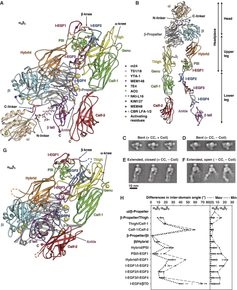
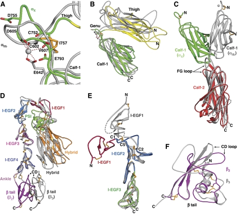
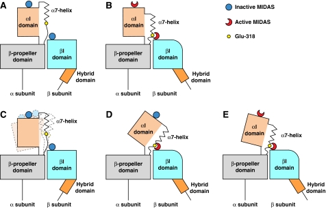
We report the structure of an integrin with an alphaI domain, alpha(X)beta(2), the complement receptor type 4. It was earlier expected that a fixed orientation between the alphaI domain and the beta-propeller domain in which it is inserted would be required for allosteric signal transmission. However, the alphaI domain is highly flexible, enabling two betaI domain conformational states to couple to three alphaI domain states, and greater accessibility for ligand recognition. Although alpha(X)beta(2) is bent similarly to integrins that lack alphaI domains, the terminal domains of the alpha- and beta-legs, calf-2 and beta-tail, are oriented differently than in alphaI-less integrins. Linkers extending to the transmembrane domains are unstructured. Previous mutations in the beta(2)-tail domain support the importance of extension, rather than a deadbolt, in integrin activation. The locations of further activating mutations and antibody epitopes show the critical role of extension, and conversion from the closed to the open headpiece conformation, in integrin activation. Differences among 10 molecules in crystal lattices provide unprecedented information on interdomain flexibility important for modelling integrin extension and activation.


Conflict of interest statement

The authors declare that they have no conflict of interest.

Figures

Figure 1
Figure 1
Representative electron density. (A, B) The αI domain. The main chain of αI domain is shown in white. Se anomalous map is shown at 3 σ level (red). Electron density map of αI domain after multi-crystal averaging is at 1 σ level (blue). Electron density around residues 318 and 319 is shown at 0.6 σ in green. The methionine residues are shown in stick, with yellow carbon atoms and orange sulphur atoms. (B) Different view to show the electron density around the α7-helix. (C, D) Multi-crystal averaging electron density around β-tail domain (C) and around residues 765–771 and 783–786 in the calf-1 domain (D) Colours and map σ levels are as in (A). (E) The αI/β-propeller/βI domain interface. Multi-crystal averaging electron density map shown at 1 σ level around α-subunit β-propeller residue Asn-373 and its N-linked glycan (blue), the N-linker residues 127–130 and disulfide-bonded β-propeller residues C126 and C97 (green), and residues 326–327 of the C-linker and β-propeller residues 328–331 (yellow). The sidechains of residues C97, C126, F328, M332, N373 and its N-linked glycan are shown in stick. The electron density in this region shows distinct paths of the three segments, and clear connected density with the β-propeller domain. Se anomalous map is shown at 3 σ level around the sidechain of Met-332. αI domain, β-propeller domain, and βI domain are in wheat, grey, and cyan, respectively. N- and C-linkers are in pink. The oxygen atoms, nitrogen atoms, and sulphur atoms are in red, blue, and orange, respectively. The carbon atoms of the glycan are in white. (F) Stereo image of β-tail domain CD loop. The oxygen, sulphur, nitrogen, and carbon atoms are in red, orange, blue, and wheat, respectively. Multi-crystal averaging map at 1 σ is blue and Se anomalous map at 3 σ level is red.
Figure 2
Figure 2
Structure of CR4, integrin αXβ2. (A) Cartoon of αXβ2 molecule 1 in lattice A and (B) an extended model made by adjusting domain interfaces at and near the knees. Disulfides are gold sticks, and glycans are sticks with white carbons. Ca and Mg ions are gold and green spheres, respectively. Smaller spheres show Cα atoms of conformation-associated epitopes and C-terminal residues. (CF) Representative αXβ2 EM class averages. The presence (+) or absence (−) of a disulfide after the αX and β2 C-termini (CC) and subsequent linker and coiled-coil (coil) is indicated. (G) Cartoon of αIIbβ3 (Zhu et al, 2008) in same orientation and style as αXβ2 in (A). (H) Differences in interdomain angles between 10 molecules of αXβ2 and 2 molecules of αIIbβ3 (left panel), or among 10 molecules of αXβ2 (right panel). The mean values of interdomain angles are shown as bars and the maximal and minimal angles are shown in dashed or dotted lines, respectively.
Figure 3
Figure 3
The αI, βI, and β-propeller domain interface, and I domain metal-binding sites. (A, B) The interface. Representations are as in Figure 2A and B; additionally, the indicated sidechains and backbone carbonyls are shown as sticks. (C) Superposition of headpieces from αXβ2 (magenta), unliganded-closed αIIbβ3 (Zhu et al, 2008) (yellow) and liganded-open (Springer et al, 2008) αIIbβ3 (cyan, with ligand in black). (D) Enlarged view of the superposition in (C) showing only αXβ2 (with its ADMIDAS metal as cyan sphere) and the ligand and SYMBS, MIDAS, and ADMIDAS metals from liganded-open αIIbb3 (yellow). (EG) Metal coordination sites, with coordinations dashed. (E) αI domain MIDAS. (F, G) βI domain metal-binding sites in αXβ2 (F) and unliganded-closed αIIbβ3 (G).
Figure 4
Figure 4
Integrin leg domains. Domains in αXβ2 are coloured as in Figure 2A and B and in other integrins are silver with dashes for disordered loops and metals as spheres. (A) The genus. (B) The αX and αIIb thigh and calf-1 domains superimposed on calf-1. (C) The αX and αIIb calf-1 and calf-2 domains superimposed on calf-2. (D) The β2 and β3 (Zhu et al, 2008) legs superimposed on I-EGF domains 2–4. (E) Bent intact β2-leg and β2-leg fragment (Shi et al, 2007) superimposed on I-EGF2. (F) Superimposed β2 and β3 (Xiong et al, 2001)-tail domains.
Figure 5
Figure 5
Breathing of bent αXβ2. (A) Two representative diverse structures after superposition of all 10 molecules on all domains except αI (shown are chains G and H of lattice A with the αI domain from chain A and chains C and D of lattice A with the αI domain from chain G of lattice B). (B) Schematic showing maximal differences in position after superposition.
Figure 6
Figure 6
Communication of allostery between αI and βI domains. (A, B) Previous model with firm interface between the α-subunit I and β-propeller domains (Nishida et al, 2006). There should be a 1:1 correspondence between αI and βI domain conformational states according to this model (A, closed; B, open). (CE) Current model, with a flexible interface between the α-subunit I and β-propeller domains. This enables three states of the αI domain to couple to two states of the βI domain. (C) When the αI and βI domains are each in the closed state, the αI domain is flexible. (D, E) Both the intermediate state αI domain, with its MIDAS closed and α7-helix displaced one turn (D), and the open state αI domain, with its MIDAS open and α7-helix displaced two turns (E), can couple to the open βI domain. The greater tilting of the αI domain in (D) suggests greater strain than in (E). Lessening of this strain in (E) would help compensate for the higher energy of the open αI MIDAS.

References

    1. Adams PD, Grosse-Kunstleve RW, Hung LW, Ioerger TR, McCoy AJ, Moriarty NW, Read RJ, Sacchettini JC, Sauter NK, Terwilliger TC (2002) PHENIX: building new software for automated crystallographic structure determination. Acta Crystallogr D Biol Crystallogr 58: 1948–1954 - PubMed
    1. Alonso JL, Essafi M, Xiong JP, Stehle T, Arnaout MA (2002) Does the integrin αA domain act as a ligand for its βA domain? Curr Biol 12: R340–R342 - PubMed
    1. Astrof NS, Salas A, Shimaoka M, Chen JF, Springer TA (2006) Importance of force linkage in mechanochemistry of adhesion receptors. Biochemistry 45: 15020–15028 - PMC - PubMed
    1. Bailey S (1994) The CCP4 suite-programs for protein crystallography. Acta Crystallogr 50: 760–763 - PubMed
    1. Beglova N, Blacklow SC, Takagi J, Springer TA (2002) Cysteine-rich module structure reveals a fulcrum for integrin rearrangement upon activation. Nat Struct Biol 9: 282–287 - PubMed

Publication types