

A fast dynamic mode of the EF-G-bound ribosome
- PMID: 20033061
- PMCID: PMC2829159
- DOI: 10.1038/emboj.2009.384
A fast dynamic mode of the EF-G-bound ribosome
Abstract
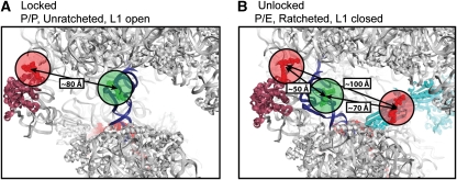
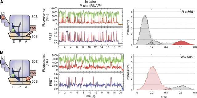
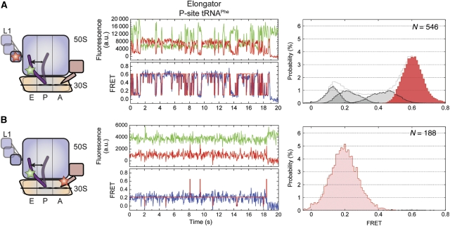
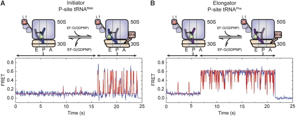
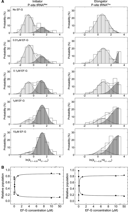
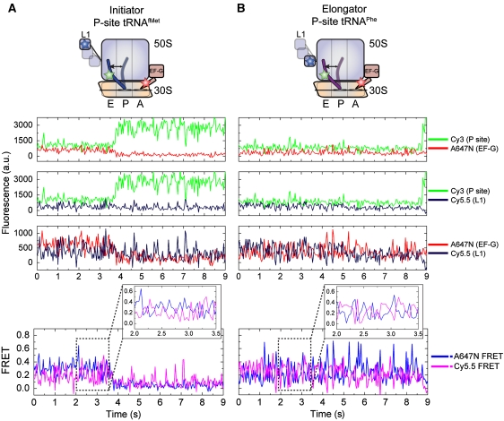
A key intermediate in translocation is an 'unlocked state' of the pre-translocation ribosome in which the P-site tRNA adopts the P/E hybrid state, the L1 stalk domain closes and ribosomal subunits adopt a ratcheted configuration. Here, through two- and three-colour smFRET imaging from multiple structural perspectives, EF-G is shown to accelerate structural and kinetic pathways in the ribosome, leading to this transition. The EF-G-bound ribosome remains highly dynamic in nature, wherein, the unlocked state is transiently and reversibly formed. The P/E hybrid state is energetically favoured, but exchange with the classical P/P configuration persists; the L1 stalk adopts a fast dynamic mode characterized by rapid cycles of closure and opening. These data support a model in which P/E hybrid state formation, L1 stalk closure and subunit ratcheting are loosely coupled, independent processes that must converge to achieve the unlocked state. The highly dynamic nature of these motions, and their sensitivity to conformational and compositional changes in the ribosome, suggests that regulating the formation of this intermediate may present an effective avenue for translational control.
Conflict of interest statement
The authors declare that they have no conflict of interest.
Figures
Similar articles
-
Coupling of ribosomal L1 stalk and tRNA dynamics during translation elongation.Mol Cell. 2008 May 9;30(3):348-59. doi: 10.1016/j.molcel.2008.03.012. Mol Cell. 2008. PMID: 18471980
-
Allosteric collaboration between elongation factor G and the ribosomal L1 stalk directs tRNA movements during translation.Proc Natl Acad Sci U S A. 2009 Sep 15;106(37):15702-7. doi: 10.1073/pnas.0908077106. Epub 2009 Aug 27. Proc Natl Acad Sci U S A. 2009. PMID: 19717422 Free PMC article.
-
Insights into the molecular determinants of EF-G catalyzed translocation.RNA. 2011 Dec;17(12):2189-200. doi: 10.1261/rna.029033.111. Epub 2011 Oct 27. RNA. 2011. PMID: 22033333 Free PMC article.
-
Synchronous tRNA movements during translocation on the ribosome are orchestrated by elongation factor G and GTP hydrolysis.Bioessays. 2014 Oct;36(10):908-18. doi: 10.1002/bies.201400076. Epub 2014 Aug 13. Bioessays. 2014. PMID: 25118068 Review.
-
Elongation factors on the ribosome.Curr Opin Struct Biol. 2005 Jun;15(3):349-54. doi: 10.1016/j.sbi.2005.05.004. Curr Opin Struct Biol. 2005. PMID: 15922593 Review.
Cited by
-
Connecting the kinetics and energy landscape of tRNA translocation on the ribosome.PLoS Comput Biol. 2013;9(3):e1003003. doi: 10.1371/journal.pcbi.1003003. Epub 2013 Mar 21. PLoS Comput Biol. 2013. PMID: 23555233 Free PMC article.
-
Fluctuations between multiple EF-G-induced chimeric tRNA states during translocation on the ribosome.Nat Commun. 2015 Jun 15;6:7442. doi: 10.1038/ncomms8442. Nat Commun. 2015. PMID: 26072700 Free PMC article.
-
Protein-guided RNA dynamics during early ribosome assembly.Nature. 2014 Feb 20;506(7488):334-8. doi: 10.1038/nature13039. Epub 2014 Feb 12. Nature. 2014. PMID: 24522531 Free PMC article.
-
Sizing up long non-coding RNAs: do lncRNAs have secondary and tertiary structure?Bioarchitecture. 2012 Nov-Dec;2(6):189-99. doi: 10.4161/bioa.22592. Bioarchitecture. 2012. PMID: 23267412 Free PMC article. Review.
-
Simulating movement of tRNA through the ribosome during hybrid-state formation.J Chem Phys. 2013 Sep 28;139(12):121919. doi: 10.1063/1.4817212. J Chem Phys. 2013. PMID: 24089731 Free PMC article.
References
-
- Andersen CB, Becker T, Blau M, Anand M, Halic M, Balar B, Mielke T, Boesen T, Pedersen JS, Spahn CM, Kinzy TG, Andersen GR, Beckmann R (2006) Structure of eEF3 and the mechanism of transfer RNA release from the E-site. Nature 443: 663–668 - PubMed
-
- Bergemann K, Nierhaus KH (1983) Spontaneous, elongation factor G independent translocation of Escherichia coli ribosomes. J Biol Chem 258: 15105–15113 - PubMed
-
- Blaha G, Nierhaus KH (2001) Features and functions of the ribosomal E site. Cold Spring Harb Symp Quant Biol 66: 135–146 - PubMed
-
- Blanchard SC, Gonzalez RL, Kim HD, Chu S, Puglisi JD (2004a) tRNA selection and kinetic proofreading in translation. Nat Struct Mol Biol 11: 1008–1014 - PubMed
Publication types
MeSH terms
Substances
Grants and funding
LinkOut - more resources
Full Text Sources
Other Literature Sources
Miscellaneous
