. 2010 Feb 17;29(4):706-16.
doi: 10.1038/emboj.2009.386. Epub 2009 Dec 24.

Position of the general transcription factor TFIIF within the RNA polymerase II transcription preinitiation complex

Affiliations

Position of the general transcription factor TFIIF within the RNA polymerase II transcription preinitiation complex

Jesse Eichner et al. EMBO J. .

Abstract

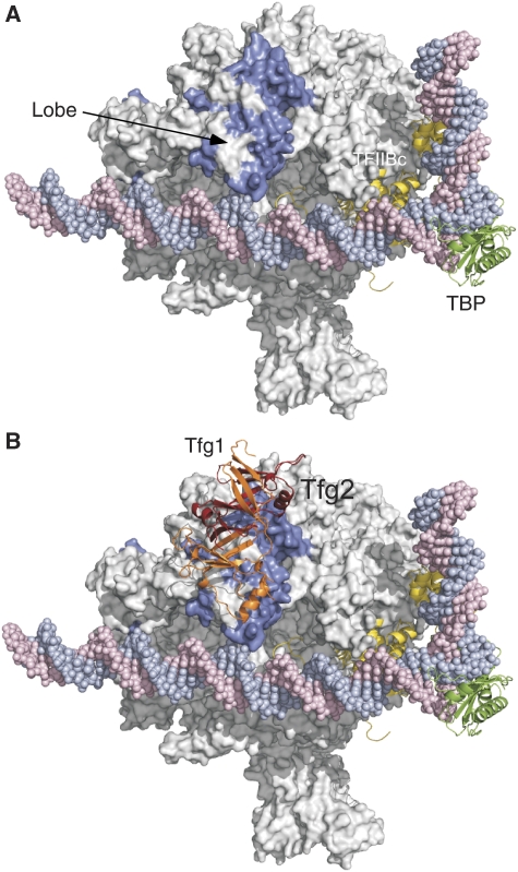
The RNA polymerase (pol) II general transcription factor TFIIF functions at several steps in transcription initiation including preinitiation complex (PIC) formation and start site selection. We find that two structured TFIIF domains bind Pol II at separate locations far from the active site with the TFIIF dimerization domain on the Pol II lobe and the winged helix domain of the TFIIF small subunit Tfg2 above the Pol II protrusion where it may interact with upstream promoter DNA. Binding of the winged helix to the protrusion is PIC specific. Anchoring of these two structured TFIIF domains at separate sites locates an essential and unstructured region of Tfg2 near the Pol II active site cleft where it may interact with flexible regions of Pol II and the general factor TFIIB to promote initiation and start site selection. Consistent with this mechanism, mutations far from the enzyme active site, which alter the binding of either structured TFIIF domains to Pol II, have similar defects in transcription start site usage.


Conflict of interest statement

The authors declare that they have no conflict of interest.

Figures

Figure 1
Figure 1
Summary of functionally important regions of conserved yeast TFIIF subunits. (A, B) The S. mikatae Tfg1 and S. cerevisiae Tfg2 subunits. Shown are the regions homologous to the human Rap74 and Rap30 dimerization domain, the yeast-specific dimerization domain insertions and the winged helix domains. Colours summarize the functionally important regions from Supplementary Table S1. Black, essential for yeast growth; orange, temperature/cold sensitive or slow growth when deleted; white, not required for normal growth. The numbers indicate the amino acid residues in which FeBABE was linked and * indicates the positions of FeBABE attachments that generate cleavage of Rpb1 and/or Rpb2 in PICs.
Figure 2
Figure 2
TFIIF-FeBABE-dependent cleavage of Rpb2 in the PIC. PICs were formed at the HIS4 promoter using nuclear extract supplemented with the indicated TFIIF-FeBABE derivatives, hydroxyl radical cleavage activated with H2O2, and the resulting cleavage products visualized by western blot. *Indicates reproducibly observed cleavage products compared with a reaction using a non-cysteine-containing TFIIF. (A) Cleavage from FeBABE inserted in the TFIIF dimerization domain and Tfg1 linker regions. Left panel uses N-terminal Flag-tagged Rpb2. All other assays use C-terminal Flag-tagged Rpb2. (B, C) Cleavage from FeBABE inserted in the Tfg2 linker and winged helix regions.
Figure 3
Figure 3
Model of the yeast TFIIF dimerization domain binding to Pol II in the PIC. (A) FeBABE cleavage data for all TFIIF-FeBABE derivatives linked to the TFIIF dimerization domain. The cleavage data are from Supplementary Table S2. Ten residues on either side of the calculated cleavage site in Rpb1 and Rpb2 are coloured blue and Pol II is shown in the PIC model (Kostrewa et al, 2009). TBP, green; TFIIB, yellow. (B) Model for the TFIIF dimerization domain binding to the Pol II lobe domain. On the basis of the cleavage data from individual TFIIF-FeBABE derivatives and manual docking, the indicated position of TFIIF can account for the observed Pol II cleavage patterns. Orange, Tfg1; Red, Tfg2.
Figure 4
Figure 4
Position of the Tfg2 winged helix domain in the PIC. (A) Combined cleavage data for all TFIIF-FeBABE derivatives linked to the Tfg2 winged helix domain. Ten residues on either side of the calculated cleavage sites in Rpb2 are coloured blue. One arrow points to α-helices 2 and 3 in the protrusion domain (Cramer et al, 2001), which cannot be assayed for cleavage because they are at the very N-terminus of Rpb2. The base pair highlighted in red and blue indicates the most upstream site of DNA melting in the human system (Miller and Hahn, 2006; Kostrewa et al, 2009). (B) Model for the position of the winged helix domain that best fits the cleavage data. α-helix 3 of the WH domain (the DNA recognition helix) is indicated.
Figure 5
Figure 5
Mapping the location of the Tfg1 and Tfg2 linkers on Pol II in the PIC. (A) FeBABE cleavage data from Supplementary Table S2A showing the positions of Rpb2 cleavage from FeBABE positioned at Tfg1 residues N480 and A541. Cleavage sites are highlighted in purple. (B) FeBABE cleavage data from Supplementary Table S2B showing the positions of Rpb2 cleavage from FeBABE positioned at Tfg2 Q239 (blue) and N274, A285 (violet). (C, D) PIC model with the combined deduced positions of the TFIIF dimerization domain, the Tfg2 winged helix domain, and the Tfg2 flexible linker represented as a dotted line.
Figure 6
Figure 6
Comparison of TFIIF binding to purified Pol II and to Pol II in PICs. TFIIF-FeBABE derivatives were mixed with purified Pol II containing a C-terminal Flag-tagged Rpb2 subunit, and hydroxyl radical production was activated and cleavage visualized by western blot. This was done in parallel with the same TFIIF derivatives incorporated in PICs. (A) FeBABE linked to the TFIIF dimerization domain. (B) FeBABE linked to the Tfg2 winged helix domain. (C) The Pol II–TFIIF dimerization domain model from Figure 3 with cleavage observed in reactions containing purified Pol II and the Tfg2 winged helix-FeBABE derivatives. The cleaved region, highlighted in blue, shows that the winged helix binds near the Pol II lobe in the Pol II–TFIIF complex. Cleavage data are from Supplementary Table S3.
Figure 7
Figure 7
Mutations in the TFIIF dimerization and winged helix domains lead to changes in transcription start site. (A) The Tfg2 winged helix contributes to Pol II–TFIIF binding. Pol II of 1 μg attached to anti-Flag beads was mixed with the indicated amounts of purified TFIIF and Pol II-associated TFIIF recovered by IP was assayed by western blot. Variable amounts of purified TFIIF in lanes 1–5 were used to quantitate TFIIF-associated Pol II in lanes 7–10. The input levels of wild type (WT) and mutant TFIIF derivatives gave equivalent signals in a western blot assay (not shown). (B) Mutation of the Tfg2 winged helix alters start site usage. Nuclear extract depleted for TFIIF was supplemented with purified TFIIF and transcription at the HIS4 promoter was visualized by primer extension. (C) Mutations in the TFIIF dimerization domain, which alter the transcription start site. The TFIIF dimerization domain-PIC model showing residues in green that when mutated shift the transcription start site upstream from the normal position (Ghazy et al, 2004; Freire-Picos et al, 2005). The Rpb9 subunit is shown in yellow and the two bound Zn atoms in Brown. (D) Primer extension analysis of in vivo ADH1 mRNAs from selected Tfg1 and Tfg2 mutations.

Similar articles

Cited by

References

    1. Burton ZF, Killeen M, Sopta M, Ortolan LG, Greenblatt J (1988) RAP30/74: a general initiation factor that binds to RNA polymerase II. Mol Cell Biol 8: 1602–1613 - PMC - PubMed
    1. Bushnell DA, Bamdad C, Kornberg RD (1996) A minimal set of RNA Pol II transcription protein interactions. J Biol Chem 271: 20170–20174 - PubMed
    1. Bushnell DA, Westover KD, Davis RE, Kornberg RD (2004) Structural basis of transcription: an RNA polymerase II-TFIIB cocrystal at 4.5 Angstroms. Science 303: 983–988 - PubMed
    1. Cairns BR, Henry NL, Kornberg RD (1996) TFG3/TAF30/ANC1, a component of the yeast SWI/SNF complex that is similar to the leukemogenic proteins ENL and AF-9. Mol Cell Biol 16: 3308–3316 - PMC - PubMed
    1. Chen HT, Hahn S (2004) Mapping the location of TFIIB within the RNA Polymerase II transcription preinitiation complex: a model for the structure of the PIC. Cell 119: 169–180 - PubMed

Publication types

MeSH terms