

Quantitative assessment of myelopathy patients using motor evoked potentials produced by transcranial magnetic stimulation
- PMID: 20033461
- PMCID: PMC2899952
- DOI: 10.1007/s00586-009-1246-8
Quantitative assessment of myelopathy patients using motor evoked potentials produced by transcranial magnetic stimulation
Abstract
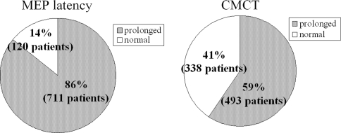
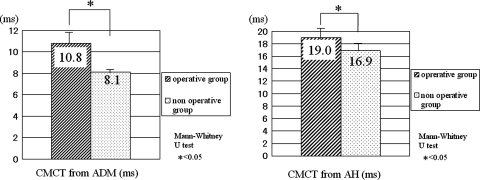
Motor evoked potentials (MEPs) study using transcranial magnetic stimulation (TMS) may give a functional assessment of corticospinal conduction. But there are no large studies on MEPs using TMS in myelopathy patients. The purpose of this study is to confirm the usefulness of MEPs for the assessment of the myelopathy and to investigate the use of MEPs using TMS as a screening tool for myelopathy. We measured the MEPs of 831 patients with symptoms and signs suggestive of myelopathy using TMS. The MEPs from the abductor digiti minimi (ADM) and abductor hallucis (AH) muscles were evoked by transcranial magnetic brain stimulation. Central motor conduction time (CMCT) is calculated by subtracting the peripheral conduction time from the MEP latency. Later, 349 patients had surgery for myelopathy (operative group) and 482 patients were treated conservatively (nonoperative group). CMCTs in the operative group and nonoperative group were assessed. MEPs were prolonged in 711 patients (86%) and CMCTs were prolonged in 493 patients (59%) compared with the control patients. CMCTs from the ADM and AH in the operative group were significantly more prolonged than that in the nonoperative group. All patients in the operative group showed prolongation of MEPs or CMCTs or multiphase of the MEP wave. MEP abnormalities are useful for an electrophysiological evaluation of myelopathy patients. Moreover, MEPs may be effective parameters in spinal pathology for deciding the operative treatment.
Figures
References
-
- Linden RD, Zhang YP, Burke DA, et al. Magnetic motor evoked potential monitoring in the rat. J Neurosurg. 1999;91:205–210. - PubMed
-
- Staudt M, Krageloh-Mann I, Holthausen H, et al. Searching for motor functions in dysgenic cortex: a clinical transcranial magnetic stimulation and functional magnetic resonance imaging study. J Neurosurg. 2004;101:69–77. - PubMed
MeSH terms
LinkOut - more resources
Full Text Sources
Medical
