

Effect of brain shift on the creation of functional atlases for deep brain stimulation surgery
- PMID: 20033503
- PMCID: PMC3114430
- DOI: 10.1007/s11548-009-0391-1
Effect of brain shift on the creation of functional atlases for deep brain stimulation surgery
Abstract
Purpose: In the recent past many groups have tried to build functional atlases of the deep brain using intra-operatively acquired information such as stimulation responses or micro-electrode recordings. An underlying assumption in building such atlases is that anatomical structures do not move between pre-operative imaging and intra-operative recording. In this study, we present evidences that this assumption is not valid. We quantify the effect of brain shift between pre-operative imaging and intra-operative recording on the creation of functional atlases using intra-operative somatotopy recordings and stimulation response data.
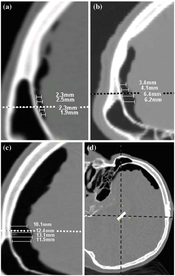
Methods: A total of 73 somatotopy points from 24 bilateral subthalamic nucleus (STN) implantations and 52 eye deviation stimulation response points from 17 bilateral STN implantations were used. These points were spatially normalized on a magnetic resonance imaging (MRI) atlas using a fully automatic non-rigid registration algorithm. Each implantation was categorized as having low, medium or large brain shift based on the amount of pneumocephalus visible on post-operative CT. The locations of somatotopy clusters and stimulation maps were analyzed for each category.
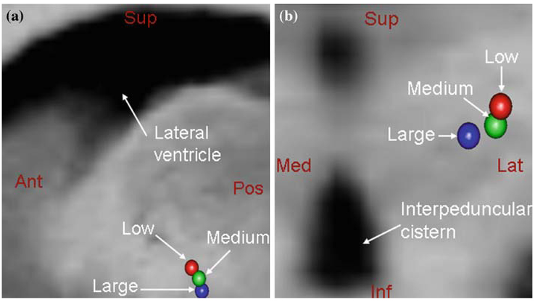
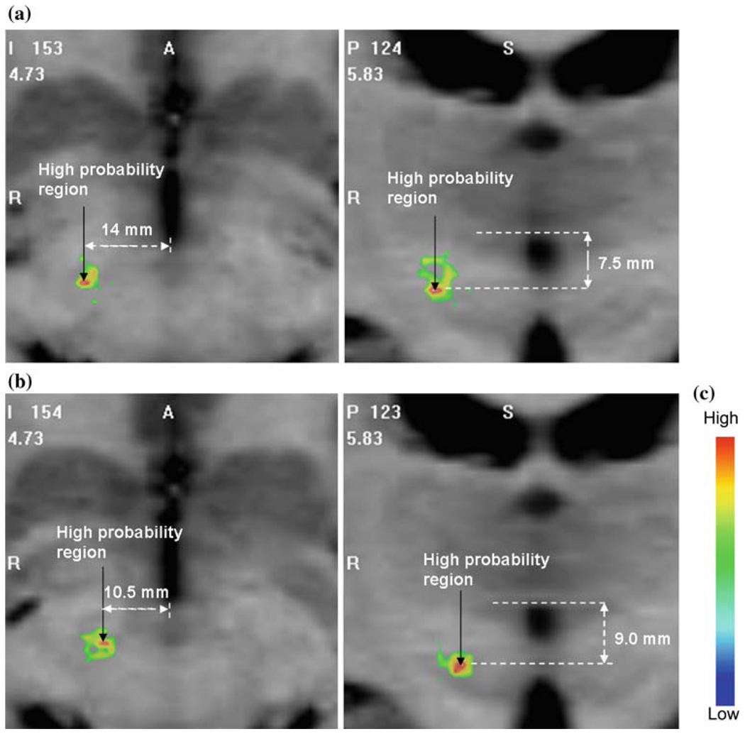
Results: The centroid of the large brain shift cluster of the somatotopy data (posterior, lateral, inferior: 3.06, 11.27, 5.36 mm) was found posterior, medial and inferior to that of the medium cluster (2.90, 13.57, 4.53 mm) which was posterior, medial and inferior to that of the low shift cluster (1.94, 13.92, 3.20 mm). The coordinates are referenced with respect to the mid-commissural point. Euclidean distances between the centroids were 1.68, 2.44 and 3.59 mm, respectively for low-medium, medium-large and low-large shift clusters. We found similar trends for the positions of the stimulation maps. The Euclidian distance between the highest probability locations on the low and medium-large shift maps was 4.06 mm.
Conclusion: The effect of brain shift in deep brain stimulation (DBS) surgery has been demonstrated using intra-operative somatotopy recordings as well as stimulation response data. The results not only indicate that considerable brain shift happens before micro-electrode recordings in DBS but also that brain shift affects the creation of accurate functional atlases. Therefore, care must be taken when building and using such atlases of intra-operative data and also when using intra-operative data to validate anatomical atlases.
Figures
References
-
- Talairach J, Tournoux P. Co-planar stereotaxic atlas of the human brain. New York: Thieme Publishing Group; 1988.
-
- Schaltenbrand G, Wahren W. New York: Thieme Publishing Group; 1977. Atlas for stereotaxy of the human brain.
-
- Yelnik J, Bardinet E, Dormont D, Malandain G, Ourselin S, Tandé D, Karachi C, Ayache N, Cornu P, Agid Y. A three-dimensional, histological and deformable atlas of the human basal ganglia. I. Atlas construction based on immunohistochemical and MRI data. Neuroimage. 2007;34:618–638. - PubMed
-
- Bardinet E, Bhattacharjee M, Dormont D, Pidoux B, Malandain G, Schüpbach M, Ayache N, Cornu P, Agid Y, Yelnik J. A three-dimensional histological atlas of the human basal ganglia. II. Atlas deformation strategy and evaluation in deep brain stimulation for Parkinson disease. J Neurosurg. 2009;110:208–219. - PubMed
-
- Chakravarty MM, Bertrand G, Hodge CP, Sadikot AF, Collins DL. The creation of a brain atlas for image guided neurosurgery using serial histological data. Neuroimage. 2006;30(2):359–376. - PubMed
Publication types
MeSH terms
Grants and funding
LinkOut - more resources
Full Text Sources
Miscellaneous
