

Comparison of diagnostic quality and accuracy in color-coded versus gray-scale DCE-MR imaging display
- PMID: 20033528
- PMCID: PMC8375561
- DOI: 10.1007/s11548-009-0356-4
Comparison of diagnostic quality and accuracy in color-coded versus gray-scale DCE-MR imaging display
Abstract
Purpose: The purpose of this study was to evaluate the diagnostic value and tumor-vascular display properties (microcirculation) of two different functional MRI post-processing and display (color and gray-scale display) techniques used in oncology.
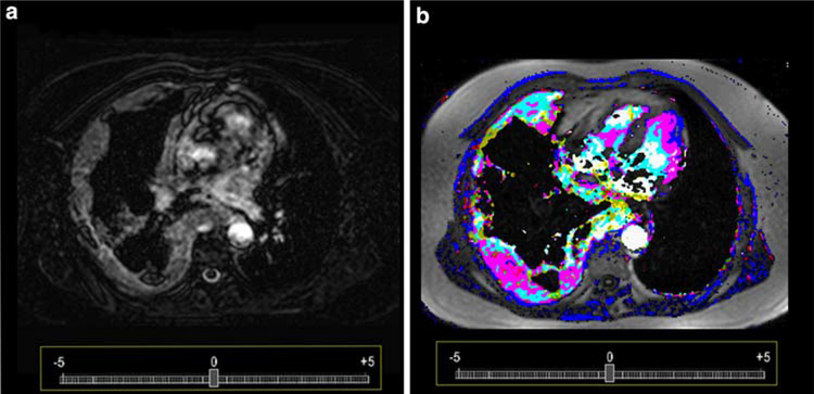
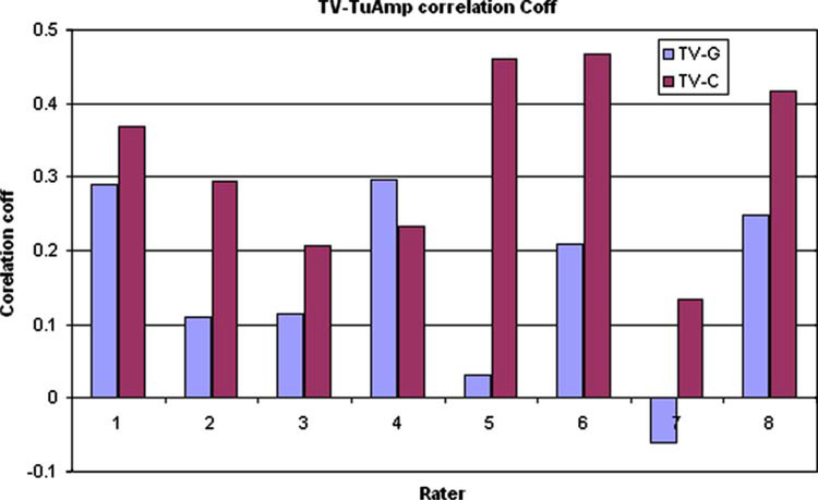
Materials and methods: The study protocol was approved by the IRB and written informed consent was obtained from all patients. 38 dynamic contrast enhanced magnetic resonance imaging (DCE-MRI) data sets of patients with malignant pleural-mesothelioma were acquired and post-processed. DCE-MRI was performed at 1.5 tesla with a T1-weighted 2D gradient-echo-sequence (TR 7.0 ms, TE 3.9 ms, 15 axial slices, 22 sequential repetitions), prior and during chemotherapy. Subtracting first image of contrast-enhanced-dynamic series from the last, produced gray-scale images. Color images were produced using a pharmacokinetic two-compartment model. Eight raters, blinded to diagnosis, by visual assessment of post-processed images evaluated both diagnostic quality of the images and vasculature of the tumor using a rating scale ranging from -5 to +5. The scores for vasculature were assessed by correlating with the maximum amplitude of the total-tumor-ROI for accuracy.
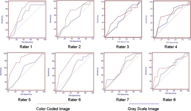
Results: Color coded images were rated as significantly higher in diagnostic quality and tumor vascular score than gray-scale images (p < 0.001, 0.005). ROI signal amplitude analysis and vascular ratings on color coded images were better correlated compared to gray-scale images rating (p < 0.05).
Conclusion: Color coded images were shown to have higher diagnostic quality and accuracy with respect to tumor vasculature in DCE-MRI, therefore their implementation in clinical assessment and follow-up should be considered for wider application.
Figures
References
-
- Giesel FL, Bischoff H, von Tengg-Kobligk H et al. (2006) Dynamic contrast-enhanced MRI of malignant pleural mesothelioma: a feasibility study of noninvasive assessment, therapeutic follow-up, and possible predictor of improved outcome. Chest 129:1570–1576. doi: 10.1378/chest.129.6.1570 - DOI - PubMed
Publication types
MeSH terms
Substances
Grants and funding
LinkOut - more resources
Full Text Sources
Medical
