. 2010:612:335-52.
doi: 10.1007/978-1-60761-362-6_23.

Functional analysis of human NK cells by flow cytometry

Affiliations

Functional analysis of human NK cells by flow cytometry

Yenan T Bryceson et al. Methods Mol Biol. 2010.

Abstract

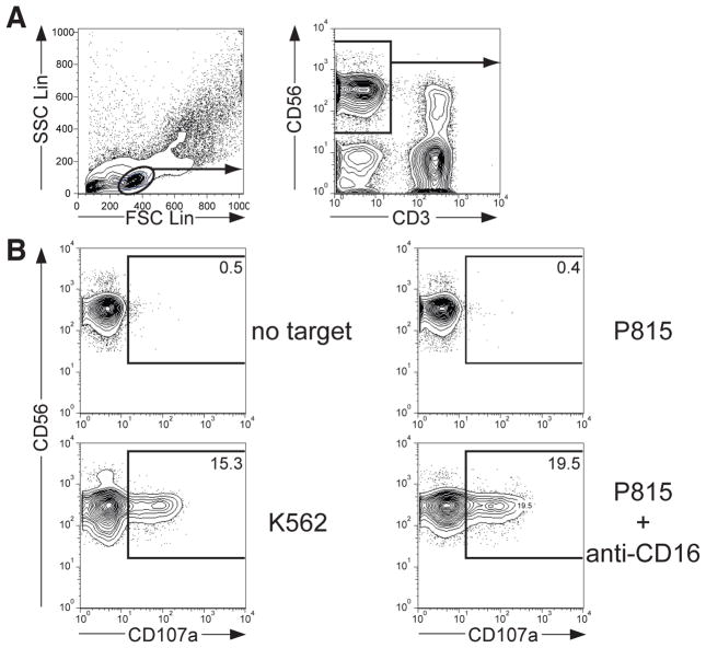
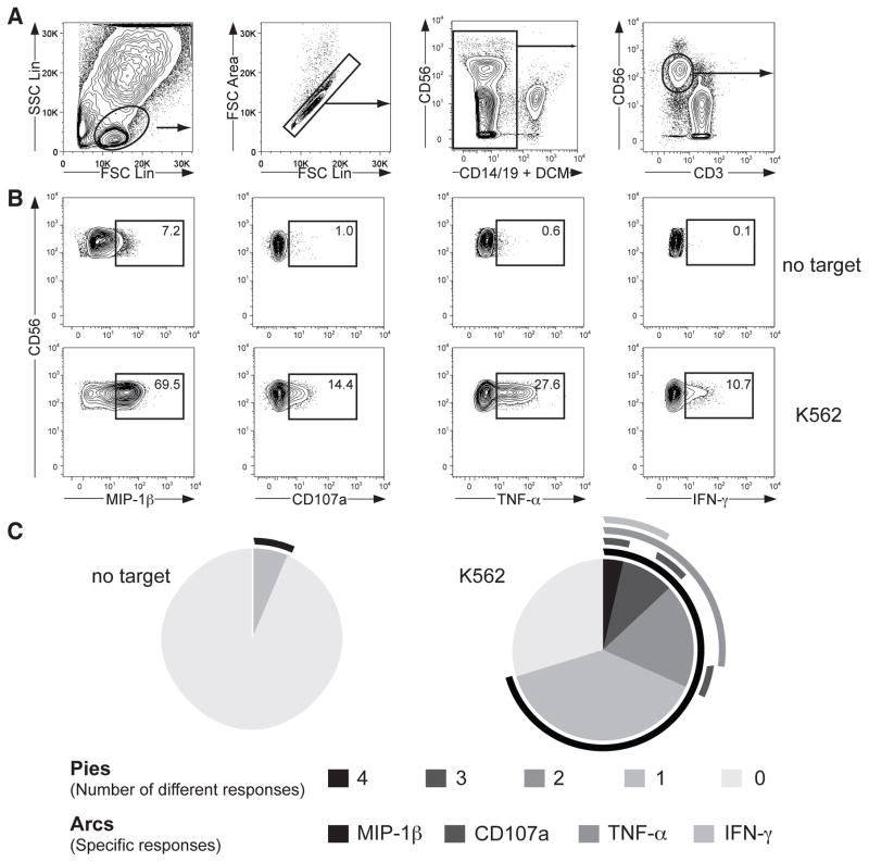
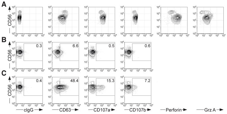
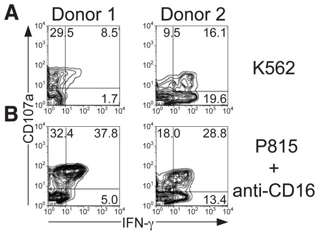
Natural killer (NK) cells are a subset of lymphocytes that contribute to innate immunity through cytokine secretion and target cell lysis. NK cell function is regulated by a multiplicity of activating and inhibitory receptors. The advance in instrumentation for multi-color flow cytometry and the generation of specific mAbs for different epitopes related to phenotypic and functional parameters have facilitated our understanding of NK cell responses. Here, we provide protocols for flow cytometric evaluation of degranulation and cytokine production by human NK cells from peripheral blood at the single-cell level. In addition to offering insight into the regulation of human NK cell responses, these techniques are applicable to the assessment of various clinical conditions, including the diagnosis of immunodeficiency syndromes.


Figures

Figure 1
Figure 1
Analysis of human NK cell degranulation by flow cytometric analysis. PBMC were incubated with target cells for 2 hours at 37°C, surface stained with fluorochrome-conjugated anti-CD3, anti-CD56, and anti-CD107a mAbs. (A) Profiles demonstrate the gating strategy [side scatter (SSC) vs. forward scatter (FCS) and CD56 vs. CD3] for identification of CD3 CD56+ NK cells with FlowJo software. (B) Profiles show CD56 versus CD107a staining on CD3 CD56+ NK cells for one representative donor after incubation of PBMC with indicated target cells.
Figure 2
Figure 2
Analysis of multiple human NK cell responses by flow cytometric analysis. PBMC were incubated with target cells for 6 hours at 37°C, surface stained with fluorochrome-conjugated anti-CD3, anti-CD14, anti-CD19, anti-CD56, and anti-CD107a mAbs, followed by intracellular staining with fluorochrome-conjugated anti-IFN-γ, anti-MIP-1β, and anti-TNF-α mAbs. (A) Gating strategy for identification of CD3 CD56dim NK cells. (B) Profiles show staining for multiple responses on CD3 CD56dim NK cells from one representative donor after incubation with target cells as indicated for 6 hours at 37°C. One representative donor is shown. (C) Pies represent the distribution of cells responding with different numbers of distinct responses, as indicated. Surrounding the pies, lines indicate the nature of individual responses.
Figure 3
Figure 3
Comparsion of different lysosomal markers for the identification of degranulating NK cells. (A) PBMC were surface stained with fluorochrome-conjugated anti-CD3 and anti-CD56 mAbs, followed by intracellular staining with fluorochrome-conjugated anti-CD63, anti-CD107a, anti-CD107b, anti-perforin, or anti-granzyme A mAbs, as indicated. Profiles show CD56 versus intracellular lysosomal protein staining, as indicated on CD3 CD56+ NK cells. PBMC were incubated either alone (B), or with K562 cells (C) for 2 hours at 37°C, surface stained with fluorochrome-conjugated anti-CD3 and anti-CD56 mAbs, in addition to anti-CD63, anti-CD107a, or anti-CD107b mAbs, as indicated. Profiles show CD56 versus surface staining of lysosomal proteins, as indicated on CD3 CD56+ NK cells.
Figure 4
Figure 4
Donor variation in NK cell responses. NK cells were stimulated with (A) K562 cells or (B) P815 cells supplemented with anti-CD16 mAb and incubated for 6 hours at 37°C. Profiles show CD107a versus IFN-γ staining on CD3 CD56dim NK cells from two different donor.

Similar articles

Cited by

References

    1. Vivier E, Tomasello E, Baratin M, Walzer T, Ugolini S. Functions of natural killer cells. Nat Immunol. 2008;9:503–10. - PubMed
    1. Raulet DH. Interplay of natural killer cells and their receptors with the adaptive immune response. Nat Immunol. 2004;5:996–1002. - PubMed
    1. Strowig T, Brilot F, Munz C. Noncytotoxic functions of NK cells: direct pathogen restriction and assistance to adaptive immunity. J Immunol. 2008;180:7785–91. - PMC - PubMed
    1. Bryceson YT, Long EO. Line of attack: NK cell specificity and integration of signals. Curr Opin Immunol 2008 - PMC - PubMed
    1. Peters PJ, Borst J, Oorschot V, Fukuda M, Krahenbuhl O, Tschopp J, Slot JW, Geuze HJ. Cytotoxic T lymphocyte granules are secretory lysosomes, containing both perforin and granzymes. J Exp Med. 1991;173:1099–109. - PMC - PubMed

Publication types

LinkOut - more resources