

Unusual bone formation in the anterior rim of foramen magnum: cause, effect and treatment
- PMID: 20033741
- PMCID: PMC2899649
- DOI: 10.1007/s00586-009-1250-z
Unusual bone formation in the anterior rim of foramen magnum: cause, effect and treatment
Abstract
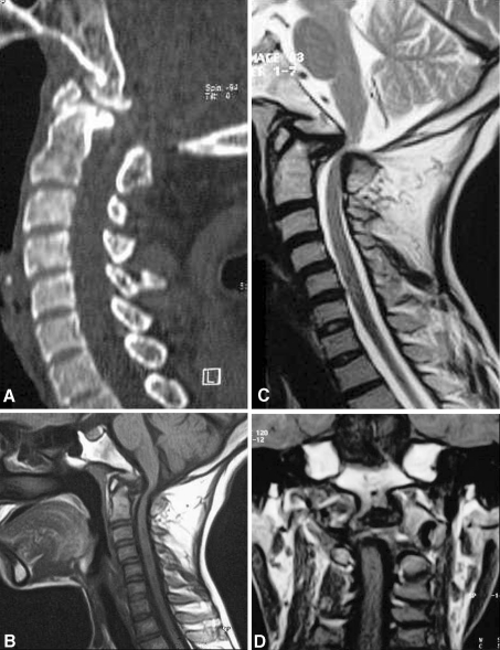
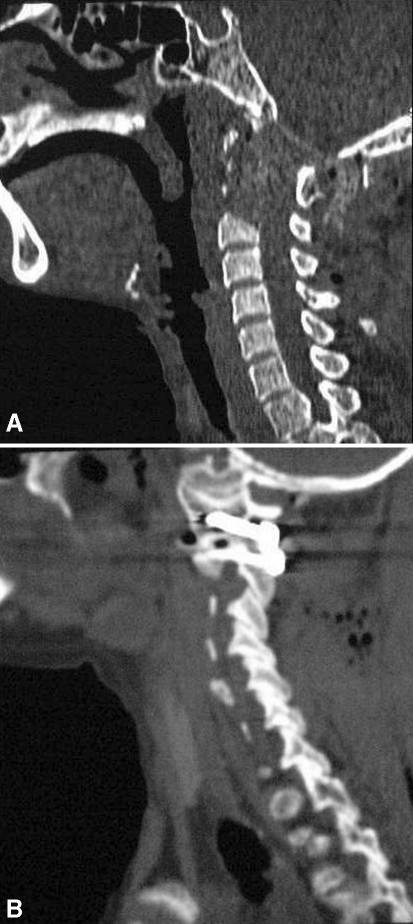
A rare case of proatlas segmental abnormality resulting in a bony mass in the anterior rim of the foramen magnum is studied. Case report of a 19-year-old female showed a progressive weakness of all four limbs for about 3 years. When admitted she could not perform any useful activities by herself. Investigations revealed an unusual bone growth in the region of the anterior rim of foramen magnum that resulted in severe cord compression. The abnormal bone formation involved the lower end of clivus, the tip of the odontoid process and the posterior arch of the atlas. Dynamic imaging did not reveal any clear evidence of instability. Following transoral decompression and posterior fixation, the patient showed dramatic and lasting clinical recovery. Conclusions were drawn as follows. Anomalies of the most caudal part of the occipital sclerotomes due to the failure of proatlas segmentation can be the cause of an abnormal bone mass in the anterior rim of foramen magnum. Transoral decompression, followed by posterior atlantoaxial fixation, results in neurological recovery and provides lasting cure from the problem.
Figures
Similar articles
-
The proatlas: a comprehensive review with clinical implications.Childs Nerv Syst. 2012 Mar;28(3):349-56. doi: 10.1007/s00381-012-1698-8. Epub 2012 Jan 27. Childs Nerv Syst. 2012. PMID: 22282080 Review.
-
Elongated Clivus with Deficient Anterior Atlantal Arch and Congenital Posterior Atlantooccipital Dislocation: Pathoembryology and Management Nuances of a Rare Form of Proatlas Segmentation Anomaly.World Neurosurg. 2019 Jun;126:286-290. doi: 10.1016/j.wneu.2019.03.091. Epub 2019 Mar 18. World Neurosurg. 2019. PMID: 30898752
-
Unilateral atlantal lateral mass hypertrophy associated with atlanto-occipital fusion.Eur Spine J. 2013 May;22 Suppl 3(Suppl 3):S429-33. doi: 10.1007/s00586-012-2574-7. Epub 2012 Nov 19. Eur Spine J. 2013. PMID: 23161418 Free PMC article.
-
Morphometry of the outlet of the foramen magnum in crania with atlantooccipital fusion.J Neurosurg Spine. 2011 Jul;15(1):55-9. doi: 10.3171/2011.3.SPINE10828. Epub 2011 Apr 8. J Neurosurg Spine. 2011. PMID: 21476797
-
A case of occipitalization in the human skull.Folia Morphol (Warsz). 2010 Aug;69(3):134-7. Folia Morphol (Warsz). 2010. PMID: 21154282 Review.
Cited by
-
Condylus tertius with atlanto-axial rotatory fixation: an unreported association.Skeletal Radiol. 2014 Apr;43(4):535-9. doi: 10.1007/s00256-013-1747-8. Epub 2013 Oct 23. Skeletal Radiol. 2014. PMID: 24150830
-
The proatlas: a comprehensive review with clinical implications.Childs Nerv Syst. 2012 Mar;28(3):349-56. doi: 10.1007/s00381-012-1698-8. Epub 2012 Jan 27. Childs Nerv Syst. 2012. PMID: 22282080 Review.
-
Proatlas segmentation anomalies: Surgical management of five cases and review of the literature.J Pediatr Neurosci. 2016 Jan-Mar;11(1):14-9. doi: 10.4103/1817-1745.181246. J Pediatr Neurosci. 2016. PMID: 27195027 Free PMC article.
References
Publication types
MeSH terms
LinkOut - more resources
Full Text Sources
