

Comparison of artery organ culture and co-culture models for studying endothelial cell migration and its effect on smooth muscle cell proliferation and migration
- PMID: 20033777
- PMCID: PMC2852582
- DOI: 10.1007/s10439-009-9877-9
Comparison of artery organ culture and co-culture models for studying endothelial cell migration and its effect on smooth muscle cell proliferation and migration
Abstract
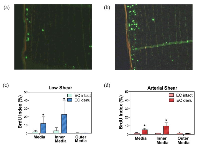
Arterial restenosis associated with intimal hyperplasia is the major cause of long-term failure of vascular interventions. Endothelium injury and the proliferation and migration of smooth muscle cells (SMC) are key events in the development of intimal hyperplasia. The objectives of this study were to develop an ex vivo artery injury model for studying endothelial cell (EC) migration and to compare it with an in vitro co-culture arterial wall injury model in terms of the effect of flow on EC migration and its effect on SMC migration and proliferation. Our results demonstrated that shear flow improves reendothelialization in the injured area by promoting EC migration. The migration distance of ECs is much smaller in the arteries than in an in vitro cell culture model (3.57+/-1.29 mm vs. 5.2+/-1.4 cm, p<0.001). SMC proliferation was significantly less in the EC intact and reendothelialization areas than in the EC denuded areas indicating that reendothelialization suppresses SMC proliferation. Our models provide a new approach to study techniques to enhance endothelium healing.
Figures


Similar articles
-
Pressure alters endothelial effects upon vascular smooth muscle cells by decreasing smooth muscle cell proliferation and increasing smooth muscle cell apoptosis.Surgery. 2004 Aug;136(2):282-90. doi: 10.1016/j.surg.2004.04.033. Surgery. 2004. PMID: 15300192
-
Regulation of vascular smooth muscle cell turnover by endothelial cell-secreted microRNA-126: role of shear stress.Circ Res. 2013 Jun 21;113(1):40-51. doi: 10.1161/CIRCRESAHA.113.280883. Epub 2013 Apr 19. Circ Res. 2013. PMID: 23603512 Free PMC article.
-
Normal shear stress and vascular smooth muscle cells modulate migration of endothelial cells through histone deacetylase 6 activation and tubulin acetylation.Ann Biomed Eng. 2010 Mar;38(3):729-37. doi: 10.1007/s10439-009-9896-6. Ann Biomed Eng. 2010. PMID: 20069369
-
[Advance in study of vascular endothelial cell and smooth muscle cell co-culture system].Zhongguo Zhong Yao Za Zhi. 2012 Feb;37(3):265-8. Zhongguo Zhong Yao Za Zhi. 2012. PMID: 22568220 Review. Chinese.
-
Cellular Crosstalk between Endothelial and Smooth Muscle Cells in Vascular Wall Remodeling.Int J Mol Sci. 2021 Jul 6;22(14):7284. doi: 10.3390/ijms22147284. Int J Mol Sci. 2021. PMID: 34298897 Free PMC article. Review.
Cited by
-
Biomechanical regulation of vascular smooth muscle cell functions: from in vitro to in vivo understanding.J R Soc Interface. 2013 Oct 23;11(90):20130852. doi: 10.1098/rsif.2013.0852. Print 2014 Jan 6. J R Soc Interface. 2013. PMID: 24152813 Free PMC article. Review.
-
Effects of Axial Stretch on Cell Proliferation and Intimal Thickness in Arteries in Organ Culture.Cell Mol Bioeng. 2010 Sep 1;3(3):286-295. doi: 10.1007/s12195-010-0128-9. Cell Mol Bioeng. 2010. PMID: 21116478 Free PMC article.
-
Artery buckling stimulates cell proliferation and NF-κB signaling.Am J Physiol Heart Circ Physiol. 2014 Aug 15;307(4):H542-51. doi: 10.1152/ajpheart.00079.2014. Am J Physiol Heart Circ Physiol. 2014. PMID: 24929858 Free PMC article.
-
Alterations in Pulse Pressure Affect Artery Function.Cell Mol Bioeng. 2012 Dec 1;5(4):474-487. doi: 10.1007/s12195-012-0251-x. Cell Mol Bioeng. 2012. PMID: 23243477 Free PMC article.
-
Endothelial-Smooth Muscle Cell Interactions in a Shear-Exposed Intimal Hyperplasia on-a-Dish Model to Evaluate Therapeutic Strategies.Adv Sci (Weinh). 2022 Oct;9(28):e2202317. doi: 10.1002/advs.202202317. Epub 2022 Aug 15. Adv Sci (Weinh). 2022. PMID: 35971167 Free PMC article.
References
-
- Albuquerque ML, Waters CM, Savla U, Schnaper HW, Flozak AS. Shear stress enhances human endothelial cell wound closure in vitro. Am J Physiol. 2000;279(1):H293–302. - PubMed
-
- Bailey SR. Endovascular stents: update on stents in practice. J Long Term Eff Med Implants. 2000;10(1-2):7–18. - PubMed
-
- Bardy N, Karillon GJ, Merval R, Samuel JL, Tedgui A. Differential effects of pressure and flow on DNA and protein synthesis and on fibronectin expression by arteries in a novel organ culture system. Circ Res. 1995;77(4):684–94. - PubMed
-
- Chesler NC, Ku DN, Galis ZS. Transmural pressure induces matrix-degrading activity in porcine arteries ex vivo. Am J Physiol. 1999;277(5 Pt 2):H2002–9. - PubMed
-
- Chien S, Li S, Shiu YT, Li YS. Molecular basis of mechanical modulation of endothelial cell migration. Front Biosci. 2005;10:1985–2000. - PubMed
Publication types
MeSH terms
Grants and funding
LinkOut - more resources
Full Text Sources
