

Newly established cell lines from mouse oral epithelium regenerate teeth when combined with dental mesenchyme
- PMID: 20033791
- PMCID: PMC2862945
- DOI: 10.1007/s11626-009-9265-7
Newly established cell lines from mouse oral epithelium regenerate teeth when combined with dental mesenchyme
Abstract
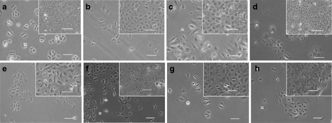
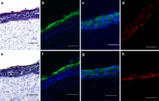
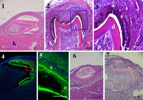
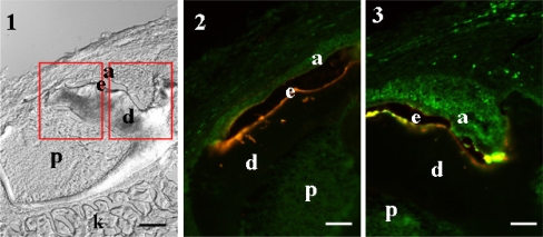
The present study attempted to examine whether clonal cell lines of the oral epithelium can differentiate into ameloblasts and regenerate tooth when combined with dental germ mesenchyme. Clonal cell lines with a distinct morphology were established from the oral epithelium of p53-deficient fetal mice at embryonic day 18 (E18). The strain of mouse is shown to be a useful source for establishing clonal and immortalized cell lines from various tissues and at various stages of development. Tooth morphogenesis is almost completed and the oral epithelium is segregated from the dental epithelium at E18. In RT-PCR analysis of cell lines, mucosal epithelial markers (cytokeratin 14) were detected, but ameloblast markers such as amelogenin and ameloblastin were not detected when cells were cultured on plastic dish. They formed stratified epithelia and expressed a specific differentiation marker (CK13) in the upper layer when cultured on feeder layer or on collagen gel for 1-3 wk, demonstrating that they are of oral mucosa origin. Next, bioengineered tooth germs were prepared with cell lines and fetal molar mesenchymal tissues and implanted under kidney capsule for 2-3 wk. Five among six cell lines regenerated calcified structures as seen in natural tooth. Our results indicate that some oral epithelial cells at E18 possess the capability to differentiate into ameloblasts. Furthermore, cell lines established in the present study are useful models to study processes in tooth organogenesis and tooth regeneration.
Figures


Similar articles
-
Tooth regeneration from newly established cell lines from a molar tooth germ epithelium.Biochem Biophys Res Commun. 2007 Apr 13;355(3):758-63. doi: 10.1016/j.bbrc.2007.02.039. Epub 2007 Feb 15. Biochem Biophys Res Commun. 2007. PMID: 17321500
-
Inductive ability of human developing and differentiated dental mesenchyme.Cells Tissues Organs. 2013;198(2):99-110. doi: 10.1159/000353116. Epub 2013 Jul 30. Cells Tissues Organs. 2013. PMID: 24192998
-
Conserved odontogenic potential in embryonic dental tissues.J Dent Res. 2014 May;93(5):490-5. doi: 10.1177/0022034514523988. Epub 2014 Feb 19. J Dent Res. 2014. PMID: 24554539
-
From Pluripotent Stem Cells to Organoids and Bioprinting: Recent Advances in Dental Epithelium and Ameloblast Models to Study Tooth Biology and Regeneration.Stem Cell Rev Rep. 2024 Jul;20(5):1184-1199. doi: 10.1007/s12015-024-10702-w. Epub 2024 Mar 18. Stem Cell Rev Rep. 2024. PMID: 38498295 Free PMC article. Review.
-
Contributions of heterospecific tissue recombinations to odontogenesis.Int J Dev Biol. 1995 Feb;39(1):291-7. Int J Dev Biol. 1995. PMID: 7626419 Review.
Cited by
-
Establishment of Hertwig's epithelial root sheath/epithelial rests of Malassez cell line from human periodontium.Mol Cells. 2014 Jul;37(7):562-7. doi: 10.14348/molcells.2014.0161. Epub 2014 Jul 31. Mol Cells. 2014. PMID: 25081036 Free PMC article.
-
Cell culture models of oral mucosal barriers: A review with a focus on applications, culture conditions and barrier properties.Tissue Barriers. 2018;6(3):1479568. doi: 10.1080/21688370.2018.1479568. Epub 2018 Sep 25. Tissue Barriers. 2018. PMID: 30252599 Free PMC article. Review.
-
Adult human gingival epithelial cells as a source for whole-tooth bioengineering.J Dent Res. 2013 Apr;92(4):329-34. doi: 10.1177/0022034513481041. Epub 2013 Mar 4. J Dent Res. 2013. PMID: 23458883 Free PMC article.
-
Molecular factors resulting in tooth agenesis and contemporary approaches for regeneration: a review.Eur Arch Paediatr Dent. 2012 Dec;13(6):297-304. doi: 10.1007/BF03320830. Eur Arch Paediatr Dent. 2012. PMID: 23235129 Review.
-
A clonal stem cell line established from a mouse mammary placode with ability to generate functional mammary glands.In Vitro Cell Dev Biol Anim. 2019 Dec;55(10):861-871. doi: 10.1007/s11626-019-00406-8. Epub 2019 Sep 16. In Vitro Cell Dev Biol Anim. 2019. PMID: 31529417
References
-
- Byrne C, Tainsky M, Fuchs E. Programming gene expression in developing epidermis. Development. 1994;120:2369–2383. - PubMed
Publication types
MeSH terms
Substances
LinkOut - more resources
Full Text Sources
Other Literature Sources
Research Materials
Miscellaneous
