. 2010 Mar 1;518(5):622-35.
doi: 10.1002/cne.22223.

Synaptic development of the mouse dorsal lateral geniculate nucleus

Affiliations

Synaptic development of the mouse dorsal lateral geniculate nucleus

Martha E Bickford et al. J Comp Neurol. .

Abstract

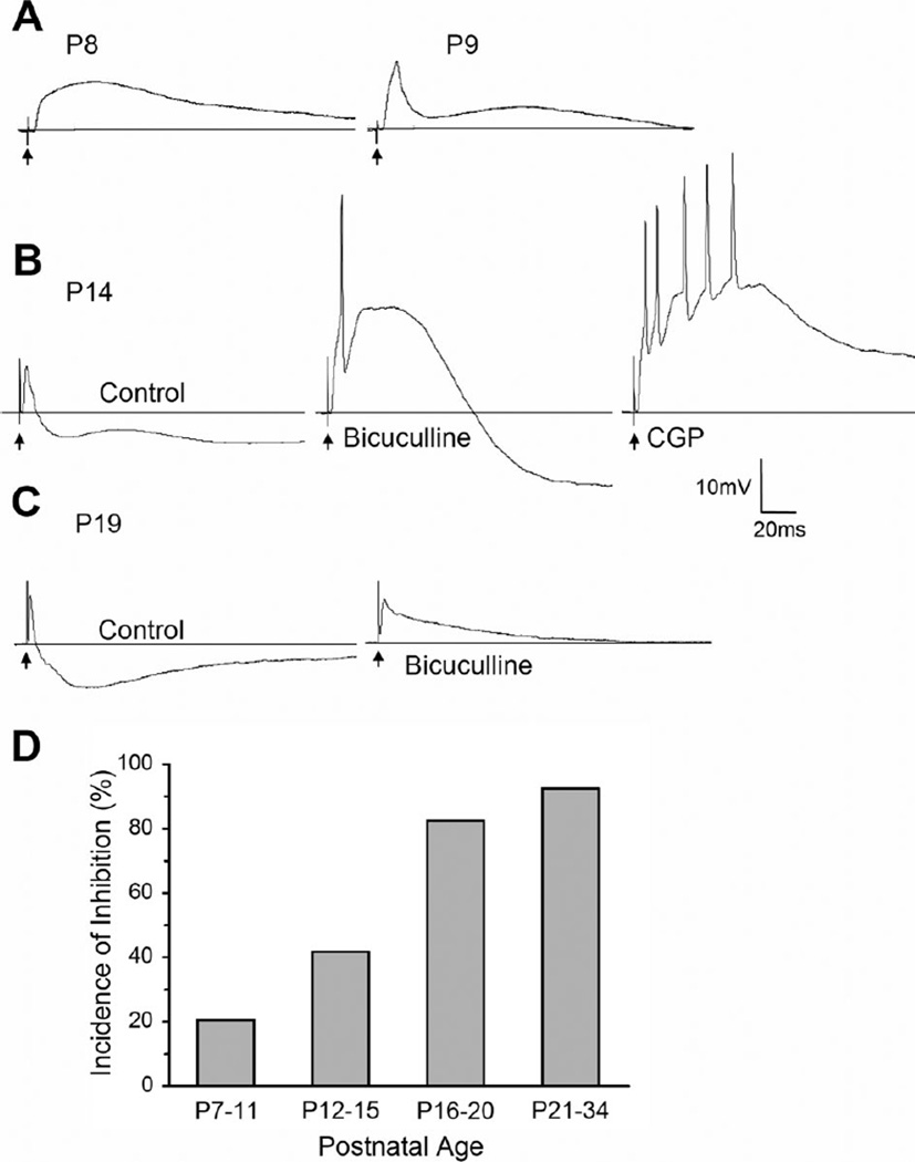
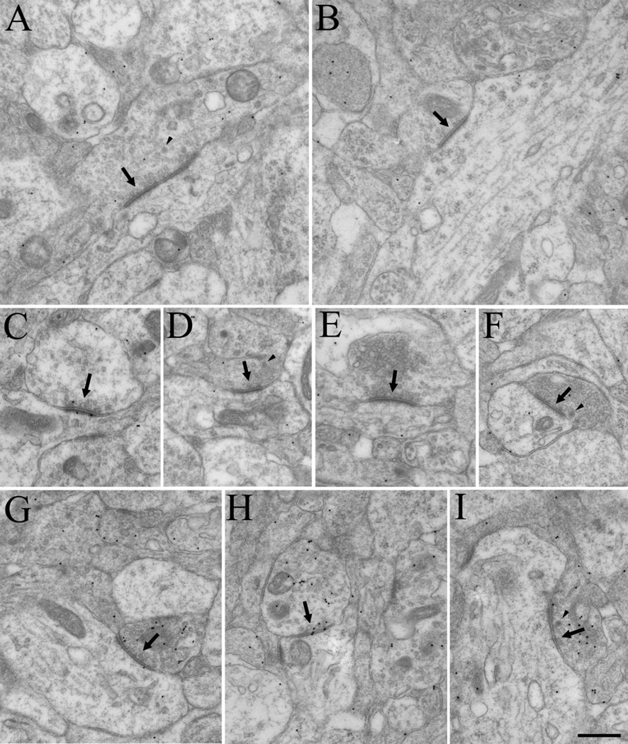
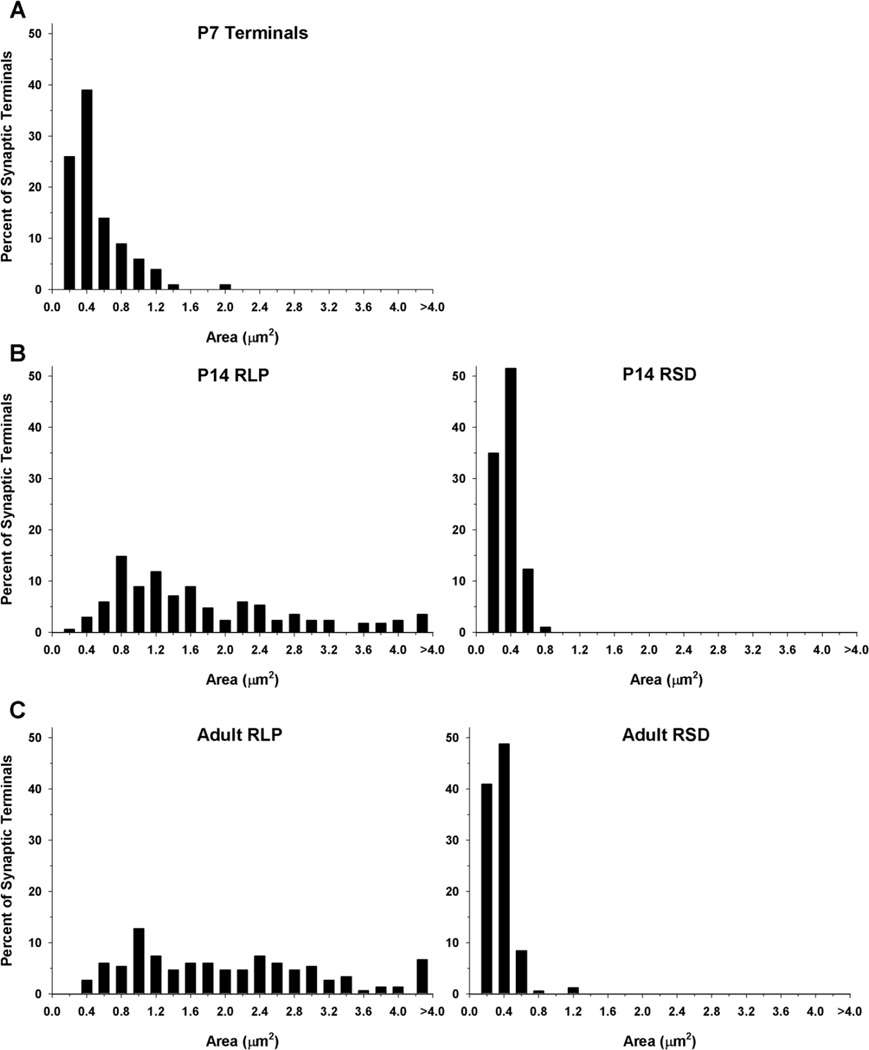
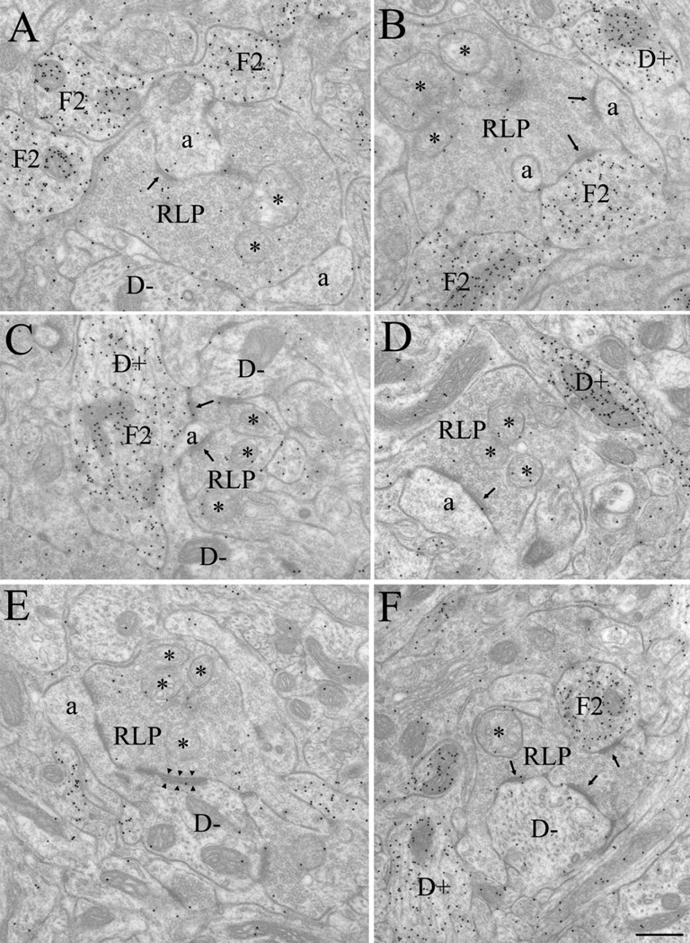
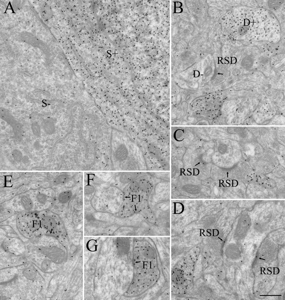
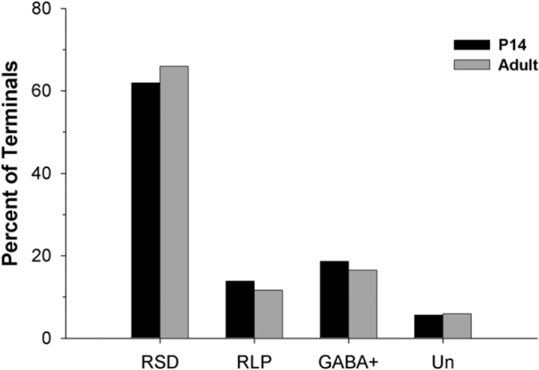
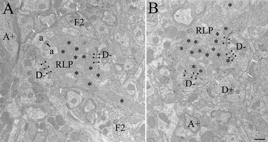
The dorsal lateral geniculate nucleus (dLGN) of the mouse has emerged as a model system in the study of thalamic circuit development. However, there is still a lack of information regarding how and when various types of retinal and nonretinal synapses develop. We examined the synaptic organization of the developing mouse dLGN in the common pigmented C57/BL6 strain, by recording the synaptic responses evoked by electrical stimulation of optic tract axons, and by investigating the ultrastructure of identified synapses. At early postnatal ages (<P12), optic tract evoked responses were primarily excitatory. The full complement of inhibitory responses did not emerge until after eye opening (>P14), when optic tract stimulation routinely evoked an excitatory postsynaptic potential/inhibitory postsynaptic potential (EPSP/IPSP) sequence, with the latter having both a GABA(A) and GABA(B) component. Electrophysiological and ultrastructural observations were consistent. At P7, many synapses were present, but synaptic profiles lacked the ultrastructural features characteristic of the adult dLGN, and little gamma-aminobutyric acid (GABA) could be detected by using immunocytochemical techniques. In contrast, by P14, GABA staining was robust, mature synaptic profiles of retinal and nonretinal origin were easily distinguished, and the size and proportion of synaptic contacts were similar to those of the adult. The emergence of nonretinal synapses coincides with pruning of retinogeniculate connections, and the transition of retinal activity from spontaneous to visually driven. These results indicate that the synaptic architecture of the mouse dLGN is similar to that of other higher mammals, and thus provides further support for its use as a model system for visual system development.


Figures

Figure 1
Figure 1
Postsynaptic responses evoked by optic tract stimulation in the developing mouse dLGN. A–C: Examples of synaptic responses recorded at different postnatal ages (P). A: Between P7 and 11, synaptic responses are largely excitatory. Left: Example of a pure EPSP recorded at P8. Right: Example of an EPSP followed by a small IPSP at P9. B: At older ages (>P12) inhibition was more pronounced and contained a GABAA and GABAB component. Example of synaptic responses recorded at P14 before (control ACSF, left) and after bath application of the GABAA antagonist bicuculline (25 µM, middle) and GABAB antagonist CGP (10 µM, right). Initially, the response contained an EPSP followed by IPSP activity that had two hyperpolarizing components: an early fast one (GABAA) followed by a somewhat slower long-duration one (GABAB). Bicuculline blocked the early hyperpolarizing response and unmasked a high-amplitude, long-duration EPSP. CGP blocked the additional slower, long-lasting hyperpolarization, leading to a further increase in the duration of the preceding EPSP. C: Example of a response at P19 before (left) and after (right) bath-applied bicuculline. The IPSP activity greatly curtailed the duration of the initial EPSP D: Summary plot showing the incidence of synaptic responses containing inhibition at different postnatal ages. There was an age-related increase in the incidence of inhibition. The number of cells recorded at each age group were: P7–11, n = 24; P12–15, n = 31; P16–20, n = 23; P21–34, n = 13. In A–C, responses were recorded at −60–mV, and arrows indicate stimulus artifacts.
Figure 2
Figure 2
Electron micrographs depict the synaptic architecture of dLGN at P7. Although synapses could be easily identified (arrows, A–I), specific types of terminals could not be categorized based on ultrastructure. Pale mitochondria could not be definitively identified, and vesicles were in general, sparsely distributed (A–F). Dense-core vesicles (arrowheads) were frequently observed, but did not appear to correlate with other ultrastructural features. Although there was some specificity in the distribution of gold particles overlying presynaptic profiles (G–I), the staining for GABA was sparse and indistinct. Scale bar = 0.5 µm in I (applies to A–I).
Figure 3
Figure 3
Frequency histogram illustrates the distribution of terminal sizes in dLGN at P7 (A), P14 (B), and adult (C). The sizes of RLP (large profiles that contain round vesicles and pale mitochondria) and RSD profiles (small profiles that contain round vesicles and dark mitochondria) were similar in the P14 and adult dLGN. Synaptic terminals types cannot be distinguished at P7, and were smaller than RLP profiles identified at later ages. At P7, n = 100 terminals; P14, n = 97 RSD and n = 168 RLP; adult, n = 166 RSD and n = 149 RLP profiles.
Figure 4
Figure 4
Electron micrographs illustrate the synaptic architecture of the P14 dLGN. At this age, retinal terminals were readily identified as RLP profiles (A–F), i.e., large profiles that contain round vesicles and pale mitochondria (*). RLP profiles contacted (arrows) non-GABAergic dendrites (D−), identified by a low density of gold particles, the presence of microtubules and occasional mitochondria, and the absence of synaptic vesicles (A,C,E,F). RLP profiles also contacted non-GABAergic dendritic appendages (a), identified as small profiles that contained diffusely organized neurofilaments, no microtubules, few mitochondria, a low density of gold particles, and no synaptic vesicles (A–E). Appendages were often observed to invaginate RLP profiles (B). RLP profiles also contacted F2 profiles (identified as profiles that contain loosely packed synaptic vesicles and a high density of gold particles (A–C,F). In C, an F2 profile originated from a GABAergic dendrite (D+), identified by the presence of microtubules and occasional mitochondria, a high density of gold particles, and few or no synaptic vesicles (C,D,F). RLP profiles also formed occasional adherent contacts with adjacent profiles (arrowheads, illustrated in E). Scale bar = 0.5 µm in F (applies to A–F).
Figure 5
Figure 5
Electron micrographs illustrate GABAergic and non-GABAergic profiles at P14. These were distinguished based on differences in the density of overlying gold particles. A: Adjacent GABAergic (S+) and non-GABAergic (S−) somata. B: GABAergic (D+) and non-GABAergic (D−) dendrites. RSD profiles were identified as small non-GABAergic terminals that contained densely packed vesicles and formed asymmetric synapses (arrows; B–D). F1 profiles were identified as GABAergic terminals that contained densely packed vesicles and formed symmetric synapses (arrows; E–G) Scale bar = 0.5 µm in D (applies to A–G).
Figure 6
Figure 6
Plot shows the proportion of RSD, RLP, GABAergic, and unclassified terminals at P14 (n = 585) and in adult (n = 558) mouse dLGN. At both ages, the majority of synaptic contacts (61.88 – 65.95%) were made by RSD profiles; 16.49–18.63% were classified as GABAergic (GABA+) based on immunocytochemical staining, whereas only 11.65–13.85% comprised RLP profiles. The remaining 5.64–5.91% could not be classified (Un).
Figure 7
Figure 7
Plot shows the proportion of retinogeniculate terminals at P14, P21, and adult that contact GABAergic (GABA+) and non-GABAergic (GABA−) profiles. At each age, the large majority (>90%) of terminals made contacts with non-GABAergic profiles, but a small percentage also contacted GABAergic F2 profiles. At P14, n = 104, P21, n = 217; and adult, n = 101 RLP profiles.
Figure 8
Figure 8
Electron micrographs illustrate retinal terminals in the adult dLGN, identified as RLP profiles: large profiles that contain round vesicles and pale mitochondria (*). In the adult, RLP profiles primarily contacted non-GABAergic dendrites (D−, identified as profiles that contain microtubules and occasional mitochondria, a low density of gold particles, and no synaptic vesicles) via puncta adherentia (arrowheads). Most synaptic contacts (black arrow) were made on dendritic appendages (a, identified as small profiles that contain diffusely organized neurofilaments, no microtubules, few mitochondria, a low density of gold particles, and no synaptic vesicles (as illustrated in A), which were often observed to invaginate RLP profiles. RLP profiles also contacted F2 profiles (identified as profiles that contain loosely packed synaptic vesicles and a high density of gold particles (as illustrated in A). In the adult, RLP synaptic arrangements were often surrounded by glial lamellae (white arrows). GABAergic dendrites (D+, identified as profiles that contain microtubules and occasional mitochondria, a high density of gold particles, and no synaptic vesicles, as illustrated in B) and GABAergic axons (A+, identified as profiles that contain a high density of gold particles and are surrounded by myelin, as illustrated in A and B) could also be identified. Scale bar = 0.5 µm in B (applies to A,B).

Similar articles

Cited by

References

    1. Arcelli P, Frassoni C, Regondi MC, De Biasi S, Spreafico R. GABAergic neurons in mammalian thalamus: a marker of thalamic complexity? Brain Res Bull. 1997;42:27–37. - PubMed
    1. Bentivoglio M, Spreafico R, Alvarez-Bolado G, Sanchez MP, Fairen A. Differential expression of the GABAA receptor complex in the dorsal thalamus and reticular nucleus: an immunohistochemical study in the adult and developing rat. Eur J Neurosci. 1991;3:118–125. - PubMed
    1. Bickford ME, Wei H, Eisenback MA, Chomsung RD, Slusarczyk AS, Dankowsi AB. Synaptic organization of thalamocortical axon collaterals in the perigeniculate nucleus and dorsal lateral geniculate nucleus. J Comp Neurol. 2008;508:264–285. - PMC - PubMed
    1. Blitz DM, Regehr WG. Timing and specificity of feed-forward inhibition within the LGN. Neuron. 2005;45:917–928. - PubMed
    1. Butts DA, Kanold PO, Shatz CJ. A burst-based “Hebbian” learning rule at retinogeniculate synapses links retinal waves to activity-dependent refinement. PLoS Biol. 2007;5:e61. - PMC - PubMed

Publication types

MeSH terms