. 2010 Mar 1;518(5):699-721.
doi: 10.1002/cne.22240.

Peripheral facial nerve axotomy in mice causes sprouting of motor axons into perineuronal central white matter: time course and molecular characterization

Affiliations

Peripheral facial nerve axotomy in mice causes sprouting of motor axons into perineuronal central white matter: time course and molecular characterization

Milan Makwana et al. J Comp Neurol. .

Erratum in

  • J Comp Neurol. 2012 Jun 1;520(8):ii. Pararajasingham, Abirami [corrected to Pararajasingam, Abirami]

Abstract

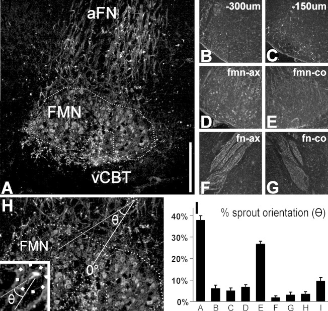
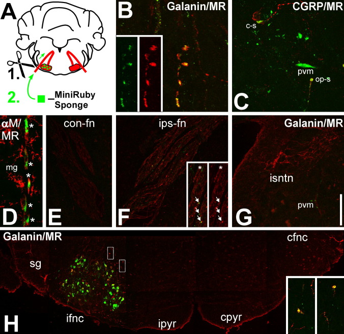
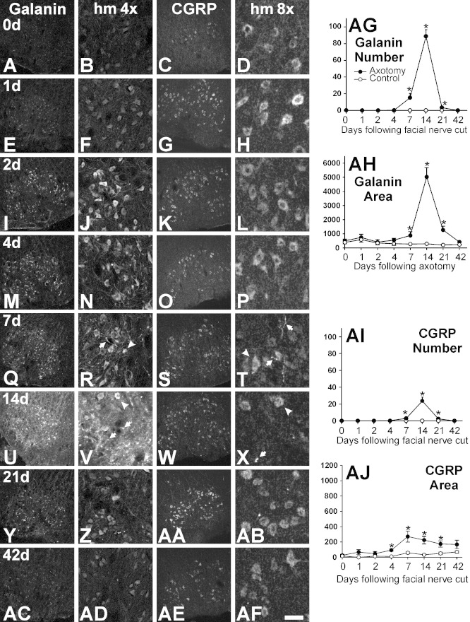
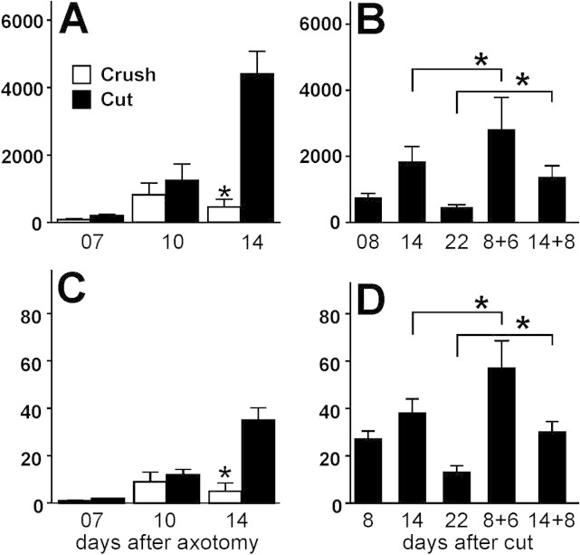
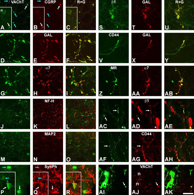
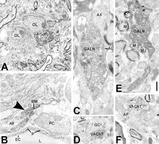
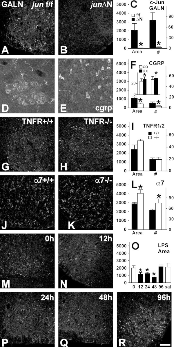
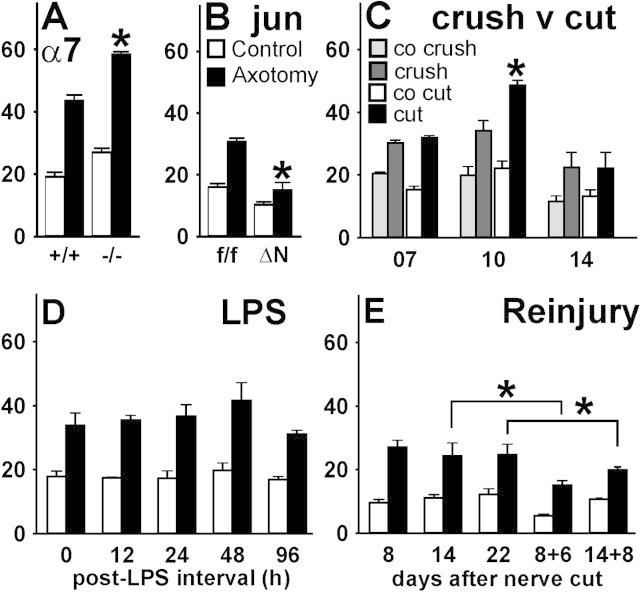
Generation of new axonal sprouts plays an important role in neural repair. In the current study, we examined the appearance, composition and effects of gene deletions on intrabrainstem sprouts following peripheral facial nerve axotomy. Axotomy was followed by the appearance of galanin(+) and calcitonin gene-related peptide (CGRP)(+) sprouts peaking at day 14, matching both large, neuropeptide(+) subpopulations of axotomized facial motoneurons, but with CGRP(+) sprouts considerably rarer. Strong immunoreactivity for vesicular acetylcholine transporter (VAChT) and retrogradely transported MiniRuby following its application on freshly cut proximal facial nerve stump confirmed their axotomized motoneuron origin; the sprouts expressed CD44 and alpha7beta1 integrin adhesion molecules and grew apparently unhindered along neighboring central white matter tracts. Quantification of the galanin(+) sprouts revealed a stronger response following cut compared with crush (day 7-14) as well as enhanced sprouting after recut (day 8 + 6 vs. 14; 14 + 8 vs. 22), arguing against delayed appearance of sprouting being the result of the initial phase of reinnervation. Sprouting was strongly diminished in brain Jun-deficient mice but enhanced in alpha7 null animals that showed apparently compensatory up-regulation in beta1, suggesting important regulatory roles for transcription factors and the sprout-associated adhesion molecules. Analysis of inflammatory stimuli revealed a 50% reduction 12-48 hours following systemic endotoxin associated with neural inflammation and a tendency toward more sprouts in TNFR1/2 null mutants (P = 10%) with a reduced inflammatory response, indicating detrimental effects of excessive inflammation. Moreover, the study points to the usefulness of the facial axotomy model in exploring physiological and molecular stimuli regulating central sprouting.


Figures

Figure 1
Figure 1
Distribution of galanin-immunoreactive sprouts in and around the facial motor nucleus (FMN), 14 days following facial nerve cut. A: Composite of the facial nucleus and surrounding areas; the nucleus outline is indicated by the dotted line. Note the numerous immunoreactive sprouts in the white matter surrounding the lesioned facial motor nucleus, particularly in the ventral corticobasal tract (vCBT) but also in the ascending part of the intracerebral portion of the facial nerve (aFN), and to the medial and lateral of the facial nucleus. B–G: Sprout distribution in the rostral direction, from the same ventrolateral brainstem position but 300 μm (B) and 150 μm (C) caudal to the axotomized facial nucleus, the axotomized nucleus (D), and the descending part of the intracerebral facial nerve (F). Note the paucity of strongly stained neurites in the contralateral, uninjured facial nucleus (E) and nerve (G). H: The orientation of central sprouts was determined by drawing two lines, one from the center of the terminal bulb to that of the nucleus (dashed line, 0°) and the second from the bulb center to that of the farthest visible part of the stalk and extending it farther into the image (dotted line) and then measuring the angle between the two lines as shown in H. The inset in H shows the terminal bulb, the attached stalk, and the orientation of the two lines at higher magnification. I: Distribution of the orientation angles for the galanin-immunoreactive sprouts outside the axotomized facial motor nucleus with respect to the nucleus. Percentage of the total number of sprouts, with orientation angles of 0–20°(A), 20–40°(B), 40–60°(C), etc., with 0° pointing directly away from and 180° pointing directly toward the nucleus. Mean ± SEM, n = 4 mice. Note that the most common orientation is the one facing away (A), with the second largest running approximately parallel to the facial nucleus (E). Scale bar = 0.5 mm in A; 0.8 mm for B–G; 0.36 mm for H; 0.1 mm for inset.
Figure 2
Figure 2
Demonstration of central axonal sprout origin using Mini-Ruby, a dual anterograde/retrograde tracer. A: Schematic summary. A gelfoam sponge soaked in 1% Mini-Ruby solution was applied onto the fresh, proximal cut end of the facial nerve, followed by retrograde transport to axotomized motoneurons and a 14-day survival. B,C: High magnification of Mini-Ruby (green) colocalization with the immunoreactivity (IR) for the neuropeptides galanin (B) and CGRP (C) (red) in axonal sprouts just outside the axotomized facial motor nucleus. The double-labeled sprout in B and the bottom sprout in C are outward pointing (op-s); the top sprout in C is oriented in parallel to the center of the nucleus (“cruising,” c-s). The insets in B show the individual red and green fluorescence channels. In C, Mini-Ruby is also incorporated by perivascular macrophages (pvm). D: Mini-Ruby uptake in a string of alphaMbeta2 integrin (aM)-positive (red) perivascular macrophages (*) lining a cerebral blood vessel. The neighboring aM-positive and ramified microglia (mg) are devoid of Mini-Ruby. E–G: Double fluorescence for Mini-Ruby and galanin-IR in the descending intracerebral part of the contralateral facial nerve (E); the axotomized, ipsilateral facial nerve (F); and the ipsilateral spinal nucleus of trigeminal nerve (G, isntn). The insets in F show a higher magnification of the facial nerve (left, red and green; right, red only fluorescence). Note the double-labeled sprouts in the axotomized nerve (arrows) and their absence in the neighboring trigeminal nerve nucleus and contralateral nerve. The asterisk points to a Mini-Ruby+ but galanin sprout. As in C, Mini-Ruby is frequently present in the populations of perivascular macrophages associated with larger blood vessels (D, pvm). The micrographs in E and F show the same galanin labeling motif as in Figure 1E,F but combine it with the Mini-Ruby fluorescence. H: Composite of Mini-Ruby and galanin-IR fluorescence in the ventral brainstem across the ipsilateral substantia gelatinosa, the ipsilateral and contralateral facial motor nuclei (ifnc, left; cfnc, right, respectively), and the ipsilateral and contralateral pyramidal tracts (ipyr and cpyr). Mini-Ruby neuronal cell body labeling is strictly limited to the axotomized facial motor nucleus, with a high density of galanin-positive sprouts in the surrounding tissue. Note the absence of both in the pyramidal tracts and the contralateral facial nucleus. The insets show higher magnifications for galanin/Mini-Ruby double labeling (yellow) of two sprouts just dorsal of the axotomized facial nucleus; their positions in the composite are indicated by the rectangles in H. A magenta/green version of Figure 2 is available as Supporting Information Figure 1. Scale bar = 10 μm in B, 45 μm in C and D, 270 μm in E–G; 350 μm in F insets.
Figure 3
Figure 3
A–AJ: Time course of the appearance (A–AF) and quantification (AG–AJ) of galanin- and CGRP-immunoreactive sprouts in the facial motor nucleus, 1–42 days after facial nerve axotomy; day 0 are uninjured controls. A–AF show the immunofluorescence in injured facial nucleus at low magnification (A,E,I,M,Q,U,Y,AC for galanin, and C,G,K,O,S,W,AA,AE for CGRP) and at ×4 or ×8 higher magnification for galanin (B,F,J,N,R,V,Z,AD) and CGRP (D,H,L,P,T,X,AB,AF). Note the massive increase in the neuropeptide-labeled sprouts (arrows) at days 7–21; arrowheads point to neighboring motoneuron cell bodies. AG–AJ: The graphs at right show the total number of galanin-and CGRP-immunoreactive sprouts per section (AG and AI, respectively) and the quantification of the area taken by the galanin-and CGRP-immunoreactive sprouts (AH, AJ) in parts per million (ppm). Mean ± SEM, n = 3 animals per group in AG and AI, n = 4 in AH and AJ). *P < 0.05, Student's t-test compared with the unoperated, contralateral side. In the case of galanin, both parameters, number (AG) and area (AH), show a sharp peak at day 14. The same also holds true for the CGRP+ sprout number (AI), but the CGRP+ area recognized by the Optimas GCD algorithm (AJ) shows a broader, elevated plateau between day 4 and day 21. Scale bar = 250 μm in first and third columns; 0.063 μm in the second column; 0.032 μm in fourth column.
Figure 4
Figure 4
A–D: Central axonal sprouting depends on the mode of injury. A,C: After facial nerve crush, the sprout area in ppm (A) and the number of sprouts per section (C) reach a moderate peak at day 10 and after cut a much higher peak at day 14. *P < 0.05, Student's t-test for crush vs. cut (n = 3 animals per group, mean ± SEM). B,D: Additional injury exacerbates central axonal sprouting following facial nerve cut—both in total area (B) and in the number of sprouts (D)—compared with the same total cut period (8 + 6 vs. 14 days, 14 + 8 vs. 22 days). *P < 0.05, one-way ANOVA and post-hoc Tukey, n = 4–5 animals per group.
Figure 5
Figure 5
Molecular characterization of growth cones in the facial nucleus 14 days following facial nerve cut. A–R: Immunoreactivity for vesicular acetylcholine transporter (VAChT), double labeling with CGRP (A–C), galanin (D–F), alpha7 integrin subunit (G–I), neurofilament heavy (NFH) isoform (J–L), microtubule-associated protein-2/MAP2 (M–O), and synaptophysin/SynPh (P–R) immunoreactivities. VAChT+ sprouts were very frequently positive for galanin, frequently also for alpha 7 and synaptophysin, and more rarely for CGRP immunoreactivity (white arrows in A–C, blue arrows mark single-labeled VAChT+ sprouts). Note the absence of double labeling for NFH and MAP2. A–C and P–R are inside the facial motor nucleus, D–L in the adjacent ventral white matter, and M–O at the gray/white matter interface. A–C and P–R are composite micrographs, to illustrate the colocalization of some but not all VAChT+ sprouts with CGRP and synaptophysin. S–Y: Colocalization of galanin-positive sprouts with the beta1 integrin subunit (S–U) and with CD44 (V–Y). Z–AK: Colocalization of Mini-Ruby-labeled growth cones with the alpha7 (Z–AB) and beta 1 (AC–AE) integrin subunits, CD44 (AF–AH), and VAChT (AI–AK). White arrows point to double-labeled sprouts in AC,AD, in AF,AG, and in AI,AJ). Asterisks in AC and AD label a Mini-Ruby+, perivascular macrophage. Micrographs in AI–AK are from the border region between facial nucleus (left) and medial white matter and also show two adjacent, Mini-Ruby-labeled motoneurons (n) at left, surrounded by large, VAChT+ synapses. Z–AH are inside the dorsal white matter, next to the facial nucleus. A magenta/green version of Figure 5 is available as Supporting Information Figure 2. Scale bar = 50 μm for A–C,J–L; 27 μm for Z–AB,AI–AK; 12.5 μm for P–R; 40 μm in all other micrographs.
Figure 6
Figure 6
Ultrastructure of axonal growth cones and their cellular contacts using alpha7 (A,B), galanin (C,E), and VAChT (D,F) immunoreactivity in the white matter surrounding the facial motor nucleus, 14 days following facial nerve cut in WT animals. A: Intensely cytoplasmically stained axonal growth cones (alpha7), containing numerous (unstained) mitochondria and synaptic vesicles and contacting adjacent oligodendrocytes (OL). B: Strong alpha7 immunoreactivity in submembranous astrocyte cytoplasm (arrowhead) next to the finger-like protrusions of the basal membrane (BM). Some alpha7 immunoreactivity was also present in blood vessel pericytes (P). L, blood vessel lumen; EC, endothelial cell; PC, perivascular cell; AF, astrocyte fibrils. C,E: Galanin (GALN)-immunoreactive growth cones, here contacting astrocyte lamella (AL; C), unmyelinated axons (AX; C), and numerous myelinated axons (E). D,F: VAChT-immunoreactive axonal sprouts growing alongside a VAChT-negative (GC*) growth cone (D) and an astrocyte fibril (AF)-containing process (F). Scale bar = 1 μm for A,B,F; 0.35 μm for C; 1.2 μm for D; 0.6 μm in E.
Figure 7
Figure 7
Effects of neural c-Jun (junΔN; A–F), TNFR1/2 (G–I), and alpha7 (J–L) gene deletions and LPS-induced inflammation (M–R) on galanin (A–C,G–R)-and CGRP (D–F)-immunoreactive central axonal sprouting in the facial motor nucleus 14 days after cut. Left row, littermate controls; center, mutants; right, quantification of the effects on the area (left Y-axis, parts per million) and number (#, right Y-axis) of galanin-or CGRP-positive sprouts (mean ± SEM, n = 3–6 animals per group). Solid bars, littermate controls; open bars, mutants. In the case of endotoxin (O), single intraperitoneal injection of 1 mg E. coli LPS /kg caused a transient decrease of sprouting 12–48 hours after injection (solid bars). Uninjected animals (0) and mice 24 hours after saline (sal) injection served as controls (open bars). *P < 5%, Student's t-test (C,I) or ANOVA followed by post hoc Tukey test (O). The inset in F shows the overall CGRP immunoreactivity (in OLV values) in the axotomized (ax, solid bars) and contralateral (co, open bars) for the junf/f controls (left) and junΔN mutants (right). Scale bar = 200 μm in R (applies to A,C,G–R); 80 μm for D,E.
Figure 8
Figure 8
Effects of alpha7 (A) and neural jun (B) deletions, crush vs. cut (C), application of LPS (D), and additional nerve injury (E) on beta1 integrin subunit levels in the axotomized and contralateral facial motor nuclei. *P < 5%, Student t-test (A–C) or ANOVA and post hoc Tukey test (E). Beta1 immunoreactivity was quantified by using the Mean-SD algorithm and is shown in OLV values, n = 3–6 animals per group, as in Figure 7. A,B,D show the results from facial motor nuclei 14 days after nerve cut; C compares crush with cut 7–14 days after axotomy; E shows the effects of second (cut) injury. co (C), contralateral side to the lesion.

Similar articles

Cited by

References

    1. Behrens A, Sibilia M, David JP, Möhle-Steinlein U, Tronche F, Schütz G, Wagner EF. Impaired postnatal hepatocyte proliferation and liver regeneration in mice lacking c-jun in the liver. EMBO J. 2002;21:1782–1790. - PMC - PubMed
    1. Berkelaar M, Clarke DB, Wang YC, Bray GM, Aguayo AJ. Axotomy results in delayed death and apoptosis of retinal ganglion cells in adult rats. J Neurosci. 1994;14:4368–4374. - PMC - PubMed
    1. Blue ML, Davis G, Conrad P, Kelley K. Specific cleavage of the α4 integrin associated with activation of peripheral T lymphocytes. Immunol. 1993;78:80–85. - PMC - PubMed
    1. Bohatschek M, Werner A, Raivich G. Systemic LPS injection leads to granulocyte influx into normal and injured brain: effects of ICAM-1 deficiency. Exp Neurol. 2001;172:137–152. - PubMed
    1. Bohatschek M, Kloss CU, Hristova M, Pfeffer K, Raivich G. Microglial major histocompatibility complex glycoprotein-1 in the axotomized facial motor nucleus: regulation and role of tumor necrosis factor receptors 1 and 2. J Comp Neurol. 2004;470:382–399. - PubMed

Publication types

MeSH terms

Substances