. 2010 Mar 1;518(5):567-85.
doi: 10.1002/cne.22256.

Altered balance of gamma-aminobutyric acidergic and glutamatergic afferent inputs in rostral ventrolateral medulla-projecting neurons in the paraventricular nucleus of the hypothalamus of renovascular hypertensive rats

Affiliations

Altered balance of gamma-aminobutyric acidergic and glutamatergic afferent inputs in rostral ventrolateral medulla-projecting neurons in the paraventricular nucleus of the hypothalamus of renovascular hypertensive rats

Vinicia Campana Biancardi et al. J Comp Neurol. .

Abstract

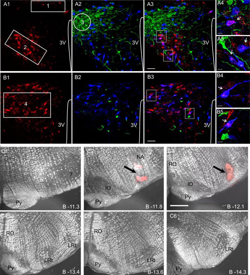
An imbalance of excitatory and inhibitory functions has been shown to contribute to numerous pathological disorders. Accumulating evidence supports the idea that a change in hypothalamic gamma-aminobutyric acid (GABA)-ergic inhibitory and glutamatergic excitatory synaptic functions contributes to exacerbated neurohumoral drive in prevalent cardiovascular disorders, including hypertension. However, the precise underlying mechanisms and neuronal substrates are still not fully elucidated. In the present study, we combined quantitative immunohistochemistry with neuronal tract tracing to determine whether plastic remodeling of afferent GABAergic and glutamatergic inputs into identified RVLM-projecting neurons of the hypothalamic paraventricular nucleus (PVN-RVLM) contributes to an imbalanced excitatory/inhibitory function in renovascular hypertensive rats (RVH). Our results indicate that both GABAergic and glutamatergic innervation densities increased in oxytocin-positive, PVN-RVLM (OT-PVN-RVLM) neurons in RVH rats. Despite this concomitant increase, time-dependent and compartment-specific differences in the reorganization of these inputs resulted in an altered balance of excitatory/inhibitory inputs in somatic and dendritic compartments. A net predominance of excitatory over inhibitory inputs was found in OT-PVN-RVLM proximal dendrites. Our results indicate that, along with previously described changes in neurotransmitter release probability and postsynaptic receptor function, remodeling of GABAergic and glutamatergic afferent inputs contributes as an underlying mechanism to the altered excitatory/inhibitory balance in the PVN of hypertensive rats.


Figures

Figure 1
Figure 1
Identification of various PVN subnuclei, RVLM-projecting neurons, and OT-and VP-ir neurons in the PVN. A: Representative confocal photomicrographs obtained at the medial level of PVN (~1.8–2.1 mm caudal to bregma). Retrogradely labeled PVN-RVLM neurons (A1, red) and OT-and VP-ir neurons (A2, blue and green, respectively) are shown. In A3, both images were superimposed, and the areas contained within the white squares were reimaged at higher magnification using different thresholds and are displayed in A4 and A5. The numbered regions in A1 and A2 depict the dorsal cap (1, DC), ventromedial parvocellular (2, VM), and lateral magnocellular (3, LM) subnuclei. B: Representative confocal photomicrographs obtained at the caudal level of PVN (~2.1–2.3 mm caudal to bregma). Retrogradely labeled PVN-RVLM neurons (B1, red) and OT-and VP-ir neurons (B2, blue and green, respectively) are shown in B1 and B2. In B3, both images were superimposed, and the areas contained within the white squares were reimaged at higher magnification using different thresholds and are displayed in B4 and B5. Arrows point to examples of double-labeled OT-PVN-RVLM neurons. C: Representative example of a rhodamine bead injection site in the RVLM, displayed at various rostrocaudal levels in the ventral medulla (C1–C6). The arrows in C2,3 point to the center of the injection site. 3V, third ventricle; Py, pyramidal tract; IO, inferior olive; RO, nucleus raphe obscurus; LRt, lateral reticular nucleus. A magenta-green copy of this figure is available as Supporting Information Figure 1. Scale bars = 50 μm in A,B; 12 μm in insets; 100 μm in C.
Figure 2
Figure 2
Triple immunofluorescent staining for oxytocin (A; green), GAD67 (B; red), and VGLUT2 (C; white) in the caudal PVN. In D, all images are superimposed. In E, the region contained within the rectangular region in D is shown at higher magnification. Note the characteristic neuropile punctuate staining of GAD67 and VGLUT2. 3V, third ventricle. A blue-green copy of this figure is available as Supporting Information Figure 2. Scale bars == 50 μm in A; 50 μm in D (applies to B–D); 25 μm in E.
Figure 3
Figure 3
GAD67 immunoreactivity in the PVN of normotensive sham and RVH rats. A: Representative confocal photomicro-graphs obtained at the medial level of PVN (1.8 –2.1 mm caudal to bregma) in sham (A1) and late-stage RVH (A2) rats, showing retrogradely labeled PVN-RVLM neurons (red) and GAD67 immunore-activity (white). For better comparisons, representative regions of A1 and A2 are displayed at higher magnification in A3 and A4, respectively. B: Representative confocal photomicrographs obtained at the caudal level of PVN (2.1–2.3 mm caudal to bregma) in sham (B1) and RVH (B2) rats, showing retrogradely labeled PVN-RVLM neurons (red) and GAD67 immunoreactivity (white). For better comparisons, representative regions of B1 and B2 are displayed at higher magnification in B3 and B4, respectively. C: Summary showing a significant increase in the relative GAD67 immunoreactivity density in RVH rats in all PVN subnuclei analyzed. 3V, third ventricle. *P < 0.05 vs. sham, n = 6 in each group. Scale bars = 50 μm in A (applies to A,B); 10 μm in insets.
Figure 4
Figure 4
VGLUT2 immunoreactivity in the PVN of normotensive sham and RVH rats. A: *P < 0.05 vs. sham, n = 6 in each group. Representative confocal photomicrographs obtained at the medial level of PVN (1.8–2.1 mm caudal to bregma) in sham (A1) and RVH (A2) rats, showing retrogradely labeled PVN-RVLM neurons (red) and VGLUT2 immunoreactivity (white). For better comparisons, representative regions of A1 and A2 are displayed at higher magnification in A3 and A4, respectively. B: Representative confocal photomicro-graphs obtained at the caudal level of PVN (2.1–2.3 mm caudal to bregma) in sham (B1) and RVH (B2) rats, showing retrogradely labeled PVN-RVLM neurons (red) and VGLUT2 immunoreactivity (white). For better comparisons, representative regions of B1 and B2 are displayed at higher magnification in B3 and B4, respectively. C: Summary showing a significant increase in the relative VGLUT2 immunoreactivity density in the PaPo subnucleus. 3V, third ventricle. *P < 0.05 vs. sham, n = 6 in each group. Scale bars = 50 μm in A (applies to A,B); 10 μm in insets.
Figure 5
Figure 5
Three-dimensional rendered images of OT-positive, retrogradely labeled PVN-RVLM neurons and associated GAD67-and VGLUT2-ir boutons. A: Representative example of an OT-ir (A1, green), PVN-RVLM retrogradely labeled (rhodamine beads, white, A2) neuron located in the PaPo subnucleus. In A3, the two images were superimposed. B: Example of a PVN-RVLM retrogradely labeled, OT-positive neuron located in the PaPo subnucleus and associated GAD67-ir boutons, shown in two different spatial orientations. For better display, only the OT staining (green) is shown. Boutons in red and white colors represent those overlapping and those that are close to, but not overlapping, the OT-positive PVN-RVLM neuron, respectively. C: Example of a different PVN-RVLM retrogradely labeled, OT-positive neuron located in the PaPo subnucleus and associated VGLUT2-ir boutons, shown in two different spatial orientations. For better display, only the OT staining is shown. Boutons in red and white colors represent those overlapping and those that are close to, but not overlapping, the OT-positive PVN-RVLM neuron, respectively. Insets show representative overlapping GAD67 and VGLUT2 boutons, respectively (white boxes), at a higher magnification. A magenta-green copy of this figure is available as Supporting Information Figure 3. Scale bars = 10 μm in A; 10 μm in B (applies to B,C); 1.5 μm in insets.
Figure 6
Figure 6
Summary data showing differences in GAD67-ir bouton densities in OT-positive, retrogradely labeled PVN-RVLM neurons in the PaPo subnucleus from sham rats (n = 18 neurons) and early-stage (e-RVH, n = 6 neurons) and late-stage (l-RVH, n = 13 neurons) hypertensive rats. Differences were quantified at the whole-cell level (A1) and somatic (A2) and proximal dendritic (A3) compartments. *P < 0.05, **P < 0.0001, Bonferroni post hoc test.
Figure 7
Figure 7
Summary data showing differences in VGLUT2-ir bouton densities in OT-positive, retrogradely labeled PVN-RVLM neurons in the PaPo subnucleus from sham rats (n = 19 neurons) and early-stage (e-RVH, n = 7 neurons) and late-stage (l-RVH, n = 13 neurons) hypertensive rats. Differences were quantified at the whole-cell level (A1) and somatic (A2) and proximal dendritic (A3) compartments. *P < 0.05, **P < 0.0001, Bonferroni post hoc test.
Figure 8
Figure 8
Summary data showing the proximal dendritic/somatic relative GAD67 (A) and VGLUT2 (B) immunoreactivity densities in OT-positive, retrogradely labeled PVN-RVLM neurons in the PaPo subnucleus from sham (n = 18 and 19 for GAD67 and VGLUT2, respectively) and early-stage (e-RVH, n = 6 and 7 for GAD67 and VGLUT2, respectively) and late-stage (l-RVH, n = 13 for both GAD67 and VGLUT2) hypertensive rats. Note that, in all groups, GAD67-and VGLUT2-ir bouton densities were more prominent in proximal dendrites vs. cell bodies (i.e., proximal dendritic/somatic ratio > 1), although a tendency for a diminished dendritic/somatic GAD67 ratio was observed in RVH rats.
Figure 9
Figure 9
Summary data comparing VGLUT2-and GAD67-ir boutons in somatic (A) and proximal dendritic (B) compartments in OT-positive, retrogradely labeled PVN-RVLM neurons in the PaPo subnucleus from sham (n = 18 and 19 for GAD67 and VGLUT2, respectively) and early-stage (e-RVH, n = 6 and 7 for GAD67 and VGLUT2, respectively) and late-stage (l-RVH, n = 13 for both GAD67 and VGLUT2) hypertensive rats. *P < 0.05, **P < 0.01 compared with GAD67 within the same experimental group, Bonferroni post hoc test.

References

    1. Aguado F, Carmona MA, Pozas E, Aguilo A, Martinez-Guijarro FJ, Alcantara S, Borrell V, Yuste R, Ibanez CF, Soriano E. BDNF regulates spontaneous correlated activity at early developmental stages by increasing synaptogenesis and expression of the K+/Cl− co-transporter KCC2. Development. 2003;130:1267–1280. - PubMed
    1. Al-Noori S, Sanders NM, Taborsky GJ, Jr, Wilkinson CW, Zavosh A, West C, Sanders CM, Figlewicz DP. Recurrent hypoglycemia alters hypothalamic expression of the regulatory proteins FosB and synaptophysin. Am J Physiol Regul Integr Comp Physiol. 2008;295:R1446–1454. - PMC - PubMed
    1. Allen AM. Inhibition of the hypothalamic paraventricular nucleus in spontaneously hypertensive rats dramatically reduces sympathetic vasomotor tone. Hypertension. 2002;39:275–280. - PubMed
    1. Alvarez FJ, Villalba RM, Zerda R, Schneider SP. Vesicular glutamate transporters in the spinal cord, with special reference to sensory primary afferent synapses. J Comp Neurol. 2004;472:257–280. - PubMed
    1. Armstrong WE, Warach S, Hatton GI, McNeill TH. Subnuclei in the rat hypothalamic paraventricular nucleus: a cytoarchitectural, horseradish peroxidase and immunocytochemical analysis. Neuroscience. 1980;5:1931–1958. - PubMed

Publication types

MeSH terms