. 2010 Mar 1;518(5):647-67.
doi: 10.1002/cne.22235.

Initial loss but later excess of GABAergic synapses with dentate granule cells in a rat model of temporal lobe epilepsy

Affiliations

Initial loss but later excess of GABAergic synapses with dentate granule cells in a rat model of temporal lobe epilepsy

Khushdev K Thind et al. J Comp Neurol. .

Abstract

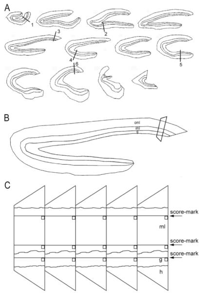
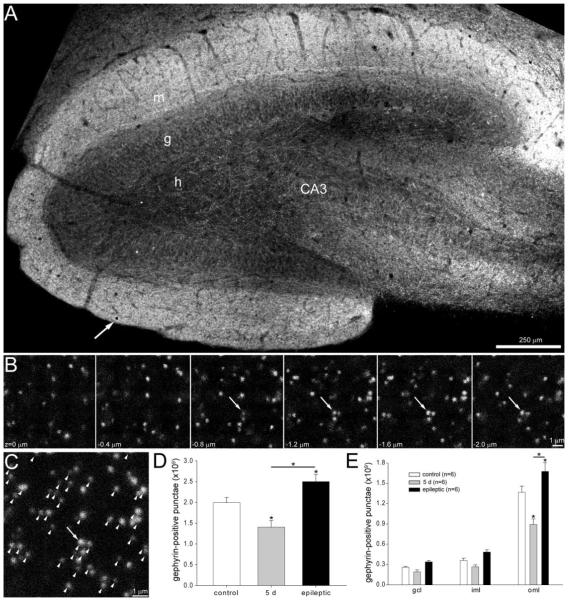
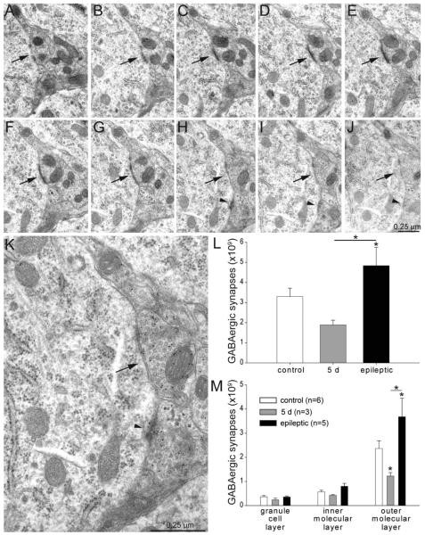
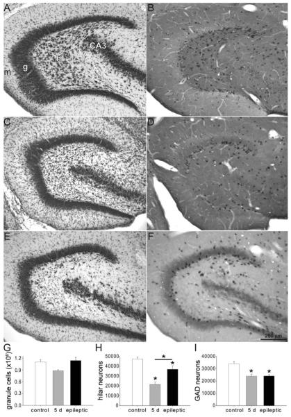
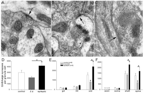
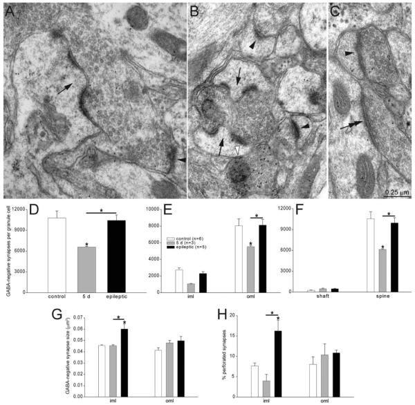
Many patients with temporal lobe epilepsy display neuron loss in the dentate gyrus. One potential epileptogenic mechanism is loss of GABAergic interneurons and inhibitory synapses with granule cells. Stereological techniques were used to estimate numbers of gephyrin-positive punctae in the dentate gyrus, which were reduced short-term (5 days after pilocarpine-induced status epilepticus) but later rebounded beyond controls in epileptic rats. Stereological techniques were used to estimate numbers of synapses in electron micrographs of serial sections processed for postembedding GABA-immunoreactivity. Adjacent sections were used to estimate numbers of granule cells and glutamic acid decarboxylase-positive neurons per dentate gyrus. GABAergic neurons were reduced to 70% of control levels short-term, where they remained in epileptic rats. Integrating synapse and cell counts yielded average numbers of GABAergic synapses per granule cell, which decreased short-term and rebounded in epileptic animals beyond control levels. Axo-shaft and axo-spinous GABAergic synapse numbers in the outer molecular layer changed most. These findings suggest interneuron loss initially reduces numbers of GABAergic synapses with granule cells, but later, synaptogenesis by surviving interneurons overshoots control levels. In contrast, the average number of excitatory synapses per granule cell decreased short-term but recovered only toward control levels, although in epileptic rats excitatory synapses in the inner molecular layer were larger than in controls. These findings reveal a relative excess of GABAergic synapses and suggest that reports of reduced functional inhibitory synaptic input to granule cells in epilepsy might be attributable not to fewer but instead to abundant but dysfunctional GABAergic synapses.


Figures

Figure 1
Figure 1
Sampling scheme for stereological analysis of synapse numbers in the dentate gyrus. A: Starting from a random section near the septal pole, a 1-in-12 series of 40-μm-thick transverse sections was embedded. Cumulative length of the granule cell layer was measured and divided by 6. Sample sites along the granule cell layer were identified, starting at a random site in the first interval and at equal distances thereafter, designated by lines 1–6. B: Enlarged view of the section that includes sample site 3 in panel A. Areas of the granule cell layer (g), inner third of the molecular layer (iml), and outer two-thirds of the molecular layer (oml) were measured. Tissue at each sample site was trimmed into a trapezoid and mounted for ultrathin sectioning. C: Ribbons of ultrathin sections included the hilus (h), granule cell layer (g), and molecular layer (ml). Randomly located score-marks and edges of sections were used to place counting frames (squares) across aligned regions of serial sections.
Figure 2
Figure 2
Gephyrin-immunoreactive punctae numbers in the dentate gyrus decrease shortly after status epilepticus but later rebound and exceed control levels in epileptic rats. A: Gephyrin immunoreactivity in the dentate gyrus of an epileptic rat. h, hilus; g, granule cell layer; m, molecular layer; CA3, CA3 pyramidal cell layer. An arrow indicates one of the 9.2 × 9.2 μm counting frames (black square) in this section. B: A typical series of optical sections collected at the sample point indicated by the arrow and square in panel A. Punctae were counted if they were not visible in the most superficial section (z = 0 μm) and did not touch top or left borders. Arrows indicate a cluster of punctae that were counted. C: A composite image of the stack of optical sections shown in B. Arrow indicates the cluster of punctae identified in panel B. Arrowheads indicate all counted punctae. D: Numbers of gephyrin-positive punctae per dentate gyrus (not hilus) were reduced 5 days after status epilepticus and increased later in epileptic rats. Asterisks indicate differences from the control value, unless specified by a bar, which indicates a difference between rats 5 days after status epilepticus and epileptic animals (P < 0.05, ANOVA, Student–Newman–Keuls method). Values indicate mean ± SEM. E: Numbers of gephyrin-positive punctae by strata of dentate gyrus. gcl, granule cell layer; iml, inner molecular layer; oml, outer molecular layer.
Figure 3
Figure 3
GABA-immunoreactive synapse numbers per dentate gyrus (not hilus) decrease shortly after status epilepticus but later rebound and exceed control levels in epileptic rats. AJ: A series of consecutive electron micrographs showing an axo-somatic synapse (arrow) in the granule cell layer of a control rat. A second axo-somatic synapse becomes apparent in panel H (arrowhead). K: High-magnification view of panel H reveals 10-nm-diameter colloidal gold particles over GABA-immunoreactive axon terminals. L: Numbers of GABA-immunoreactive synapses per dentate gyrus (not hilus) were reduced 5 days after status epilepticus and increased later in epileptic rats. Asterisks indicate differences from the control value, unless specified by a bar, which indicates a difference between rats 5 days after status epilepticus and epileptic animals (P < 0.05, ANOVA, Student–Newman–Keuls method). Values indicate mean ± SEM. M: Numbers of GABA-immunoreactive synapses by strata of dentate gyrus. Largest changes were in the outer molecular layer.
Figure 4
Figure 4
Dentate gyrus stained with thionin (A,C,E) and processed for in situ hybridization for glutamic acid decarboxylase (B,D,F) in a control rat (A,B), a rat 5 days after status epilepticus (C,D), and an epileptic rat (E,F). h, hilus; g, granule cell layer; m, molecular layer; CA3, CA3 pyramidal cell layer. Numbers of granule cells (G), hilar neurons (H), and GABAergic interneurons (I) per dentate gyrus in controls, rats 5 days after status epilepticus, and epileptic rats. Asterisks indicate differences from the control value, unless specified by a bar, which indicates a difference between rats 5 days after status epilepticus and epileptic animals (P < 0.05, ANOVA, Student–Newman–Keuls method). Values indicate mean ± SEM n = 6, 3, and 5 rats for control, 5 days after status epilepticus, and epileptic groups, respectively.
Figure 5
Figure 5
GABA-immunoreactive synapses with putative granule cells. A: An axo-shaft GABAergic synapse (arrow) in the outer molecular layer of an epileptic rat. GABA-immunoreactivity is evident by 10-nm-diameter colloidal gold particles. B: An axo-spinous GABAergic synapse (arrow) in the outer molecular layer of an epileptic rat. C: An axo-axonic GABAergic synapse (arrow) in the granule cell layer of an epileptic rat. The axon initial segment is recognized by microtubules and dense membrane undercoating. D: Numbers of GABAergic synapses per granule cell were decreased 5 days after status epilepticus but later rebounded and exceeded control levels in epileptic rats. Asterisks indicate differences from the control value, unless specified by a bar, which indicates a difference between rats 5 days after status epilepticus and epileptic animals (P < 0.05, ANOVA, Student–Newman–Keuls method). Values indicate mean ± SEM. E: Numbers of GABAergic synapses per granule cell by strata of the dentate gyrus reveal largest reductions and later proliferations in the outer molecular layer. gcl = granule cell layer; iml = inner molecular layer; oml = outer molecular layer. F: Numbers of GABAergic synapses per granule cell by subcellular targets reveal most synapses with dendritic shafts and spines. Five days after status epilepticus, axo-spinous GABAergic synapse numbers were reduced most. In epileptic rats, axo-spinous and axo-shaft GABAergic synapses were increased most.
Figure 6
Figure 6
GABA-negative synapses with putative granule cell dendrites in the molecular layer. A: A GABA-negative axon bouton forming a perforated synapse (arrow) with a GABA-negative dendritic spine in the inner molecular layer of an epileptic rat. A simple GABA-negative synapse with a spine is indicated by an arrowhead. B: A GABA-negative axon bouton forming perforated synapses (arrows) with GABA-negative dendritic spines in the inner molecular layer of an epileptic rat. Simple GABA-negative synapses with spines are indicated by black arrowheads. A GABA-positive synapse with a dendritic spine is indicated by a white arrowhead. GABA immunoreactivity is evident as concentrated 10-nm-diameter colloidal gold particles. C: A GABA-negative axon bouton synapsing with a GABA-negative dendritic shaft (double arrow) in the outer two-thirds of the molecular layer in a rat 5 days after status epilepticus. Another GABA-negative axon bouton forms a simple synapse with a dendritic spine (arrowhead). D: Numbers of GABA-negative synapses per granule cell were decreased 5 days after status epilepticus but later recovered to control levels in epileptic rats. iml = inner molecular layer; oml = outer molecular layer. Asterisks indicate differences from the control value, unless specified by a bar, which indicates a difference between rats 5 days after status epilepticus and epileptic animals (P < 0.05, ANOVA, Student–Newman–Keuls method). Values indicate mean ± SEM. E: Numbers of GABA-negative synapses per granule cell in the inner third (iml) and outer molecular layer (oml). F: Numbers of GABA-negative synapses per granule cell by subcellular targets reveal most synapses were with dendritic spines. Five days after status epilepticus, axo-spinous GABA-negative synapse numbers were reduced but recovered to control levels in epileptic animals. G: GABA-negative synapse size is increased in the inner molecular layer of epileptic rats. H: Percent of perforated synapses is increased in the inner molecular layer of epileptic rats.

Similar articles

Cited by

References

    1. André V, Marescaux C, Nehlig A, Fritschy JM. Alterations of hippocampal GABAergic system contribute to development of spontaneous recurrent seizures in the rat lithium-pilocarpine model of temporal lobe epilepsy. Hippocampus. 2001;11:452–468. - PubMed
    1. Andrioli A, Alonso-Nanclares L, Arellano JI, DeFelipe J. Quantitative analysis of parvalbumin-immunoreactive cells in the human epileptic hippocampus. Neuroscience. 2007;149:131–143. - PubMed
    1. Ang CW, Carlson GC, Coulter DA. Massive and specific dysregulation of direct cortical input to the hippocampus in temporal lobe epilepsy. J Neurosci. 2006;26:11850–11856. - PMC - PubMed
    1. Arellano JI, Muñoz A, Ballesteros-Yáñnez I, Sola RG, DeFelipe J. Histopathology and reorganization of chandelier cells in the human epileptic sclerotic hippocampus. Brain. 2004;127:45–64. - PubMed
    1. Austin JE, Buckmaster PS. Recurrent excitation of granule cells with basal dendrites and low interneuron density and inhibitory postsynaptic current frequency in the dentate gyrus of macaque monkeys. J Comp Neurol. 2004;476:205–218. - PubMed

Publication types

MeSH terms