. 2010 Feb;239(2):548-58.
doi: 10.1002/dvdy.22201.

Churchill and Sip1a repress fibroblast growth factor signaling during zebrafish somitogenesis

Affiliations

Churchill and Sip1a repress fibroblast growth factor signaling during zebrafish somitogenesis

Fatma O Kok et al. Dev Dyn. 2010 Feb.

Abstract

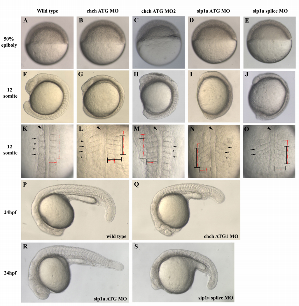
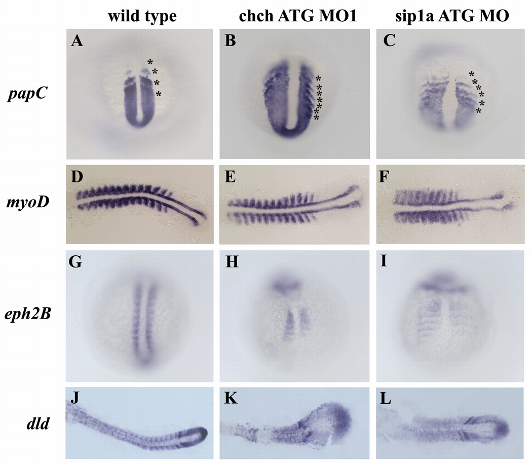
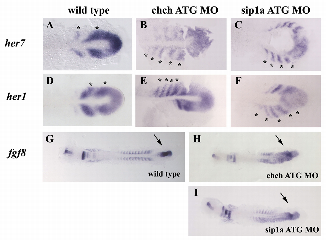
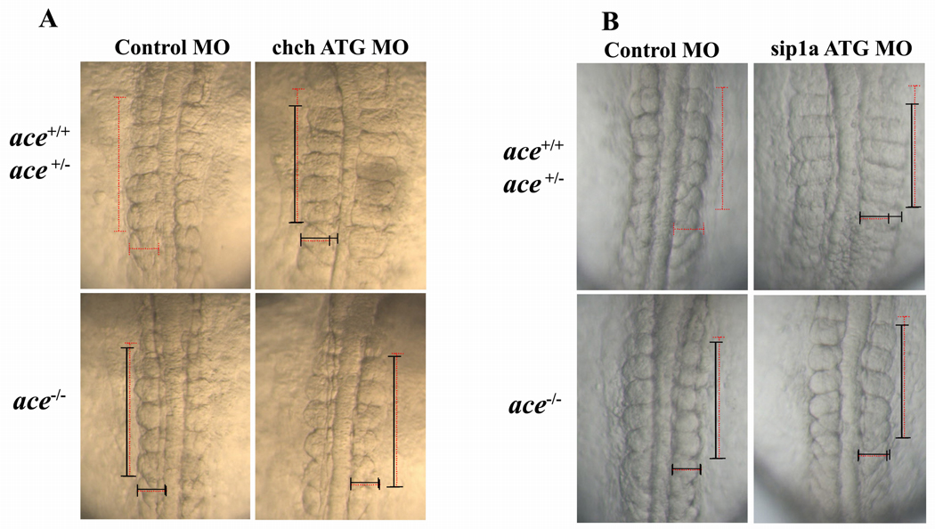
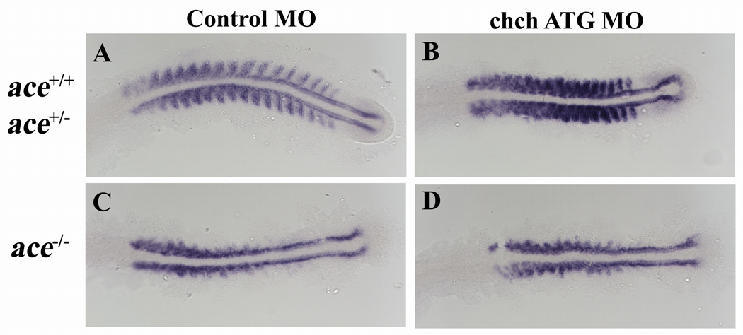
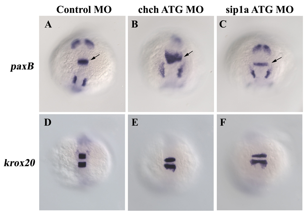
Cell-type specific regulation of a small number of growth factor signal transduction pathways generates diverse developmental outcomes. The zinc finger protein Churchill (ChCh) is a key effector of fibroblast growth factor (FGF) signaling during gastrulation. ChCh is largely thought to act by inducing expression of the multifunctional Sip1 (Smad Interacting Protein 1). We investigated the function of ChCh and Sip1a during zebrafish somitogenesis. Knockdown of ChCh or Sip1a results in misshapen somites that are short and narrow. As in wild-type embryos, cycling gene expression occurs in the developing somites in ChCh and Sip1a compromised embryos, but expression of her1 and her7 is maintained in formed somites. In addition, tail bud fgf8 expression is expanded anteriorly in these embryos. Finally, we found that blocking FGF8 restores somite morphology in ChCh and Sip1a compromised embryos. These results demonstrate a novel role for ChCh and Sip1a in repression of FGF activity.


Figures

Figure 1
Figure 1. ChCh and Sip1a are required for somitogenesis
Living wild-type, ChCh and Sip1a-compromised embryos. Early epiboly movements appear normal in chch or sip1a morphants (A–E). By 12s misshapen somites are apparent in both chch and sip1 morpholino treated embryos (F–J). In these embryos, somites are less extended thru anterior-posterior axis and over-extended thru mediolateral axis (K–O). Horizontal and vertical red dotted lines span the width and length of the first four wild type somites for comparison to the first four somites of morphant embryos, which are marked with a black line (K–O). At 24hpf, somites are enlarged and misshapen in both ChCh and Sip1a compromised embryos (P–S). Arrowheads denote notochord and black arrows denote somites. (A–J, P–S) are lateral views; (K–O), dorsal views.
Figure 2
Figure 2. Patterning of the presomitic and somitic mesoderm is disrupted in ChCh and Sip1a compromised embryos
Whole mount (A–C, G–I) and flat mount (D–F, J–L) RNA in situ hybridization of somite markers in wild type, ChCh and Sip1a compromised embryos. All views are dorsal; anterior to the top (A–C, G–I) and anterior to the left (D–F, J–L)). The expression domains of the PSM marker, papC is broader mediolaterally in ChCh and Sip1a compromised embryos than the wild type siblings (A–C). The number of the papC expression stripes corresponding to the prospective and formed somites at the segmentation plate in chch and sip1a MO is higher than in wild type siblings (A–C, compare asterisks number). The myogenic regulatory factor, myoD, is expressed in the posterior somite compartment in chch and sip1a MO injected embryos as in wild-type embryos (D–F). ephB2 and dld expression at the anterior half of the somites (G–L) is reduced and diffuse. Asterisks denote (prospective) somites.
Figure 3
Figure 3. Inhibition of ChCh and Sip1a affects the components of the “clock and wavefront model”
Flat mount RNA in situ hybridization of 10 somite stage wild type, ChCh and Sip1a compromised embryos(A–I). All views are dorsal; anterior to the left. In wildtype embryos, periodic activation of Notch signaling provides cycling gene expression of Notch pathway genes such as her1 and her7 (A–F). The number of the her7 expressing stripes in chch and sip1a ATG MO injected embryos ranges from 4 to 5 (B,C; E,F). fgf8 expression domain at tailbud is expanded anteriorly in ChCh and Sip1a compromised embryos (G–I). Asterisks denote each her1 or her7 expressing premesoderm stripe (A–F). Arrows denotes fgf8 tailbud expression domain (G–I).
Figure 4
Figure 4. Somite malformation in ChCh and Sip1a compromised embryos can be rescued by reduction of FGF8
(A) Dorsal views of 10 somite stage and (B) 12 somite stage living embryos, anterior to the top. Somites are narrower at A/P axis, broader at mediolateral axis in wild type and ace heterozygous chch morphants, but not ace homozygous chch morphants (A). ace homozygous sip1a ATG morphants does not have somite phenotype but wild type and ace heterozygous sip1a morphants still have the somite defects. (B). Horizontal and vertical red dotted line spans the first five somites of the control injected embryo width and length for comparison to the first five somite of morphant embryos which is indicated with a black line. Embryos were genotyped for the ace allele after imaging.
Figure 5
Figure 5. Somite defects in ace mutants are unaltered by ChCh knockdown
Flat mount RNA in situ hybridization of myoD in 17 somite stage ChCh compromised wild type, ace+/− and ace−/− embryos(A–D). All views are dorsal; anterior to the left. Lateral myoD expression in the somites is lost in ace mutant embryos (C). myoD expression pattern in ChCh compromised ace−/− embryos is similar to control MO injected ace−/− siblings (D).
Figure 6
Figure 6. Repression of FGF8 by ChCh is not limited to the mesoderm
Whole mount RNA in situ hybridization of pax2a and krox20 in 10-somite stage ChCh and Sip1a compromised embryos (A–F). All views are dorsal; anterior to the top. FGF target gene pax2a is expanded anteriorly in the isthmus in chch morphants, but not in sip1a morphants (A–C). The anterior extent of krox20 expression is not altered by chch or sip1a knockdown (D–F). Arrows denote isthmus.

References

    1. Amaya E, Musci TJ, Kirschner MW. Expression of a dominant negative mutant of the FGF receptor disrupts mesoderm formation in Xenopus embryos. Cell. 1991;66:257–270. - PubMed
    1. Aulehla A, Wehrle C, Brand-Saberi B, Kemler R, Gossler A, Kanzler B, Herrmann BG. Wnt3a plays a major role in the segmentation clock controlling somitogenesis. Dev Cell. 2003;4:395–406. - PubMed
    1. Bessho Y, Sakata R, Komatsu S, Shiota K, Yamada S, Kageyama R. Dynamic expression and essential functions of Hes7 in somite segmentation. Genes Dev. 2001;15:2642–2647. - PMC - PubMed
    1. Borland CZ, Schutzman JL, Stern MJ. Fibroblast growth factor signaling in Caenorhabditis elegans. Bioessays. 2001;23:1120–1130. - PubMed
    1. Bottcher RT, Niehrs C. Fibroblast growth factor signaling during early vertebrate development. Endocr Rev. 2005;26:63–77. - PubMed

Publication types