. 2010 Feb;239(2):482-95.
doi: 10.1002/dvdy.22190.

Transcriptional regulatory regions of gap43 needed in developing and regenerating retinal ganglion cells

Affiliations

Transcriptional regulatory regions of gap43 needed in developing and regenerating retinal ganglion cells

Brandon W Kusik et al. Dev Dyn. 2010 Feb.

Abstract

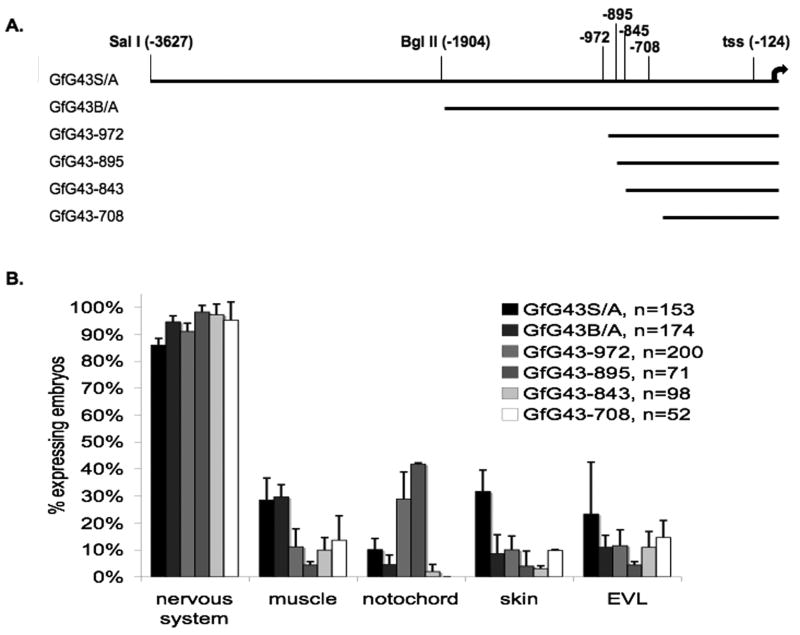
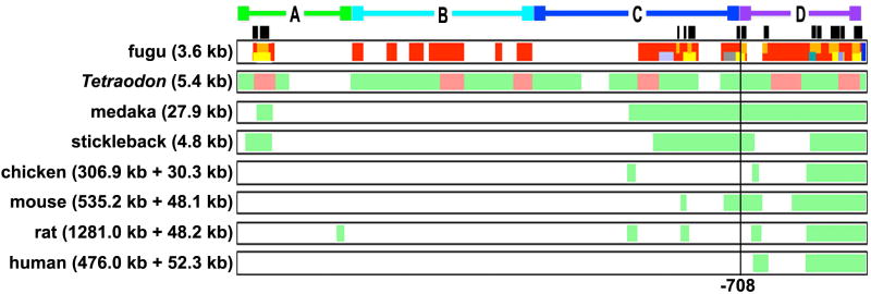
Mammals and fish differ in their ability to express axon growth-associated genes in response to CNS injury, which contributes to the differences in their ability for CNS regeneration. Previously we demonstrated that for the axon growth-associated gene, gap43, regions of the rat promoter that are sufficient to promote reporter gene expression in the developing zebrafish nervous system are not sufficient to promote expression in regenerating retinal ganglion cells in zebrafish. Recently, we identified a 3.6-kb gap43 promoter fragment from the pufferfish, Takifugu rubripes (fugu), that can promote reporter gene expression during both development and regeneration. Using promoter deletion analysis, we have found regions of the 3.6-kb fugu gap43 promoter that are necessary for expression in regenerating, but not developing, retinal ganglion cells. Within the 3.6-kb promoter, we have identified elements that are highly conserved among fish, as well as elements conserved among fish, mammals, and birds.


Figures

Fig. 1
Fig. 1. 5′-end deletions of 3.6 kb fugu gap43 regulatory sequence promote reporter gene expression preferentially in neurons
A. Full length promoter, GfG43S/A, and 5′-end deletions (GfG43B/A, GfG43-972, GfG43-895, GfG43-845, and GfG43-708) used in transient GFP reporter assays in zebrafish. B. Comparison of tissue distribution of GFP cells in 30 hpf embryos that were injected at the 1-cell stage with the various deletion constructs. Each injected embryo was scored as positive or negative for GFP-expressing cells in the nervous system, muscle, notochord, skin, or the enveloping layer (EVL = extra-embryonic peridermal tissue). Each bar represents the average value from 2-4 separate experiments. Standard errors are indicated by error bars. n = total number of fish examined for each construct.
Fig. 2
Fig. 2. 708 bp promoter is sufficient to promote expression in developing but not regenerating retina
Native GFP expression (green) and DAPI staining (blue) in transverse sections of embryonic, larval, and adult retinas. The GfG43SA and GfG43-708 constructs both express GFP in the retinal ganglion cell layer (gcl) and their axons (*) observed in the fiber layer at 48 hpf (A,E) and in the inner plexiform layer (ipl) within the retina at 96 hpf (B, F). In control and regenerating adult retina (7 days post crush) of the GfG43SA line, low levels of GFP are observed in control retina (C) and high levels in the GCL and fiber layer of regenerating retina (D). In the GfG43-708 line there is no GFP expression in the GCL of either control (G) or regenerating (H) retina. Developing retina scale bar (F), 25 μm. Adult retina scale bar (H), 50 μm.
Fig. 3
Fig. 3. Deletion mutations for stable reporter lines
The Fugu 3.6 kb gap43 promoter is represented by the black ruler at the top. Numbers on the ruler refer to distance (bp) from the translation start site (ATG), and tss refers to the transcription start site. The fugu 3.6 kb promoter was divided into 4 regions based on clusters of conserved sequence. All constructs contain the D region, which is equivalent to the 708 bp promoter that is sufficient for promoting expression in developing but not regenerating neurons. All possible combinations of regions A, B, and C were generated. Dotted lines represent deleted sequences.
Fig. 4
Fig. 4. Changes in developmental expression pattern as a result of promoter deletions
Transgene expression in stable GFP reporter fish carrying the Fugu 3.6 kb gap43 promoter (A-D), or promoter deletions: ΔA (E-H), ΔB (I-L), ΔAB (M-P), and ΔABC (Q-T). Images of 48 hpf embryos captured on the fluorescent stereoscope (A, A inset, E, I, M, Q) show that promoter deletions result in overall lower levels of GFP expression (scale bar in Q, 500 μm). The image of the WT fish in A was taken at 1/3 the exposure time, while the overexposed image in the inset was taken at the same exposure time as E, I, M, Q. Higher magnification views of the embryos (B-D, F-H, J-L, N-P, R-T; scale bar in T, 100 μm). Dorsal view of hindbrain at 24 hpf (B, F, J, N, R). Lateral view of brain at 48 hpf (C, G, K, O, S). Ventral view of head at 48 hpf (D, H, L, P, T). cb, cerebellum; hb, hindbrain; ob, olfactory bulb; oc, optic chiasm; oe, olfactory epithelium; ot, optic tectum; rgc, retinal ganglion cells.
Fig. 5
Fig. 5. Transgene expression in regenerating retina requires region C
Native GFP expression (green) and DAPI staining (blue) in transverse sections of embryonic (48 hpf; A, E, I), larval (4 dpf; B, F, J) and adult retinas (control; C, G, K; and regenerating; D, H, L) from promoter deletion GFP reporter lines: ΔA (A-D); ΔB (E-H); ΔAB (I-L). All lines displayed the same spatial and temporal pattern of expression in the developing retina (A, B, E, F, I, J) as previously observed for the full-length promoter (see Fig. 2A, B). Unlike ΔABC reporter lines, which did not express GFP in regenerating adult retina (see Fig. 2G, H), addition of the C region in the ΔAB lines was sufficient to restore regenerative expression (L). (*) Axons of RGCs in fiber layer. dpc, days post-crush; gcl, ganglion cell layer; ipl, inner plexiform layer; inl, inner nuclear layer. Developing retina scale bar (J), 25 μm. Adult retina scale bar (L), 50 μm.
Fig. 6
Fig. 6. Teleost conserved sequence elements within the gap43 promoter
Comparison of gap43 promoter regions from four different fish species (Fugu, Tetraodon, Medaka, and Stickleback) using 2-way BLAST show that there are several regions of highly conserved sequence. The conserved regions, represented by the red, orange, and yellow boxes below the ruler (Tetraodon (red), medaka (orange), and stickleback (yellow)) share >80% sequence identity with similar regions in the Fugu 3.6 kb gap43 promoter. The ruler only applies to the fugu sequence, however, the relative position of all conserved sequences, in relation to the translational start site, is also conserved. EvoPrinter (Odenwald et al., 2005) was also used to identify teleost conserved sequence elements (TCSEs, black rectangles) from Fugu, Tetraodon, and Medaka which were manually aligned with sequences from Stickleback.
Fig. 7
Fig. 7. Distal gap43 regulatory regions contain elements that are highly conserved among fish
Potential gap43 regulatory sequences from Tetraodon, medaka, stickleback, chicken, mouse, rat, and human compared to the fugu gap43 3.6 kb 5′ flanking sequence using MultiPipMaker. Lengths of 5′ flanking sequences that were compared to the 3.6 kb fugu sequence are shown in parentheses. Intron 1 sequences were also included in the comparison for chicken, mouse, rat, and human (length listed in parentheses after “+”). A, B, and C correspond to the regions involved in the deletion series. D corresponds to the 708 bp promoter that is sufficient for expression in developing, but not regenerating retina. Black stripes correspond to the TCSEs in Fig. 6. Top bar represents fugu 3.6 kb gap43 sequence. Red, orange, and yellow stripes correspond to the Tetraodon, medaka, and stickleback conserved elements as determined by 2-way BLAST and in Fig. 6. Dark blue stripe corresponds to 30 bp coding region of exon 1. Light grey, grey, teal, and mauve stripes correspond to repeat sequences detected by Repeat Masker. Green shading refers to sequences within the fugu gap43 promoter that share 60-100% identity to sequences found somewhere in the sampled region of gap43 from the indicated species; pink shading refers sequences that share 100 bp or more, without gaps and with ≥70% identity.
Fig. 8
Fig. 8. Differences in synteny around the gap43 gene locus between fish and other species
The lsamp gene (orange) lies 3′ to the gap43 gene (yellow) in fish, chicken, and mammals. However, the gene 5′ of gap43 in fish (eva1; blue) is different from that in birds and mammals (zbtb20; green). In addition, the orientation of eva1 in fish is the same as gap43, while zbtb20 in birds and mammals is in the opposite orientation. The distance from gap43 to its neighboring genes is indicated below the dotted lines to the left and right of the arrow representing gap43. The yellow, orange, green and blue boxes are not to scale and represent only the coding sequences of the genes. Note: the medaka and stickleback genomes contain gaps in these regions so distances reflect the gap size predicted by Ensembl, and thus may not be entirely accurate.

Similar articles

Cited by

References

    1. Altschul SF, Gish W, Miller W, Myers EW, Lipman DJ. Basic local alignment search tool. J Mol Biol. 1990;215:403–410. - PubMed
    1. Amsterdam A, Burgess S, Golling G, Chen W, Sun Z, Townsend K, Farrington S, Haldi M, Hopkins N. A large-scale insertional mutagenesis screen in zebrafish. Genes Dev. 1999;13:2713–2724. - PMC - PubMed
    1. Bacon A, Kerr NC, Holmes FE, Gaston K, Wynick D. Characterization of an enhancer region of the galanin gene that directs expression to the dorsal root ganglion and confers responsiveness to axotomy. J Neurosci. 2007;27:6573–6580. - PMC - PubMed
    1. Barthel LK, Raymond PA. Improved method for obtaining 3-microns cryosections for immunocytochemistry. J Histochem Cytochem. 1990;38:1383–1388. - PubMed
    1. Becker CG, Becker T. Growth and pathfinding of regenerating axons in the optic projection of adult fish. J Neurosci Res. 2007;85:2793–2799. - PubMed

Publication types

LinkOut - more resources