. 2010 Feb;239(2):446-57.
doi: 10.1002/dvdy.22183.

Survival of Hoxa13 homozygous mutants reveals a novel role in digit patterning and appendicular skeletal development

Affiliations

Survival of Hoxa13 homozygous mutants reveals a novel role in digit patterning and appendicular skeletal development

Wilma D Perez et al. Dev Dyn. 2010 Feb.

Abstract

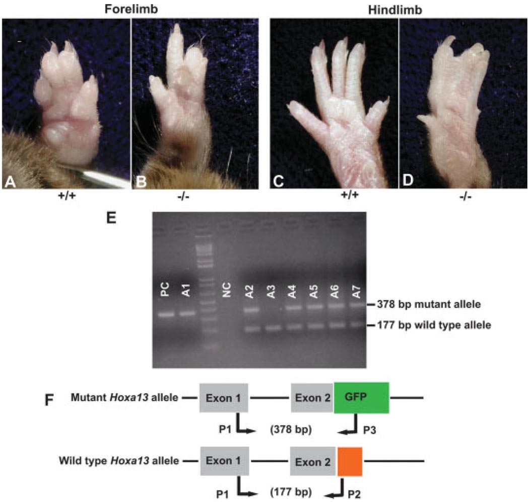
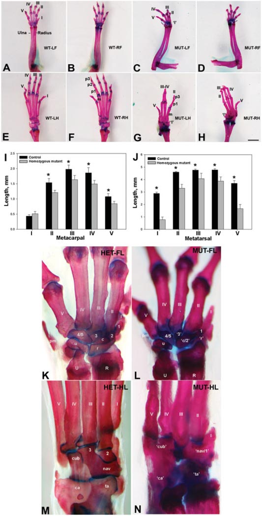
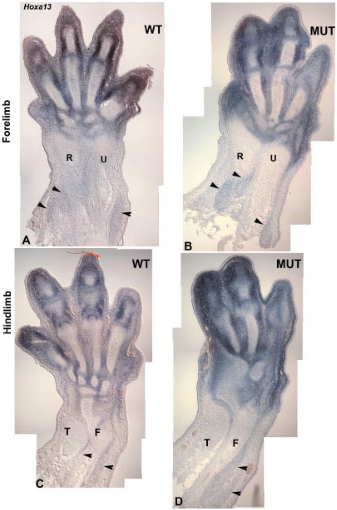
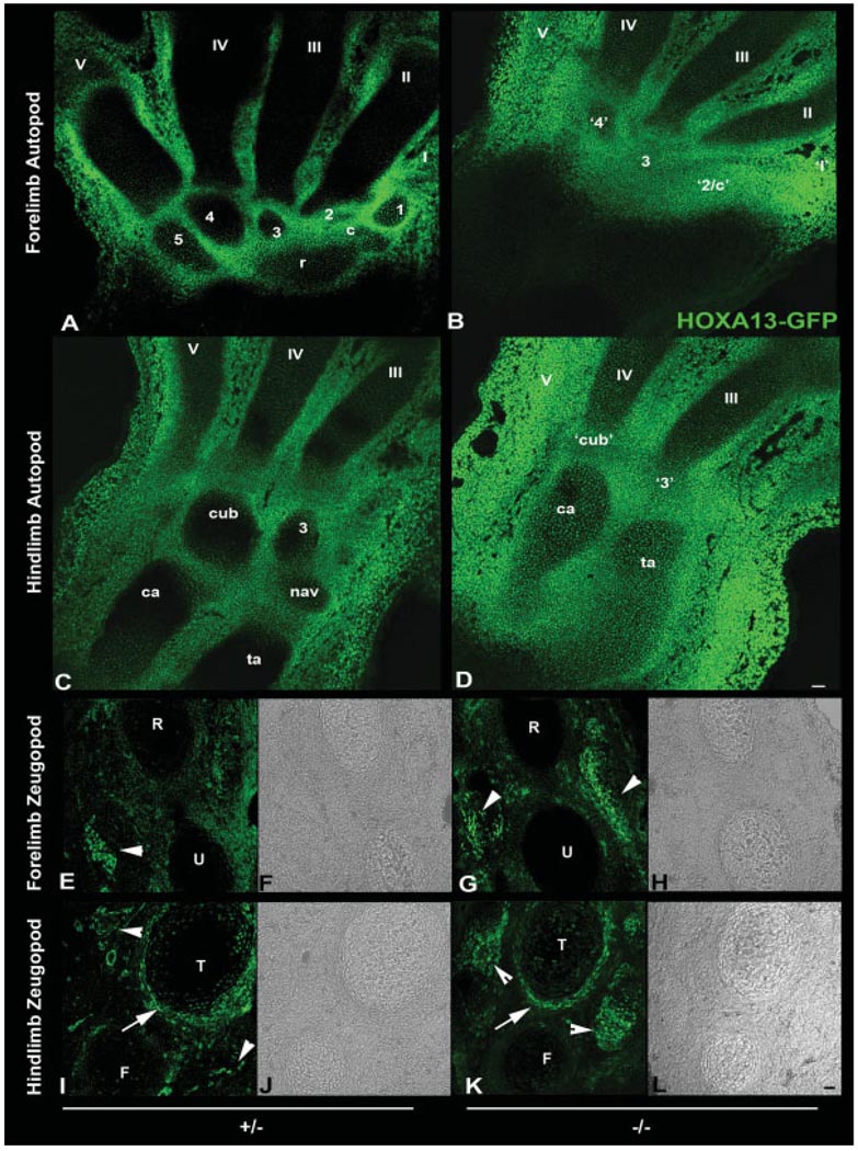
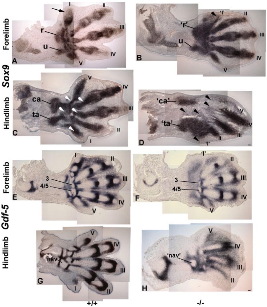
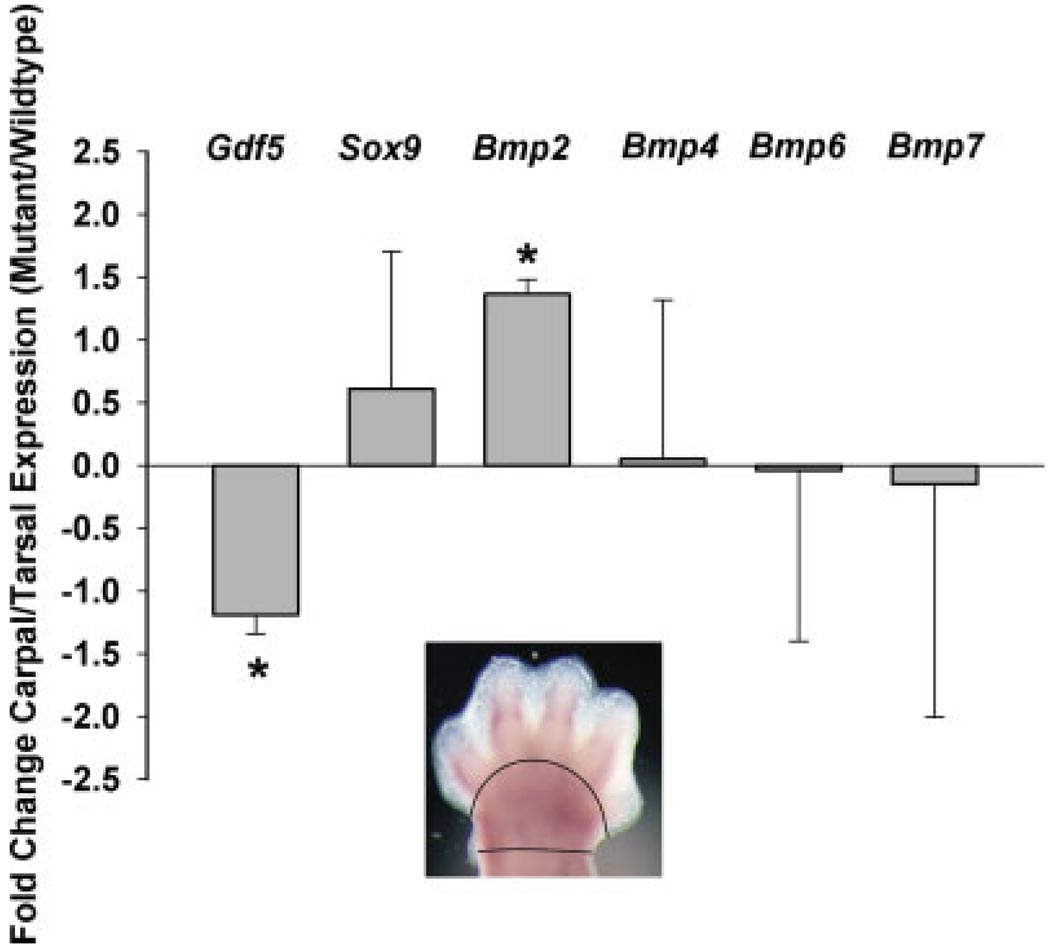
The loss of HOXA13 function severely disrupts embryonic limb development. However, because embryos lacking HOXA13 die by mid-gestation, the defects present in the mutant limb could arise as a secondary consequence of failing embryonic health. In our analysis of the mutant Hoxa13(GFP) allele, we identified a surviving cohort of homozygous mutants exhibiting severe limb defects including: missing phalanx elements, fusions of the carpal/tarsal elements, and significant reductions in metacarpal/metatarsal length. Characterization of prochondrogenic genes in the affected carpal/tarsal regions revealed significant reduction in Gdf5 expression, whereas Bmp2 expression was significantly elevated. Analysis of Gdf5 mRNA localization also revealed diffuse expression in the carpal/tarsal anlagen, suggesting a role for HOXA13 in the organization of the cells necessary to delineate individual carpal/tarsal elements. Together these results identify Gdf5 as a potential target gene of HOXA13 target gene and confirm a specific role for HOXA13 during appendicular skeletal development.


Figures

Fig. 1
Fig. 1
Surviving Hoxa13GFP homozygous mutants exhibit malformations of the distal autopod including: hypodactyly, brachydactyly, and syndactyly. A–D: Comparison of wild type and homozygous mutant forelimb and hindlimb in 6-month-old littermates. Note the syndactyly and hypodactyly in the mutant forelimb and hindlimb. E: PCR genotyping of the 6-month-old Hoxa13GFP mice confirmed that the mouse exhibiting autopod defects, sample A1, is a homozygous mutant. F: Representation of the Hoxa13GFP targeted mutation and PCR genotyping strategy. Note that the GFP expression cassette replaces the last portion of Hoxa13 exon 2, which encodes the third helix of the HOXA13 homeodomain.
Fig. 2
Fig. 2
Analysis of the appendicular skeleton in 3-month-old Hoxa13GFP littermates. A,B,E,F: Analysis of wild type forelimbs and hindlimbs. Roman numerals I–V denote the digit skeletal elements. Phalanx segments are denoted by p1, p2, and p3. m, metatarsal skeletal element. C,D,G,H: Analysis of surviving homozygous mutant forelimbs and hindlimbs. Roman numerals I–V denote the digit skeletal elements. Shortened or missing digits are denoted by roman numerals flanked by an apostrophe. m, metatarsal; p1 and p3, phalanx segments. Note that the homozygous mutant hindlimbs are missing the p2 phalanx segment and that the p1 and p3 phalanx segments for digits III and IV are fused in the homozygous mutant left hindlimb. Blue staining, Alcian blue, which detects cartilage and connective tissue; red staining, alizarin red, which detects mineralized bone. Bar represents 2 mm. I: Analysis of metacarpal lengths in control (black bars) and homozygous mutants (grey bars). Asterisks denote a significant difference in metacarpal length between control and homozygous mutant mice (P ≤ 0.01). J: Analysis of metatarsal lengths in control (black bars) and homozygous mutants (grey bars). Asterisks denote a significant difference in metatarsal length between control and homozygous mutant mice (P ≤ 0.001). K,L: Metacarpal and carpal elements present in the wild type and homozygous mutant left forelimbs. Roman numerals I–V, digit metacarpals; 1–5, distal carpal skeletal elements; U, ulna; R, radius; c, central carpal element; u, ulnare; r, radiale; pm, post-minimus bone. Malformed or fused skeletal elements are flanked by an apostrophe. M,N: Metatarsal and tarsal elements present in the wild type and homozygous mutant left hindlimb. Roman numerals I–V, digit metatarsals; 1–3, distal tarsal skeletal elements; ca, calcaneous; ta, talus; nav, naviculare; cub, cuboideum. Malformed or fused skeletal elements are flanked by an apostrophe. Blue staining, Alcian blue, which detects cartilage and connective tissue; red staining, alizarin red, which detects mineralized bone.
Fig. 3
Fig. 3
Localization of Hoxa13 mRNA in E 14.5 limbs using section in situ hybridization. A: Hoxa13 expression in wildtype forelimbs. B: Hoxa13 expression in homzoygous mutant forelimbs. C: Hoxa13 expression in wildtype hindlimbs. D: Hoxa13 expression in homozygous mutant hindlimbs. Arrowheads denote expression in the zeugopod musculature. R, radius; U, ulna; T, tibia; F, fibula.
Fig. 4
Fig. 4
Distribution of HOXA13-GFP protein in the carpal and tarsal skeletal elements of E 14.5 Hoxa13GFP embryos. A: Heterozygous control forelimb. Note the localization of HOXA13-GFP to the periphery of each skeletal element. Roman numerals I–V denote the digit metacarpals. Distal carpal elements are denoted by the numbers 1–5. r, radiale; c, central carpal bone. B: HOXA13-GFP is diffusely distributed in the carpal region of homozygous mutants, which exhibit poor delineation of the individual carpal skeletal elements. Roman numerals I–V denote the digit metacarpals. Distal carpal elements are denoted by the numbers 2–4. Fused or malformed carpal elements are flanked by an apostrophe. c, central carpal bone. C: Localization of HOXA13-GFP protein to the periphery of each tarsal and metatarsal skeletal element in a heterozygous control hindlimb. Roman numerals III–V represent the metatarsal elements. 3, distal tarsal element associated with metatarsal III; cub, cuboideum; nav, naviculare; ca, calcaneous; ta, talus. D: HOXA13-GFP is diffusely distributed in the tarsal region of homozygous mutants, which exhibit poor delineation of metatarsal IV and the cuboidal (c) element as well as poor separation of metatarsal III from the third tarsal element (3). Roman numerals III–V denote the digit metatarsals. ca, calcaneus; ta, talus. Fused or malformed tarsal elements are flanked by an apostrophe. E: HOXA13-GFP expression in the forelimb zeugopod of a heterozygous control. Arrowhead denotes expression in the zeugopod musculature. F: Bright field image of E. G: HOXA13-GFP expression in the forelimb zeugopod of a homozygous mutant. Arrowheads denote expression in the zeugopod musculature. H: Bright field image of G. I: HOXA13-GFP expression in the hindlimb zeugopod of a heterozygous control. Arrow denotes expression in the cells surrounding the tibia. Arrowheads denote HOXA13-GFP expression in the zeugopod musculature. J: Bright-field image of I. K: HOXA13-GFP expression in the hindlimb zeugopod of a homozygous mutant. Arrow denotes expression in the cells surrounding the tibia. Arrowheads denote expression in the zeugopod musculature. L: Bright-field image of K. R, radius; U, ulna; T, tibia; F, fibula. Bars = 50 µm.
Fig. 5
Fig. 5
Expression of Sox9 and Gdf-5 in the E 13.5 autopod of wild type and Hoxa13GFP homozygous mutants. A: Sox9 expression is restricted to the condensing mesenchyme contributing to digits I–V as well as in the developing carpal in the wild type forelimb. r, radiale condensation; u, ulnare condensation. Arrow denotes the detection of Sox9 expression in the condensing mesenchyme contributing to the digit I metacarpal region. B: Expression of Sox9 in the homozygous mutant forelimb. Arrowhead denotes reduced condensation of the Sox9-expressing cells in the digit I region. Poor condensation was also detected in the Sox9-expressing cells in the radiale condensation (r). u, ulnare condensation. C: Expression of Sox9 in the wild type hindlimb mesenchymal condensations contributing to digits I–V. White arrowheads denote the detection of Sox9 expression in the condensations contributing to the digit metatarsal elements. Note the condensations forming the calcaneus (ca) and talus (ta) are already established in the control limb. D: Expression of Sox9 in the homozygous mutant hindlimb. Note the diffuse expression in the regions contributing to the presumptive calcaneus (c) and talus (t). Black arrowheads denote the sites of metatarsal formation that exhibit missing or low levels of Sox9 expression. A defined boundary is also absent between the talus and the digit I region, which contains a uniform layer of Sox9-expressing cells. E: Gdf5 transcripts are localized to the joint fields of the digits as well as to the sites of carpal element delineation. Roman numerals denote the forelimb digits. Carpal skeletal elements 3 and 4/5 are also indicated. F: Homozygous mutant forelimbs exhibit reduced Gdf5 expression in the digit I region as well as diffuse expression in the developing carpal elements. Roman numerals denote the forelimb digits. Carpal skeletal elements 3 and 4/5 are indicated and represent the only distinguishable carpal elements in the mutant forelimb. G: Gdf5 expression in the wild type hindlimb corresponds to the sites of joint formation in the digits as well as the sites of tarsal element delineation. Roman numerals denote the developing digits. nav, naviculare tarsal element. H: Homozygous mutant hindlimb exhibits reduced and diffuse Gdf5 expression in digits II–V. No Gdf5 expression was detected in the digit I region or in the majority of the developing tarsal elements. nav, malformed naviculare tarsal element. Bar = 50 µm.
Fig. 6
Fig. 6
qRTPCR analysis of prochondrogenic gene expression in the carpal/tarsal region of Hoxa13GFP homozygous mutants. Grey bars represent the fold-change differences in expression (Mutant/Wildtype). Asterisks denote a significant fold-change difference in gene expression. Inset depicts the region dissected for carpal/tarsal total RNA isolation.
Fig. 7
Fig. 7
Analysis of programmed cell death and proliferation in E 11.5 Hoxa13GFP homozygous mutant and heterozygous control limbs. A,C: Fluorescent detection of programmed cell death using terminal UTP nick end labeling (TUNEL) in heterozygous control forelimbs and hindlimbs indicates a low level of programmed cell death (red signal) in the anterior autopod mesenchyme (white arrowhead) with higher levels of TUNEL-positive cells in the apical ectodermal ridge (aer; white arrow). B,D: Detection of TUNEL-positive cells in homozygous mutant forelimbs and hindlimbs reveals similar levels of programmed cell death in the aer compared to controls (white arrow) whereas the homozygous mutant hindlimb exhibited a greater amount of TUNEL-positive cells in the anterior limb bud (white arrowhead). Homozygous mutant forelimbs did not exhibit any increase in the number of TUNEL-positive cells in the anterior limb bud compared to heterozygous controls (white arrowhead). Bar = 100 µm. E,I,G,K: Immunohistochemical detection (red signal) of mitotic cells using an anti-phosphohistone H3 antibody (APH3) in cryosections of heterozygous control forelimbs and hindlimbs expressing HOXA13-GFP (green signal). F,J,H,L: APH3 localization in the homozygous mutant autopod cryosections reveals similar levels of mitotic cells (red signal) in the regions expressing HOXA13-GFP (green). Yellow cells in I–L denote mitotic cells (APH3-positive) that are also expressing HOXA13-GFP. Bar = 100 µm (A–D) and 50 µm (E–L).

Similar articles

Cited by

References

    1. Akiyama H. Control of chondrogenesis by the transcription factor Sox9. Mod Rheumatol. 2008;18:213–219. - PubMed
    1. Akiyama H, Chaboissier MC, Martin JF, Schedl A, de Crombrugghe B. The transcription factor Sox9 has essential roles in successive steps of the chondrocyte differentiation pathway and is required for expression of Sox5 and Sox6. Genes Dev. 2002;16:2813–2828. - PMC - PubMed
    1. Akiyama H, Kim JE, Nakashima K, Balmes G, Iwai N, Deng JM, Zhang Z, Martin JF, Behringer RR, Nakamura T, de Crombrugghe B. Osteo-chondroprogenitor cells are derived from Sox9 expressing precursors. Proc Natl Acad Sci USA. 2005;102:14665–14670. - PMC - PubMed
    1. Archer CW, Cottril C, Rooney P. Cellular aspects of cartilage differentiation and morphogenesis. In: Kemp R, Hinchliffe JR, editors. Matrices and differentiation. New York: A.R. Liss; 1984. p. 480. - PubMed
    1. Barna M, Niswander L. Visualization of cartilage formation: insight into cellular properties of skeletal progenitors and chondrodysplasia syndromes. Dev Cell. 2007;12:931–941. - PubMed

Publication types

LinkOut - more resources