. 2009 Dec 24:2:20.
doi: 10.1186/1757-2215-2-20.

Activation of PKA, p38 MAPK and ERK1/2 by gonadotropins in cumulus cells is critical for induction of EGF-like factor and TACE/ADAM17 gene expression during in vitro maturation of porcine COCs

Affiliations

Activation of PKA, p38 MAPK and ERK1/2 by gonadotropins in cumulus cells is critical for induction of EGF-like factor and TACE/ADAM17 gene expression during in vitro maturation of porcine COCs

Yasuhisa Yamashita et al. J Ovarian Res. .

Abstract

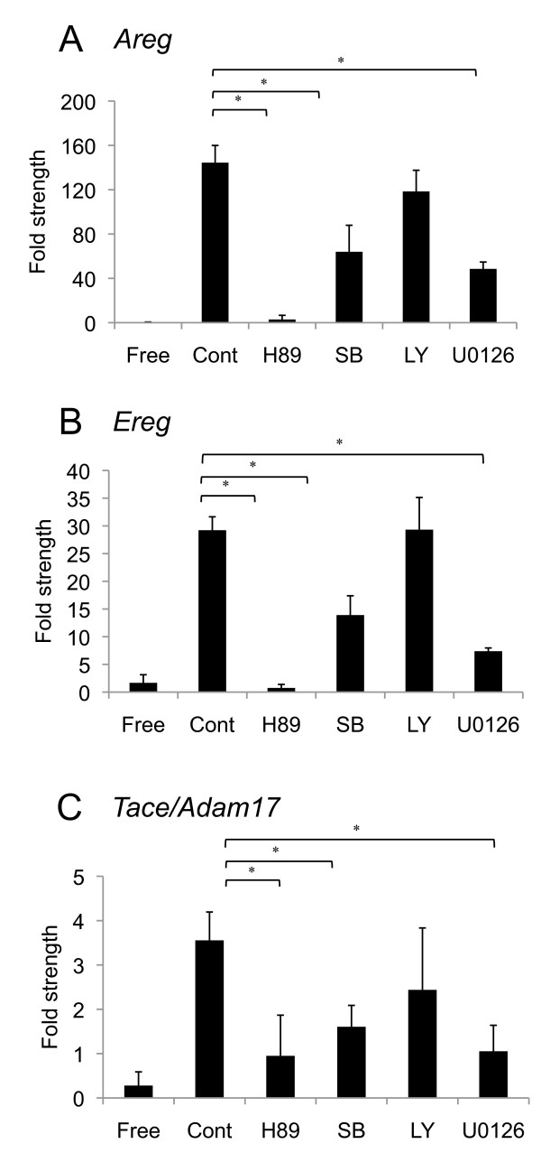
Objectives: During ovulation, it has been shown that LH stimulus induces the expression of numerous genes via PKA, p38 MAPK, PI3K and ERK1/2 in cumulus cells and granulosa cells. Our recent study showed that EGF-like factor and its protease (TACE/ADAM17) are required for the activation of EGF receptor (EGFR), cumulus expansion and oocyte maturation of porcine cumulus-oocyte complexes (COCs). In the present study, we investigated which signaling pathways are involved in the gene expression of EGF-like factor and in Tace/Adam17 expression in cumulus cells of porcine COC during in vitro maturation.

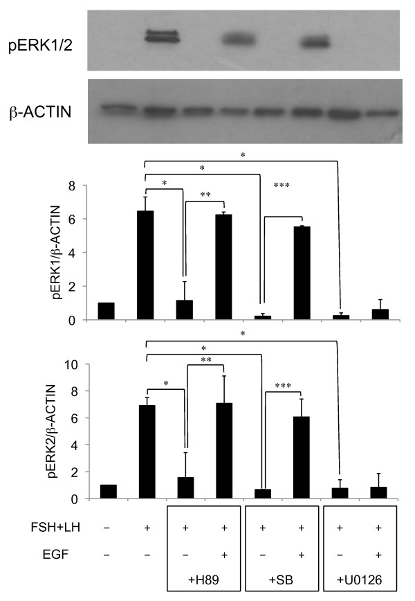
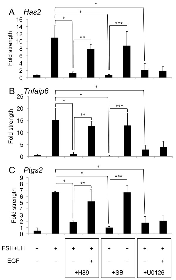
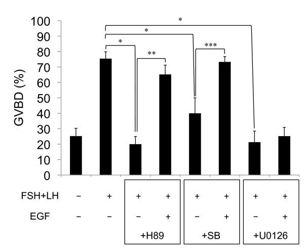
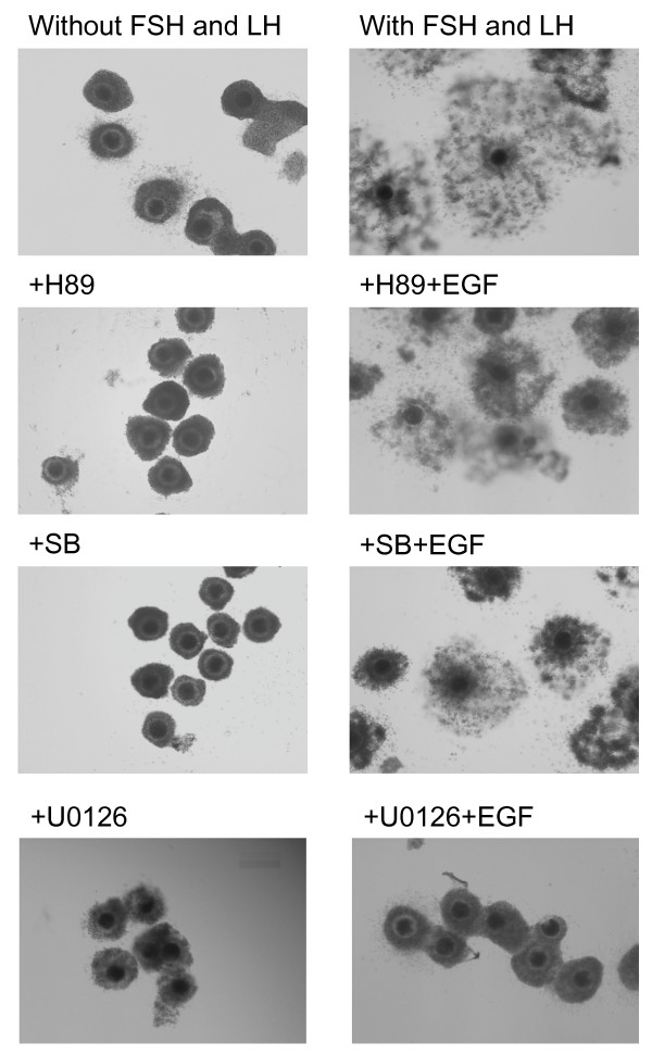
Methods: Areg, Ereg, Tace/Adam17, Has2, Tnfaip6 and Ptgs2 mRNA expressions were detected in cumulus cells of porcine COCs by RT-PCR. Protein level of ERK1/2 phosphorylation in cultured cumulus cells was analyzed by westernblotting. COCs were visualized using a phase-contrast microscope.

Results: When COCs were cultured with FSH and LH up to 2.5 h, Areg, Ereg and Tace/Adam17 mRNA were expressed in cumulus cells of COCs. Areg, Ereg and Tace/Adam17 gene expressions were not suppressed by PI3K inhibitor (LY294002), whereas PKA inhibitor (H89), p38 MAPK inhibitor (SB203580) and MEK inhibitor (U0126) significantly suppressed these gene expressions. Phosphorylation of ERK1/2, and the gene expression of Has2, Tnfaip6 and Ptgs2 were also suppressed by H89, SB203580 and U0126, however, these negative effects were overcome by the addition of EGF to the medium, but not in the U0126 treatment group.

Conclusion: The results showed that PKA, p38 MAPK and ERK1/2 positively controlled the expression of EGF-like factor and TACE/ADMA17, the latter of which impacts the cumulus expansion and oocyte maturation of porcine COCs via the EGFR-ERK1/2 pathway in cumulus cells.


Figures

Figure 1
Figure 1
Effect of H89, SB203580, LY294002 or U0126 on Areg(A), Ereg(B) or Tace/Adam17(C) mRNA. For reference, the 0 h COC value was set as 1 and the data presented as the fold strength. Values are mean +/- SEM of 3 replicates. *: The significant differences were observed as compared with that in COCs cultured with FSH and LH for 2.5 h. The respective value of among Areg, Ereg, and Adam17 mRNA were normalized according to those of β-actin mRNA to evaluate arbitrary units of the relative abundance of the targets. Free: COCs were cultured without FSH and LH for 2.5 h; Cont: COCs were cultured with FSH and LH for 2.5 h; H89: COCs were cultured with FSH, LH and H89 for 2.5 h; SB: COCs were cultured with FSH, LH and SB203580 for 2.5 h; LY: COCs were cultured with FSH, LH and LY294002 for 2.5 h; U0126: COCs were cultured with FSH, LH and U0126 for 2.5 h
Figure 2
Figure 2
Effect of H89, SB203580 or U0126 on ERK1/2 phosphorylation in cumulus cells. For reference, the COC that were cultured without FSH and LH for 5 h value was set as 1 and the data presented as the fold strength. Values are mean +/- SEM of 3 replicates. *: The significant differences were observed as compared with that in COCs cultured with FSH and LH for 5 h. **: The significant differences were observed as compared with that in COCs cultured with FSH, LH, H89 and EGF for 5 h. ***: The significant differences were observed as compared with that in COCs cultured with FSH, LH SB230580 and EGF for 5 h. The respective value of protein levels of ERK1/2 phosphorylation were normalized according to those of β-ACTIN to evaluate arbitrary units of the relative abundance. FSH(-): COCs were cultured without FSH and LH for 5 h; FSH(+): COCs were cultured with FSH and LH for 5 h; EGF(-): COCs were cultured without EGF for 5 h; EGF(+): COCs were cultured with EGF for 5 h; +H89: COCs were cultured with H89 for 5 h; +SB: COCs were cultured with SB203580 for 5 h; +U0126: COCs were cultured with U0126 for 5 h
Figure 3
Figure 3
Effect of H89, SB203580 or U0126 on expression of Has2(A), Tnfaip6(B) or Ptgs2(C) mRNA. For reference, the 0 h COC value was set as 1 and the data presented as the fold strength. Values are mean +/- SEM of 3 replicates. *: The significant differences were observed as compared with that in COCs cultured with FSH and LH for 10 h. **: The significant differences were observed as compared with that in COCs cultured with FSH, LH, H89 and EGF for 10 h. ***: The significant differences were observed as compared with that in COCs cultured with FSH, LH, SB230580 and EGF for 10 h. The respective value of among Has2, Tnfaip6 and Ptgs2 mRNA were normalized according to those of β-actin mRNA to evaluate arbitrary units of the relative abundance of the targets. FSH(-): COCs were cultured without FSH and LH for 10 h; FSH(+): COCs were cultured with FSH and LH for 10 h; EGF(-): COCs were cultured without EGF for 10 h; EGF(+): COCs were cultured with EGF for 10 h; +H89: COCs were cultured with H89 for 10 h; +SB: COCs were cultured with SB203580 for 10 h; +U0126: COCs were cultured with U0126 for 10 h
Figure 4
Figure 4
Effect of H89, SB203580 or U0126 on rate of oocyte exhibiting GVBD. Values are mean +/- SEM of 3 replicates. *: The significant differences were observed as compared with that in COCs cultured with FSH and LH for 20 h. **: The significant differences were observed as compared with that in COCs cultured with FSH, LH, H89 and EGF for 20 h. ***: The significant differences were observed as compared with that in COCs cultured with FSH, LH, SB230580 and EGF for 20 h. FSH(-): COCs were cultured without FSH and LH for 20 h; FSH(+): COCs were cultured with FSH and LH for 20 h; EGF(-): COCs were cultured without EGF for 20 h; EGF(+): COCs were cultured with EGF for 20 h; +H89: COCs were cultured with H89 for 20 h; +SB: COCs were cultured with SB203580 for 20 h; +U0126: COCs were cultured with U0126 for 20 h
Figure 5
Figure 5
Effect H89, SB203580 or U0126 on cumulus expansion of cultured COCs. Without FSH and LH: COCs were cultured without FSH and LH for 40 h; With FSH and LH: COCs were cultured with FSH and LH for 40 h; +H89: COCs were cultured with FSH, LH and H89 for 40 h; +H89+EGF: COCs were cultured with FSH, LH, H89 and EGF for 40 h; +SB: COCs were cultured with FSH, LH and SB203580 for 40 h; +SB+EGF: COCs were cultured with FSH, LH, SB and EGF for 40 h; +U0126: COCs were cultured with FSH, LH and U0126 for 40 h; +U0126+EGF: COCs were cultured with FSH, LH, U0126 and EGF for 40 h.

Similar articles

Cited by

References

    1. Richards JS. Ovulation: new factors that prepare the oocyte for fertilization. Mol Cell Endocrinol. 2005;234:75–79. doi: 10.1016/j.mce.2005.01.004. - DOI - PubMed
    1. Camaioni A, Hascall VC, Yanagishita M, Salustri A. Effects of exogenous hyaluronic acid and serum on matrix organization and stability in the mouse cumulus cell-oocyte complex. J Biol Chem. 1993;268:20473–20481. - PubMed
    1. Chen L, Russell PT, Larsen WJ. Functional significance of cumulus expansion in the mouse: roles for the preovulatory synthesis of hyaluronic acid within the cumulus mass. Mol Reprod Dev. 1993;34:87–93. doi: 10.1002/mrd.1080340114. - DOI - PubMed
    1. Ochsner SA, Day AJ, Rugg MS, Breyer RM, Gomer RH, Richards JS. Disrupted function of tumor necrosis factor-alpha-stimulated gene 6 blocks cumulus cell-oocyte complex expansion. Endocrinology. 2003;144:4376–4384. doi: 10.1210/en.2003-0487. - DOI - PubMed
    1. Fulop C, Szanto S, Mukhopadhyay D, Bardos T, Kamath RV, Rugg MS, Day AJ, Salustri A, Hascall VC, Glant TT, Mikecz K. Impaired cumulus mucification and female sterility in tumor necrosis factor-induced protein-6 deficient mice. Development. 2003;130:2253–2261. doi: 10.1242/dev.00422. - DOI - PubMed