. 2009 Dec 24:7:150.
doi: 10.1186/1477-7827-7-150.

Genes targeted by the estrogen and progesterone receptors in the human endometrial cell lines HEC1A and RL95-2

Affiliations

Genes targeted by the estrogen and progesterone receptors in the human endometrial cell lines HEC1A and RL95-2

Karin Tamm et al. Reprod Biol Endocrinol. .

Abstract

Background: When the steroid hormones estrogen and progesterone bind to nuclear receptors, they have transcriptional impact on target genes in the human endometrium. These transcriptional changes have a critical function in preparing the endometrium for embryo implantation.

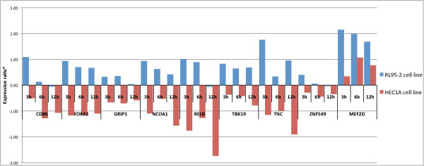
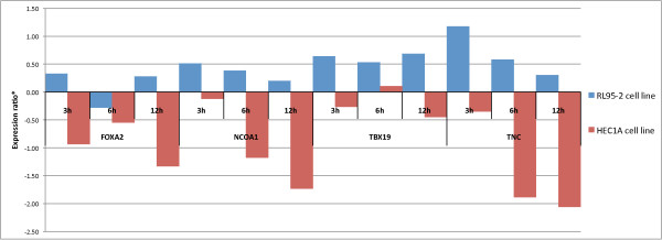
Methods: 382 genes were selected, differentially expressed in the receptive endometrium, to study their responsiveness of estrogen and progesterone. The endometrial cell lines HEC1A and RL95-2 were used as experimental models for the non-receptive and receptive endometrium, respectively. Putative targets for activated steroid hormone receptors were investigated by chromatin immunoprecipitation (ChIP) using receptor-specific antibodies. Promoter occupancy of the selected genes by steroid receptors was detected in ChIP-purified DNA by quantitative PCR (qPCR). Expression analysis by reverse transcriptase (RT)-PCR was used to further investigate hormone dependent mRNA expression regulation of a subset of genes.

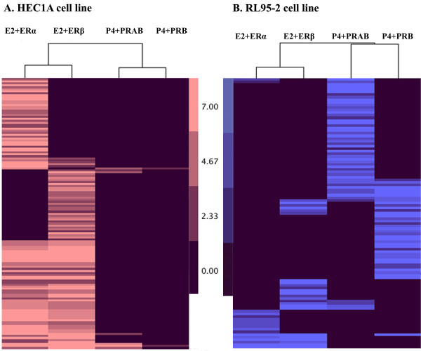
Results: ChIP-qPCR analysis demonstrated that each steroid hormone receptor had distinct group of target genes in the endometrial cell lines. After estradiol treatment, expression of estrogen receptor target genes predominated in HEC1A cells (n = 137) compared to RL95-2 cells (n = 35). In contrast, expression of progesterone receptor target genes was higher in RL95-2 cells (n = 83) than in HEC1A cells (n = 7) after progesterone treatment. RT-PCR analysis of 20 genes demonstrated transcriptional changes after estradiol or progesterone treatment of the cell lines.

Conclusions: Combined results from ChIP-qPCR and RT-PCR analysis showed different patterns of steroid hormone receptor occupancy at target genes, corresponding to activation or suppression of gene expression after hormone treatment of HEC1A and RL95-2 cell lines.


Figures

Figure 1
Figure 1
Cluster analysis of ERα, ERβ, PRAB, PRB target genes in HEC1A and RL95-2 cells after E2 or P4 treatment. Target genes revealed from ChIP-qPCR experiment were clustered with Cluster 3.0 program and visualized using Java TreeView software. A. HEC1A cell line. E2+ERα: 101 ERα target genes after E2 treatment; E2+ERβ: 96 ERβ target genes after E2 treatment; P4+PRAB: 7 PRAB target genes after P4 treatment; and P4+PRB: 2 PRB target genes after P4 treatment. B. RL95-2 cell line. E2+ERα: 17 ERα target genes after E2 treatment; E2+ERβ: 24. ERβ target genes after E2 treatment; P4+PRAB: 52 PRAB target genes after P4 treatment; and P4+PRB: 40 PRB target genes after P4 treatment.
Figure 2
Figure 2
E2 mediated time-dependant gene expression in RL95-2 (blue) and HEC1A (red) cell lines compared to non-treated samples. Expression profiles of CD86, FOXA2, GRIP1, NCOA1, RELB, TBX19, TNC, ZNF549, and MEF2D genes had significant difference (p < 0.05, two-tailed, unpaired Student's t-test with 95% confidence of log2-transformed expression ratios relative to baseline expression) in mRNA expression ratios (*) between RL95-2 and HEC1A cells after E2 treatment. E2 exposure predominantly induced mRNA level of gene expression in RL95-2 cells but down-regulated in HEC1A cells (except for MEF2D gene).
Figure 3
Figure 3
P4 mediated time-dependant gene expression in RL95-2 (blue) and HEC1A (red) cell lines compared to non-treated samples. Expression profiles of FOXA2, NCOA1, TBX19 and TNC genes had significant difference (p < 0.05, two-tailed, unpaired Student's t-test with 95% confidence of log2-transformed expression ratios relative to baseline expression) in mRNA expression ratios (*) between RL95-2 and HEC1A cells after P4 treatment. P4 mostly induced mRNA level in RL95-2 cells but down-regulated in HEC1A cells.

Similar articles

Cited by

References

    1. Graham RA, Seif MW, Aplin JD, Li TC, Cooke ID, Rogers AW, Dockery P. An endometrial factor in unexplained infertility. Bmj. 1990;300(6737):1428–1431. doi: 10.1136/bmj.300.6737.1428. - DOI - PMC - PubMed
    1. Enmark E, Pelto-Huikko M, Grandien K, Lagercrantz S, Lagercrantz J, Fried G, Nordenskjold M, Gustafsson JA. Human estrogen receptor beta-gene structure, chromosomal localization, and expression pattern. J Clin Endocrinol Metab. 1997;82(12):4258–4265. doi: 10.1210/jc.82.12.4258. - DOI - PubMed
    1. Kuiper GG, Gustafsson JA. The novel estrogen receptor-beta subtype: potential role in the cell- and promoter-specific actions of estrogens and anti-estrogens. FEBS Lett. 1997;410(1):87–90. doi: 10.1016/S0014-5793(97)00413-4. - DOI - PubMed
    1. Matsuzaki S, Fukaya T, Suzuki T, Murakami T, Sasano H, Yajima A. Oestrogen receptor alpha and beta mRNA expression in human endometrium throughout the menstrual cycle. Mol Hum Reprod. 1999;5(6):559–564. doi: 10.1093/molehr/5.6.559. - DOI - PubMed
    1. Wen DX, Xu YF, Mais DE, Goldman ME, McDonnell DP. The A and B isoforms of the human progesterone receptor operate through distinct signaling pathways within target cells. Mol Cell Biol. 1994;14(12):8356–8364. - PMC - PubMed

Publication types