. 2009 Dec;59(6):589-97.

Epidemiology of invasive Klebsiella pneumoniae with hypermucoviscosity phenotype in a research colony of nonhuman primates

Affiliations

Epidemiology of invasive Klebsiella pneumoniae with hypermucoviscosity phenotype in a research colony of nonhuman primates

Robin L Burke et al. Comp Med. 2009 Dec.

Abstract

Invasive Klebsiella pneumoniae with hypermucoviscosity phenotype (HMV K. pneumoniae) is an emerging human pathogen that, over the past 20 y, has resulted in a distinct clinical syndrome characterized by pyogenic liver abscesses sometimes complicated by bacteremia, meningitis, and endophthalmitis. Infections occur predominantly in Taiwan and other Asian countries, but HMV K. pneumoniae is considered an emerging infectious disease in the United States and other Western countries. In 2005, fatal multisystemic disease was attributed to HMV K. pneumoniae in African green monkeys (AGM) at our institution. After identification of a cluster of subclinically infected macaques in March and April 2008, screening of all colony nonhuman primates by oropharyngeal and rectal culture revealed 19 subclinically infected rhesus and cynomolgus macaques. PCR testing for 2 genes associated with HMV K. pneumoniae, rmpA and magA, suggested genetic variability in the samples. Random amplified polymorphic DNA analysis on a subset of clinical isolates confirmed a high degree of genetic diversity between the samples. Environmental testing did not reveal evidence of aerosol or droplet transmission of the organism in housing areas. Further research is needed to characterize HMV K. pneumoniae, particularly with regard to genetic differences among bacterial strains and their relationship to human disease and to the apparent susceptibility of AGM to this organism.


Figures

Figure 1.
Figure 1.
Positive string test. The HMV phenotype of K. pneumoniae is defined by a positive-string test. The test is performed by touching a colony with a bacterial loop and gently lifting. If a mucoid ‘string’ of 5 mm or more forms, the string test is considered positive.
Figure 2.
Figure 2.
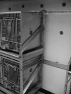
Culture plates affixed in a grid pattern around and on NHP cages. Once the plates were in place, the animal caretaker performed routine spray-down of organic material from the cages. The plates then were removed and cultured to determine the extent of environmental spread of HMV K. pneumoniae during routine cage-cleaning procedures.
Figure 3.
Figure 3.
Grid pattern configuration of culture plates. MacConkey cultures plates were taped in positions 1 through 13, and the lids were removed. Plates 8 and 9 were placed in an empty cage, directly below and beside infected NHP. The animal caretaker then followed routine procedures to spray waste material out of cage bank 1, avoiding wetting of NHP in cages. Once the gross contamination was removed from cage bank 1, all culture plates were covered and removed. A second set of MacConkey plates were taped in positions 1 through 13 and the experiment repeated for cage bank 2.
Figure 4.
Figure 4.
Room configuration for culturing for HMV K. pneumoniae after cage spray. In an effort to determine the environmental spread of HMV K. pneumoniae, covered MacConkey culture plates were placed on trays while an animal caretaker sprayed waste material from cages. As soon as the caretaker had completed the spray-down, plate covers were removed. Plates in position A (inset) remained in place from 0 to 60 min after spraying. In position B, 3 sets of plates were rotated at predetermined time points: set 1 remained in place from 0 to 5 min, set 2 from 5 to 10 min, and set 3 from 10 to 60 min after spraying.
Figure 5.
Figure 5.
RAPD-PCR banding pattern of a subset of isolates from NHP infected with HMV K. pneumoniae. Isolates were identified as being HMV K. pneumoniae based on Vitek analysis and a positive string test. “M” indicates a 1 kb plus marker, and NTC indicates a negative control. The isolate labeled AGM was an HMV K. pneumoniae culture from a fatal abdominal abscess in an African green monkey in 2006. All other isolates were from oropharyngeal or rectal swabs from asymptomatic macaques. Animal identifications correlate with those in Tables 1 and 2. Isolates labeled Cy4a and Cy4b represent 2 rectal samples taken from NHP Cy4 about 3 wk apart (Cy 4a, 2008 Apr 28; Cy4b, 2008 May 20). Animals from Vendor 1 comprised a single shipment, whereas NHP that came from Vendor 2 arrived in 3 separate shipments, as indicated. The remaining 2 macaques were transferred from 2 different Department of Defense research centers, indicated as Research Institute 1 (Res Ins 1) and Research Institute 2 (Res Ins 2). PCR results were correlated with the RAPD-PCR banding patterns. Isolates were determined to be rmpA+/magA (*), rmpA/magA+ (†), or rmpA/magA (‡). Clusters A through D indicate distinct RAPD types, which were determined based on the absence or presence of 2 or more bands.

Similar articles

Cited by

References

    1. Bolister NJ, Johnson HE, Wathes CM. 1992. The ability of airborne Klebsiella pneumoniae to colonize mouse lungs. Epidemiol Infect 109:121–131 - PMC - PubMed
    1. Brisse S, Fevre C, Passet V, Issenhuth-Jeanjean S, Tournebize R, Diancourt L, Grimont P. 2009. Virulent clones of Klebsiella pneumoniae: identification and evolutionary scenario based on genomic and phenotypic characterization. PLoS One 4:e4982. - PMC - PubMed
    1. Chan KS, Yu WL, Tsai CL, Cheng KC, Hou CC, Lee MC, Tan CK. 2007. Pyogenic liver abscess caused by Klebsiella pneumoniae: analysis of the clinical characteristics and outcomes of 84 patients. Chin Med J (Engl) 120:136–139 - PubMed
    1. Chang CM, Lu FH, Guo HR, Ko WC. 2005. Klebsiella pneumoniae fascial space infections of the head and neck in Taiwan: emphasis on diabetic patients and repetitive infections. J Infect 50:34–40 - PubMed
    1. Chuang YP, Fang CT, Lai SY, Chang SC, Wang JT. 2006. Genetic determinants of capsular serotype K1 of Klebsiella pneumoniae causing primary pyogenic liver abscess. J Infect Dis 193:645–654 - PubMed

LinkOut - more resources