

Brain substrates of learning and retention in mild cognitive impairment diagnosis and progression to Alzheimer's disease
- PMID: 20034503
- PMCID: PMC2851550
- DOI: 10.1016/j.neuropsychologia.2009.12.024
Brain substrates of learning and retention in mild cognitive impairment diagnosis and progression to Alzheimer's disease
Abstract
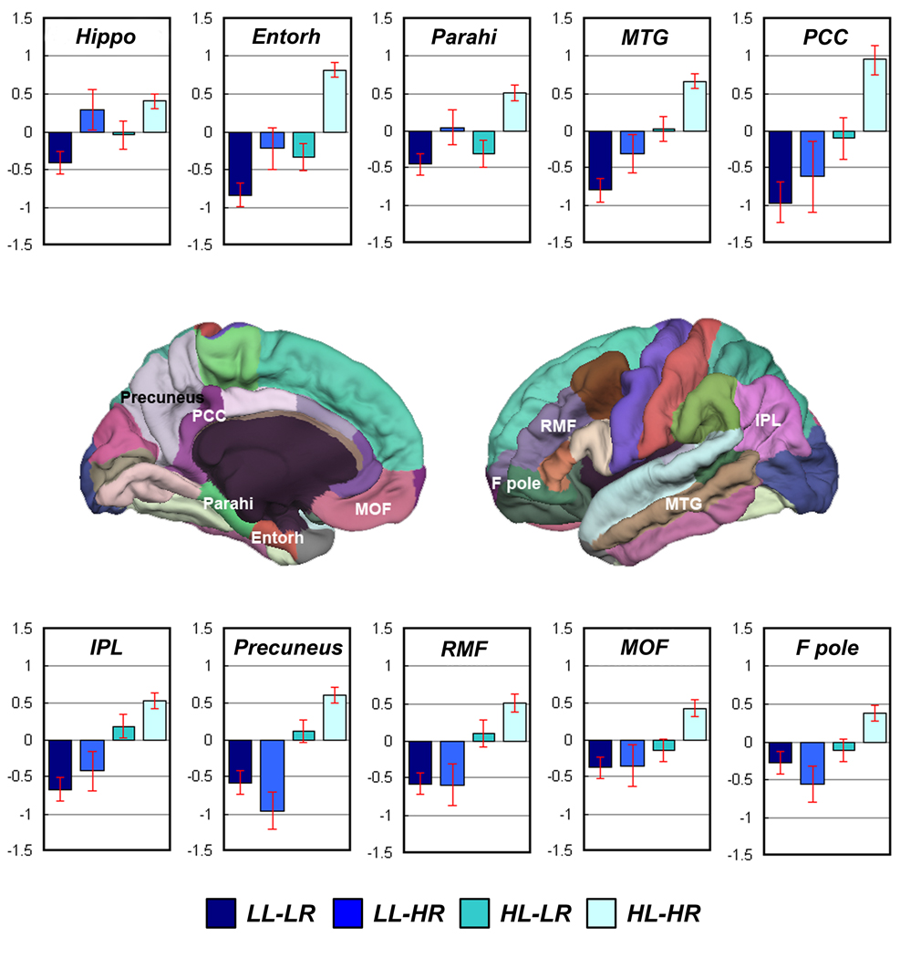
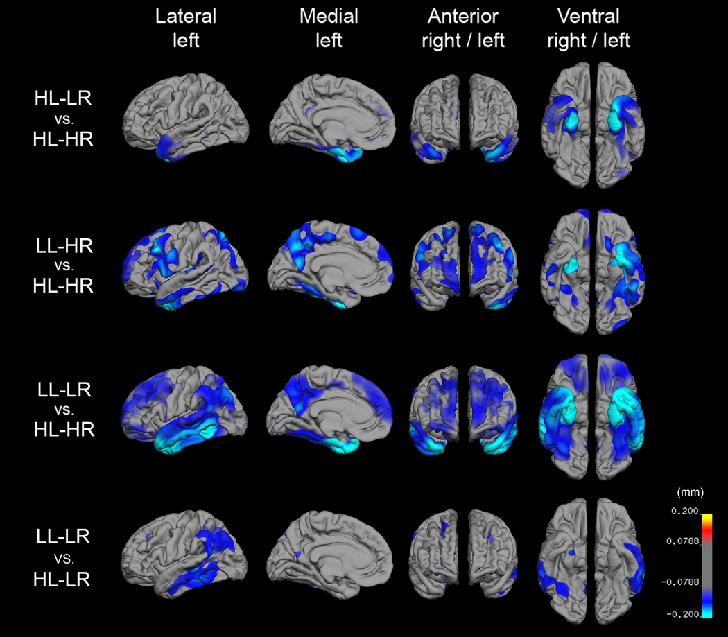
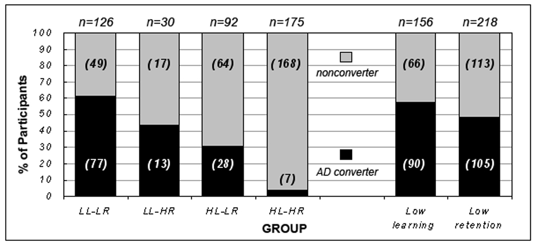
Understanding the underlying qualitative features of memory deficits in mild cognitive impairment (MCI) can provide critical information for early detection of Alzheimer's disease (AD). This study sought to investigate the utility of both learning and retention measures in (a) the diagnosis of MCI, (b) predicting progression to AD, and (c) examining their underlying brain morphometric correlates. A total of 607 participants were assigned to three MCI groups (high learning-low retention; low learning-high retention; low learning-low retention) and one control group (high learning-high retention) based on scores above or below a 1.5 SD cutoff on learning and retention indices of the Rey Auditory Verbal Learning Test. Our results demonstrated that MCI individuals with predominantly a learning deficit showed a widespread pattern of gray matter loss at baseline, whereas individuals with a retention deficit showed more focal gray matter loss. Moreover, either learning or retention measures provided good predictive value for longitudinal clinical outcome over two years, although impaired learning had modestly better predictive power than impaired retention. As expected, impairments in both measures provided the best predictive power. Thus, the conventional practice of relying solely on the use of delayed recall or retention measures in studies of amnestic MCI misses an important subset of older adults at risk of developing AD. Overall, our results highlight the importance of including learning measures in addition to retention measures when making a diagnosis of MCI and for predicting clinical outcome.
Keywords: Amnestic MCI; Early detection; Episodic memory; Longitudinal outcome; MR morphometry.
(c) 2009 Elsevier Ltd. All rights reserved.
Conflict of interest statement
Conflict of interest: Anders M. Dale is a founder and holds equity in CorTechs Labs, Inc., and also serves on the Scientific Advisory Board. The terms of this arrangement have been reviewed and approved by the University of California, San Diego in accordance with its conflict of interest policies. The other authors do not have a financial or any other conflict of interest to disclose related to this manuscript.
Figures
References
-
- Albert MS, Moss MB, Tanzi R, Jones K. Preclinical prediction of AD using neuropsychological tests. Journal of International Neuropsychological Society. 2001;7(5):631–639. - PubMed
-
- Arnaiz E, Almkvist O. Neuropsychological features of mild cognitive impairment and preclinical Alzheimer's disease. Acta Neurologica Scandinavica Supplementum. 2003;179:34–41. - PubMed
-
- Axmacher N, Schmitz DP, Weinreich I, Elger CE, Fell J. Interaction of working memory and long-term memory in the medial temporal lobe. Cerebral Cortex. 2008;18(12):2868–2878. - PubMed
Publication types
MeSH terms
Grants and funding
LinkOut - more resources
Full Text Sources
Medical
Research Materials
