

The test-retest reliability of 18F-DOPA PET in assessing striatal and extrastriatal presynaptic dopaminergic function
- PMID: 20034580
- PMCID: PMC4096947
- DOI: 10.1016/j.neuroimage.2009.12.058
The test-retest reliability of 18F-DOPA PET in assessing striatal and extrastriatal presynaptic dopaminergic function
Abstract
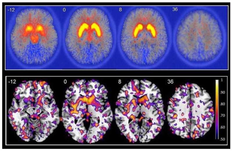
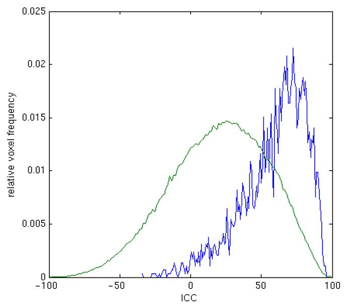
Brain presynaptic dopaminergic function can be assessed using 18F-DOPA positron emission tomography (PET). Regional 18F-DOPA utilization may be used to index dopaminergic abnormalities over time or dopaminergic response to treatment in clinical populations. Such studies require prior knowledge of the stability of the 18F-DOPA signal in the brain regions of interest. Test-retest reliability was examined in eight healthy volunteers who each received two 18F-DOPA PET scans, approximately 2 years apart. 18F-DOPA utilization (k(i)(cer)) was determined using graphical analysis relative to a reference tissue input (Patlak and Blasberg, 1985). Reproducibility (measured as the within-subjects variation) and reliability (measured as intraclass correlation coefficients, ICCs) of 18F-DOPA k(i)(cer) were assessed in the structural and functional subdivisions of the striatum and select extrastriatal brain regions. Voxel-based median ICC maps were used to visualize the distribution of 18F-DOPA k(i)(cer) reliability across the brain. The caudate and putamen, and associative and sensorimotor, striatal subdivisions showed good reliability across the two scan sessions with bilateral ICCs ranging from 0.681 to 0.944. Reliability was generally lower in extrastriatal regions, with bilateral ICCs ranging from 0.235 in the amygdala to 0.894 in the thalamus. These data confirm the utility of 18F-DOPA PET in assessing dopaminergic function in the striatum and select extrastriatal areas but highlight the limitations in using this approach to measure dopaminergic function in low uptake extrastriatal brain areas. This information can be used to optimize the experimental design of future studies investigating changes in brain dopaminergic function with 18F-DOPA.
Copyright 2009 Elsevier Inc. All rights reserved.
Figures
References
-
- Bose SK, Turkheimer FE, Howes OD, Mehta MA, Cunliffe R, Stokes PR, Grasby PM. Classification of schizophrenic patients and healthy controls using [18F] fluorodopa PET imaging. Schizophr.Res. 2008;106:148–155. - PubMed
-
- Bouchard S, Bousquet C, Roberge AG. Characteristics of dihydroxyphenylalanine/5-hydroxytryptophan decarboxylase activity in brain and liver of cat. J.Neurochem. 1981;37:781–787. - PubMed
-
- Brooks DJ. Imaging end points for monitoring neuroprotection in Parkinson’s disease. Ann.Neurol. 2003a;53(Suppl 3):S110–S118. - PubMed
-
- Brooks DJ. PET studies on the function of dopamine in health and Parkinson’s disease. Ann.N.Y.Acad.Sci. 2003b;991:22–35. - PubMed
-
- Brooks DJ, Frey KA, Marek KL, Oakes D, Paty D, Prentice R, Shults CW, Stoessl AJ. Assessment of neuroimaging techniques as biomarkers of the progression of Parkinson’s disease. Exp.Neurol. 2003;184(Suppl 1):S68–S79. - PubMed
Publication types
MeSH terms
Substances
Grants and funding
LinkOut - more resources
Full Text Sources