

Human circulating fibrocytes have the capacity to differentiate osteoblasts and chondrocytes
- PMID: 20034590
- PMCID: PMC2835809
- DOI: 10.1016/j.biocel.2009.12.011
Human circulating fibrocytes have the capacity to differentiate osteoblasts and chondrocytes
Abstract
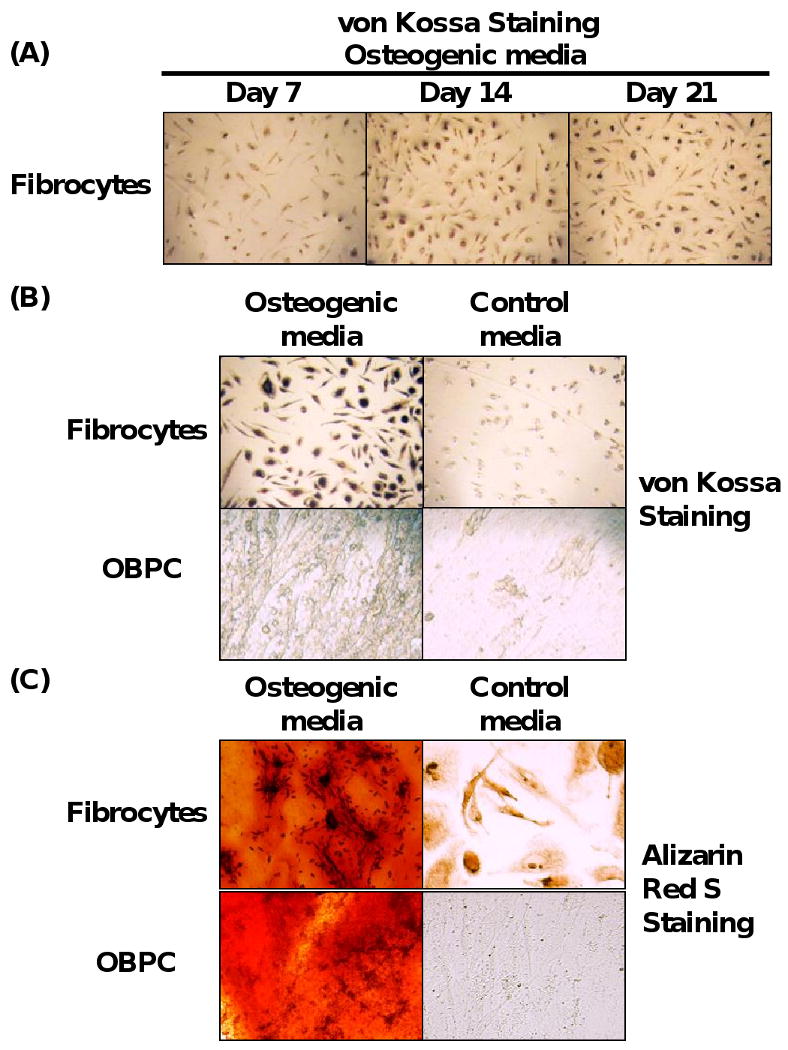
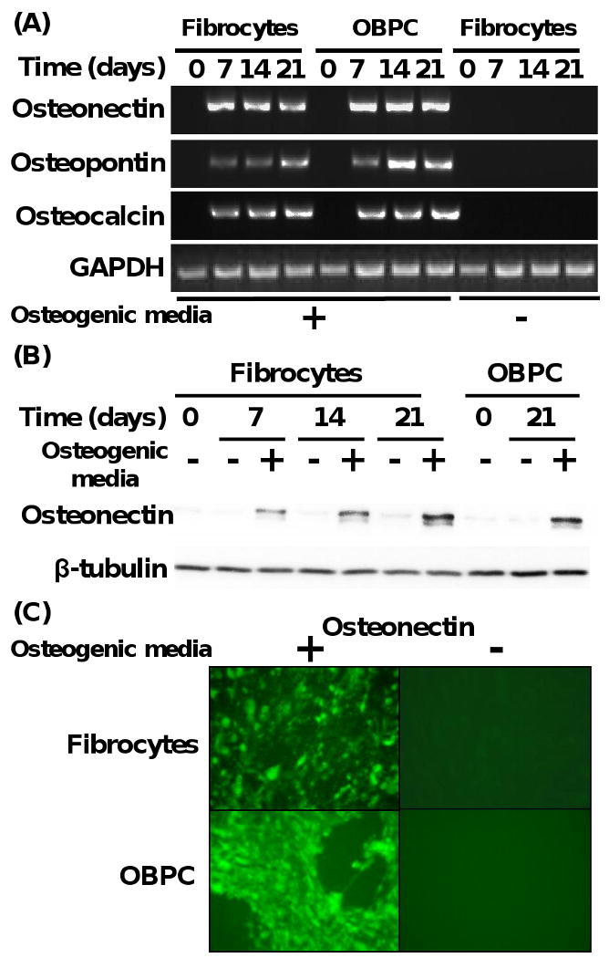
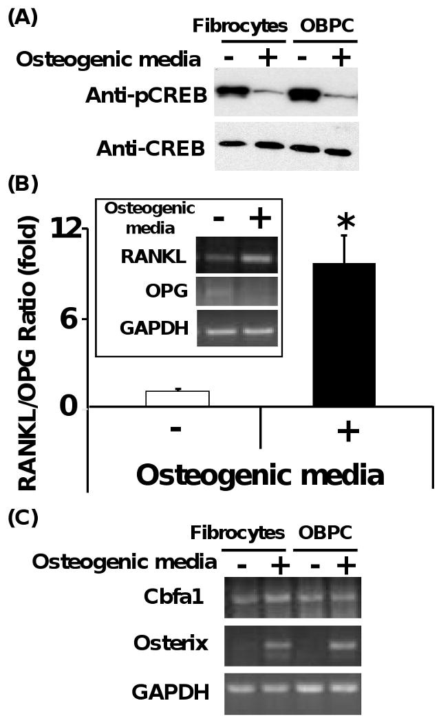
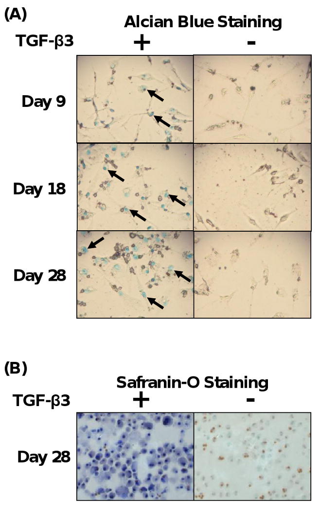
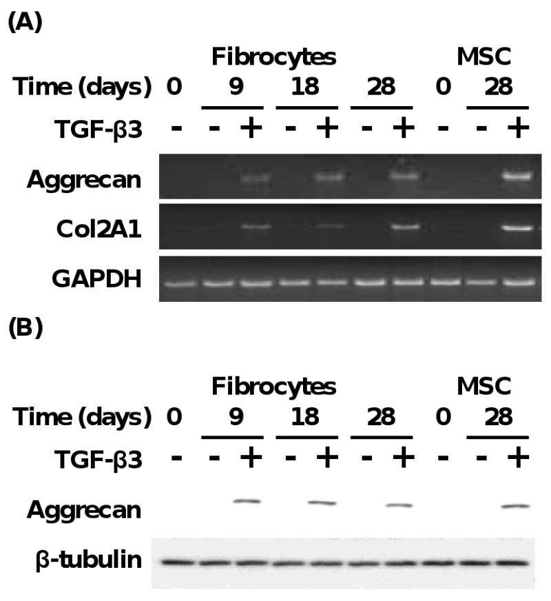
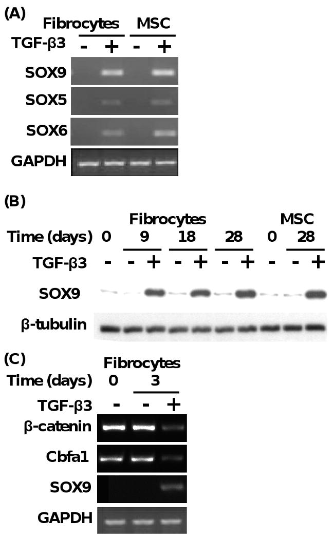
Fibrocytes are bone marrow-derived cells. Fibrocytes can differentiate into adipocyte- and myofibroblast-like cells. Since fibrocytes can behave like mesenchymal progenitor cells, we hypothesized that fibrocytes have the potential to differentiate into other mesenchymal lineage cells, such as osteoblasts and chondrocytes. In this study, we found that fibrocytes differentiated into osteoblast-like cells when cultured in osteogenic media in a manner similar to osteoblast precursor cells. Under these conditions, fibrocytes and osteoblast precursor cells displayed increased calcium deposition, and increased expression of specific osteogenic genes. In addition, dephosphorylation of cAMP-responsive element binding protein was associated with the increased ratio of receptor activator of the NF-kappaB Ligand/osteoprotegerin gene expression and enhanced gene expression of osterix in these cells under these conditions. Both events are important in promoting osteogenesis. In contrast, fibrocytes and mesenchymal stem cells cultured in chondrogenic media in the presence of transforming growth factor-beta3 were found to differentiate to chondrocyte-like cells. Fibrocytes and mesenchymal stem cells under these conditions were found to express increased levels of aggrecan and type II collagen genes. Transcription factor genes associated with chondrogenesis were also found to be induced in fibrocytes and mesenchymal stem cells under these conditions. In contrast, beta-catenin protein and the core binding factor alpha1 subunit protein transcription factor were decreased in expression under these conditions. These data indicate that human fibrocytes have the capability to differentiate into osteoblast- and chondrocyte-like cells. These findings suggest that such cells could be used in cell-based tissue-regenerative therapy.
2009 Elsevier Ltd. All rights reserved.
Figures
Similar articles
-
Adipose-derived mesenchymal stromal (stem) cells differentiate to osteoblast and chondroblast lineages upon incubation with conditioned media from dental pulp stem cell-derived osteoblasts and auricle cartilage chondrocytes.J Biol Regul Homeost Agents. 2016 Jan-Mar;30(1):111-22. J Biol Regul Homeost Agents. 2016. PMID: 27049081
-
Demineralized bone promotes chondrocyte or osteoblast differentiation of human marrow stromal cells cultured in collagen sponges.Cell Tissue Bank. 2005;6(1):33-44. doi: 10.1007/s10561-005-4253-y. Cell Tissue Bank. 2005. PMID: 15735899 Free PMC article.
-
Runx1 up-regulates chondrocyte to osteoblast lineage commitment and promotes bone formation by enhancing both chondrogenesis and osteogenesis.Biochem J. 2020 Jul 17;477(13):2421-2438. doi: 10.1042/BCJ20200036. Biochem J. 2020. PMID: 32391876 Free PMC article.
-
The role of microRNAs in the osteogenic and chondrogenic differentiation of mesenchymal stem cells and bone pathologies.Theranostics. 2021 Apr 30;11(13):6573-6591. doi: 10.7150/thno.55664. eCollection 2021. Theranostics. 2021. PMID: 33995677 Free PMC article. Review.
-
The role of circulating mesenchymal progenitor cells (fibrocytes) in the pathogenesis of fibrotic disorders.Thromb Haemost. 2009 Apr;101(4):613-8. Thromb Haemost. 2009. PMID: 19350102 Free PMC article. Review.
Cited by
-
Peripheral blood-derived mesenchymal stem cells: candidate cells responsible for healing critical-sized calvarial bone defects.Stem Cells Transl Med. 2015 Apr;4(4):359-68. doi: 10.5966/sctm.2014-0150. Epub 2015 Mar 5. Stem Cells Transl Med. 2015. PMID: 25742693 Free PMC article.
-
Mesenchymal stem cells: Molecular characteristics and clinical applications.World J Stem Cells. 2010 Aug 26;2(4):67-80. doi: 10.4252/wjsc.v2.i4.67. World J Stem Cells. 2010. PMID: 21607123 Free PMC article.
-
Obesity and Fibrosis: Setting the Stage for Breast Cancer.Cancers (Basel). 2023 May 26;15(11):2929. doi: 10.3390/cancers15112929. Cancers (Basel). 2023. PMID: 37296891 Free PMC article. Review.
-
The role of fibrocytes in fibrotic diseases of the lungs and heart.Fibrogenesis Tissue Repair. 2011 Jan 10;4:2. doi: 10.1186/1755-1536-4-2. Fibrogenesis Tissue Repair. 2011. PMID: 21219601 Free PMC article.
-
Concise review: evidence for CD34 as a common marker for diverse progenitors.Stem Cells. 2014 Jun;32(6):1380-9. doi: 10.1002/stem.1661. Stem Cells. 2014. PMID: 24497003 Free PMC article. Review.
References
-
- Alhadlaq A, Mao JJ. Mesenchymal stem cells: isolation and therapeutics. Stem Cells Dev. 2004;13:436–48. - PubMed
-
- Andersson-Sjoland A, de Alba CG, Nihlberg K, Becerril C, Ramirez R, Pardo A, Westergren-Thorsson G, Selman M. Fibrocytes are a potential source of lung fibroblasts in idiopathic pulmonary fibrosis. Int J Biochem Cell Biol. 2008;40:2129–40. - PubMed
-
- Bellini A, Mattoli S. The role of the fibrocyte, a bone marrow-derived mesenchymal progenitor, in reactive and reparative fibroses. Lab Invest. 2007;87:858–70. - PubMed
-
- Bonewald LF, Harris SE, Rosser J, Dallas MR, Dallas SL, Camacho NP, Boyan B, Boskey A. von Kossa staining alone is not sufficient to confirm that mineralization in vitro represents bone formation. Calcif Tissue Int. 2003;72:537–47. - PubMed
-
- Brittberg M, Lindahl A, Nilsson A, Ohlsson C, Isaksson O, Peterson L. Treatment of deep cartilage defects in the knee with autologous chondrocyte transplantation. N Engl J Med. 1994;331:889–95. - PubMed
Publication types
MeSH terms
Substances
Grants and funding
LinkOut - more resources
Full Text Sources
