

Abnormal calcium homeostasis in peripheral neuropathies
- PMID: 20034667
- PMCID: PMC2834846
- DOI: 10.1016/j.ceca.2009.11.008
Abnormal calcium homeostasis in peripheral neuropathies
Abstract
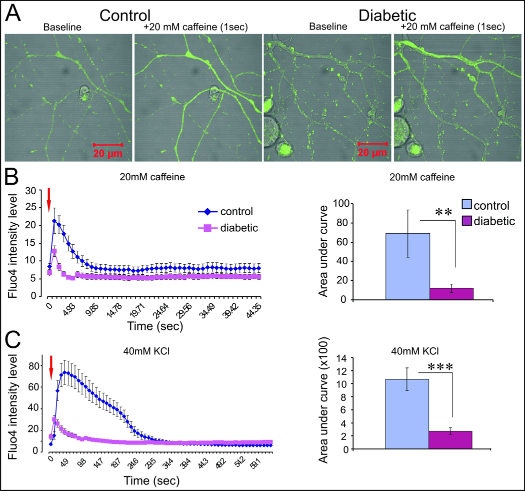
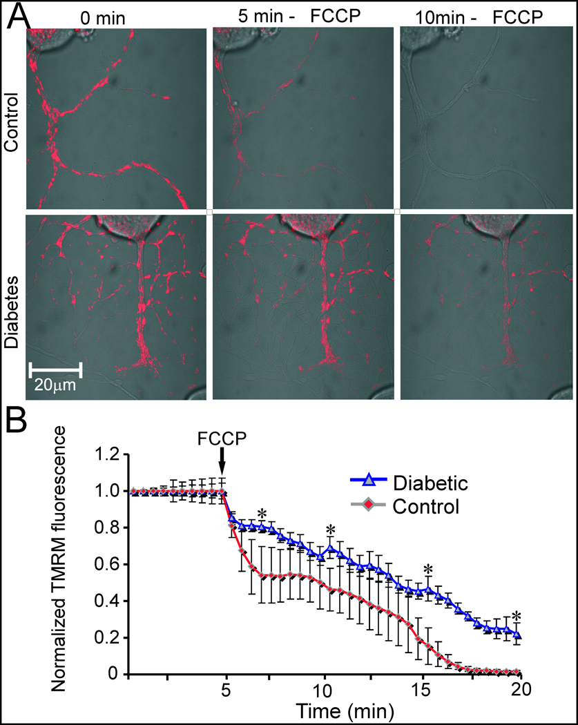
Abnormal neuronal calcium (Ca2+) homeostasis has been implicated in numerous diseases of the nervous system. The pathogenesis of two increasingly common disorders of the peripheral nervous system, namely neuropathic pain and diabetic polyneuropathy, has been associated with aberrant Ca2+ channel expression and function. Here we review the current state of knowledge regarding the role of Ca2+ dyshomeostasis and associated mitochondrial dysfunction in painful and diabetic neuropathies. The central impact of both alterations of Ca2+ signalling at the plasma membrane and also intracellular Ca2+ handling on sensory neurone function is discussed and related to abnormal endoplasmic reticulum performance. We also present new data highlighting sub-optimal axonal Ca2+ signalling in diabetic neuropathy and discuss the putative role for this abnormality in the induction of axonal degeneration in peripheral neuropathies. The accumulating evidence implicating Ca2+ dysregulation in both painful and degenerative neuropathies, along with recent advances in understanding of regional variations in Ca2+ channel and pump structures, makes modulation of neuronal Ca2+ handling an increasingly viable approach for therapeutic interventions against the painful and degenerative aspects of many peripheral neuropathies.
2009 Elsevier Ltd. All rights reserved.
Figures
Similar articles
-
Calcium signalling in sensory neurones and peripheral glia in the context of diabetic neuropathies.Cell Calcium. 2014 Nov;56(5):362-71. doi: 10.1016/j.ceca.2014.07.005. Epub 2014 Aug 1. Cell Calcium. 2014. PMID: 25149565 Review.
-
Mitochondrial malfunction and Ca2+ dyshomeostasis drive neuronal pathology in diabetes.Cell Calcium. 2008 Jul;44(1):112-22. doi: 10.1016/j.ceca.2007.11.010. Epub 2008 Jan 11. Cell Calcium. 2008. PMID: 18191198 Review.
-
Role of oxidative stress and Ca²⁺ signaling on molecular pathways of neuropathic pain in diabetes: focus on TRP channels.Neurochem Res. 2012 Oct;37(10):2065-75. doi: 10.1007/s11064-012-0850-x. Epub 2012 Jul 31. Neurochem Res. 2012. PMID: 22846968 Review.
-
Taurine replacement attenuates hyperalgesia and abnormal calcium signaling in sensory neurons of STZ-D rats.Am J Physiol Endocrinol Metab. 2005 Jan;288(1):E29-36. doi: 10.1152/ajpendo.00168.2004. Am J Physiol Endocrinol Metab. 2005. PMID: 15585600
-
[Role of Ca2+ channels in the pathogenesis of diabetic neuropathy].Nihon Rinsho. 2005 Jun;63 Suppl 6:647-51. Nihon Rinsho. 2005. PMID: 15999785 Review. Japanese. No abstract available.
Cited by
-
High Dietary Fat Consumption Impairs Axonal Mitochondrial Function In Vivo.J Neurosci. 2021 May 12;41(19):4321-4334. doi: 10.1523/JNEUROSCI.1852-20.2021. Epub 2021 Mar 30. J Neurosci. 2021. PMID: 33785643 Free PMC article.
-
Therapeutic Potential of Polyphenols in the Management of Diabetic Neuropathy.Evid Based Complement Alternat Med. 2021 May 13;2021:9940169. doi: 10.1155/2021/9940169. eCollection 2021. Evid Based Complement Alternat Med. 2021. PMID: 34093722 Free PMC article. Review.
-
Genome-wide profiling of DNA methylation and gene expression identifies candidate genes for human diabetic neuropathy.Clin Epigenetics. 2020 Aug 12;12(1):123. doi: 10.1186/s13148-020-00913-6. Clin Epigenetics. 2020. PMID: 32787975 Free PMC article.
-
Fecal transplantation and butyrate improve neuropathic pain, modify immune cell profile, and gene expression in the PNS of obese mice.Proc Natl Acad Sci U S A. 2020 Oct 20;117(42):26482-26493. doi: 10.1073/pnas.2006065117. Epub 2020 Oct 5. Proc Natl Acad Sci U S A. 2020. PMID: 33020290 Free PMC article.
-
Salvianolic acid A protects the peripheral nerve function in diabetic rats through regulation of the AMPK-PGC1α-Sirt3 axis.Molecules. 2012 Sep 20;17(9):11216-28. doi: 10.3390/molecules170911216. Molecules. 2012. PMID: 22996345 Free PMC article.
References
-
- Brownlee M. Biochemistry and molecular cell biology of diabetic complications. Nature. 2001;414:813–820. - PubMed
-
- Nishikawa T, Edelstein D, Du XL, Yamagishi S, Matsumura T, Kaneda Y, Yorek MA, Beebe D, Oates PJ, Hammes HP, Giardino I, Brownlee M. Normalizing mitochondrial superoxide production blocks three pathways of hyperglycaemic damage. Nature. 2000;404:787–790. - PubMed
-
- Dawson KG, Gomes D, Gerstein H, Blanchard JF, Kahler KH. The economic cost of diabetes in Canada, 1998. Diabetes Care. 2002;25:1303–1307. - PubMed
-
- Caro JJ, Ward AJ, O'Brien JA. Lifetime costs of complications resulting from type 2 diabetes in the U.S. Diabetes Care. 2002;25:476–481. - PubMed
Publication types
MeSH terms
Substances
Grants and funding
LinkOut - more resources
Full Text Sources
Other Literature Sources
Miscellaneous
