. 2010 Mar;298(3):L371-81.
doi: 10.1152/ajplung.00308.2009. Epub 2009 Dec 24.

Moderate oxygen augments lipopolysaccharide-induced lung injury in mice

Affiliations

Moderate oxygen augments lipopolysaccharide-induced lung injury in mice

Neil R Aggarwal et al. Am J Physiol Lung Cell Mol Physiol. 2010 Mar.

Abstract

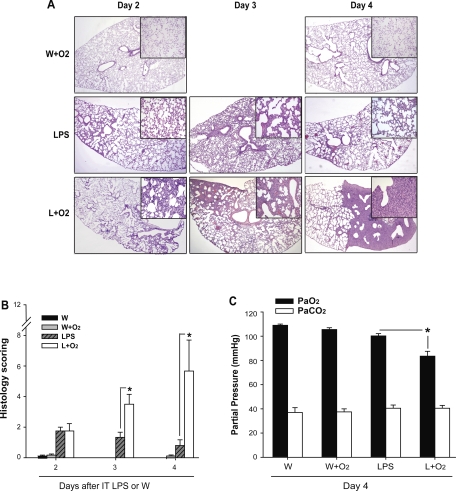
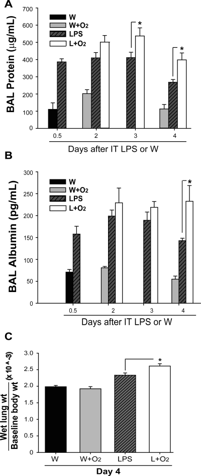
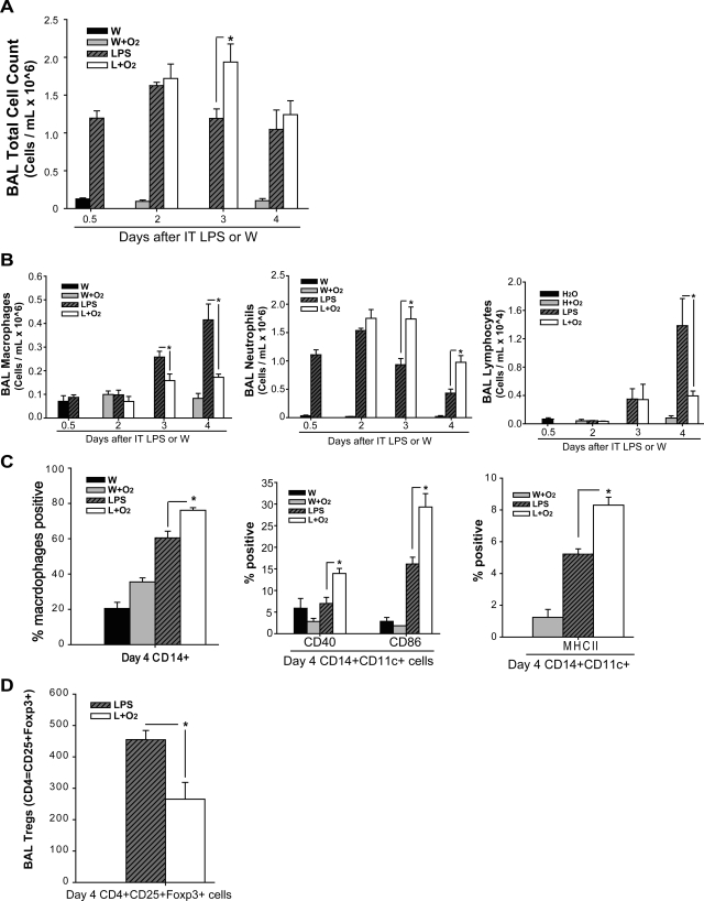
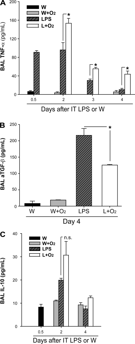
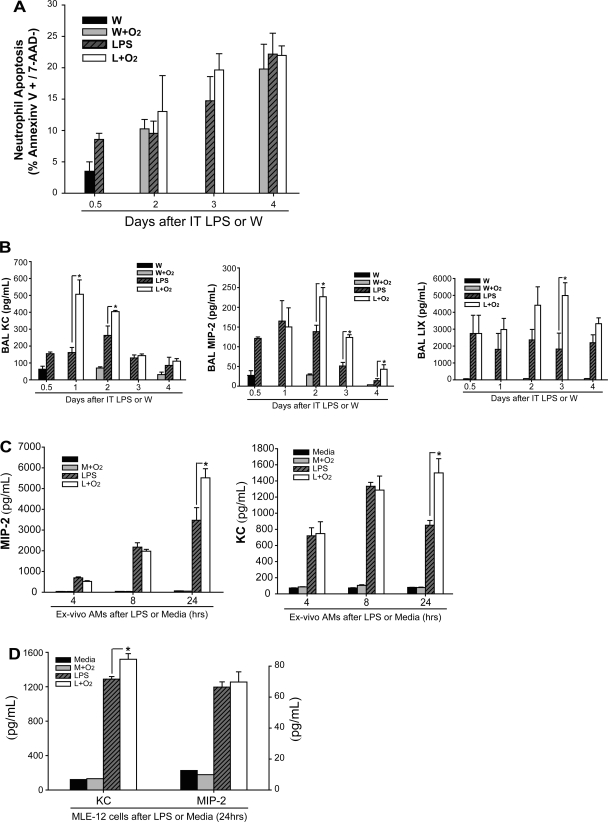
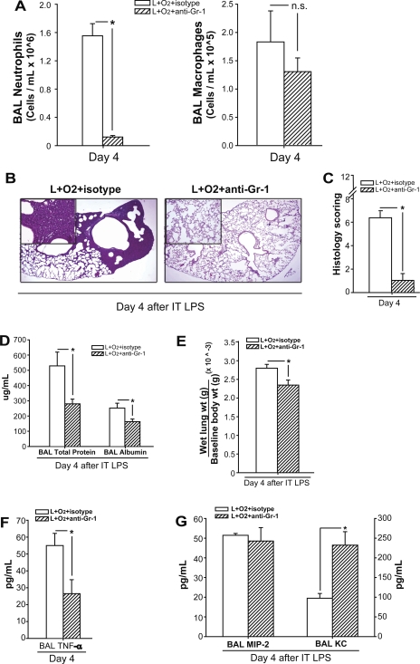
Despite the associated morbidity and mortality, underlying mechanisms leading to the development of acute lung injury (ALI) remain incompletely understood. Frequently, ALI develops in the hospital, coinciding with institution of various therapies, including the use of supplemental oxygen. Although pathological evidence of hyperoxia-induced ALI in humans has yet to be proven, animal studies involving high oxygen concentration reproducibly induce ALI. The potentially injurious role of lower and presumably safer oxygen concentrations has not been well characterized in any species. We hypothesized that in the setting of a preexisting insult to the lung, the addition of moderate-range oxygen can augment lung injury. Our model of low-dose intratracheal LPS (IT LPS) followed by 60% oxygen caused a significant increase in ALI compared with LPS or oxygen alone with increased alveolar neutrophils, histological injury, and epithelial barrier permeability. In the LPS plus oxygen group, regulatory T cell number was reduced, and macrophage activation markers were increased, compared with LPS alone. Antibody-mediated depletion of neutrophils significantly abrogated the observed lung injury for all measured factors. The enhanced presence of alveolar neutrophils in the setting of LPS and oxygen is due, at least in part, to elevated chemokine gradients signaling neutrophils to the alveolar space. We believe these results strongly support an effect of lower concentrations of oxygen to augment the severity of a mild preexisting lung injury and warrants further investigation in both animals and humans.


Figures

Fig. 1.
Fig. 1.
Supplemental oxygen augments LPS-induced lung injury. A: representative histology sections are shown at low- (2×) and high- (20×) power for days 2, 3, and 4 after IT water (W) + supplemental oxygen (W+O2) or IT LPS + supplemental oxygen. B: a semiquantitative assessment of morphological changes based on percent of involved lung with pathological interstitial changes, inflammation, and consolidation (n = 3–5/group, *P < 0.05 vs. LPS group at each time point). C: day 4 arterial blood gases were obtained via cannulation of the carotid artery in mice ventilated for 15 min at FiO2 = 0.21 (n = 3–4/group, *P < 0.05).
Fig. 2.
Fig. 2.
Supplemental oxygen increases LPS-induced permeability changes. Bronchoalveolar lavage (BAL) total protein (A) and albumin (B) were analyzed from the cell-free BAL fluid at intervals after IT LPS or IT W (n = 3–6/group, *P < 0.05). C: ratio of lung wet weight (g) to baseline body weight (g) was measured at day 4 after IT W or IT LPS with or without the addition of oxygen (n = 3–5/group, *P < 0.05).
Fig. 3.
Fig. 3.
Supplemental oxygen alters LPS-induced BAL cell profiles. BAL total cell count (A) and BAL differential cell count (B) were obtained at defined time points (n = 5–7 for each group, *P < 0.05). C: among monocyte-gated cells, CD14+ cells are labeled via flow cytometry; subsequently, among CD14+CD11c+ alveolar macrophages, flow cytometry was used to further define the presence of costimulatory molecules CD40, CD86, and MHC class II (IA/IE) in macrophages at day 4 after IT LPS or water (n = 3, *P < 0.05). D: among CD3+ lymphocytes, the number of CD4+CD25+Foxp3+ Tregs is identified at day 4 after IT LPS (n = 4, *P < 0.05).
Fig. 4.
Fig. 4.
Supplemental oxygen modifies LPS-induced cytokine profiles. The proinflammatory cytokine TNFα (A) and the anti-inflammatory cytokines TGF-β (B) and IL-10 (C) were measured by ELISA at various time points after IT LPS or IT W (n = 3–6/group, *P < 0.05).
Fig. 5.
Fig. 5.
Supplemental oxygen increases neutrophil chemokines in BAL fluid or media. A: apoptosis of alveolar neutrophils was assessed via FACS using Annexin V and 7-AAD staining at various time points. B: neutrophil-recruiting chemokines including MIP-2, KC, and LIX were assessed at intervals after IT LPS or IT W in mice receiving either 21% or 60% oxygen (n = 3–6/group, *P < 0.05). C: naïve alveolar macrophages were exposed to LPS or media ex vivo followed 3 h later by 21% or 60% oxygen, and MIP-2 and KC were measured in the supernatant at 4, 8, and 24 h (n = 5–6/group, *P < 0.05). D: MLE-12 cells, a murine alveolar epithelial cell line, were exposed to LPS or media similar to above followed 3 h later by 21 or 60% oxygen, and MIP-2 and KC were measured in the supernatant at 24 h (n = 4/group, *P < 0.05).
Fig. 6.
Fig. 6.
Neutrophil depletion abrogates the augmentation of LPS-induced lung injury by supplemental oxygen. Mice received LPS and supplemental oxygen as described, as well as concomitant treatment with neutrophil-depleting Gr-1 or isotype antibodies. Injury parameters were assessed at day 4. A: BAL neutrophils and macrophages were quantified (n = 3–5/group, *P < 0.05). B: representative lung histology sections from each group, stained with H&E. C: lung injury scores as described for each group; n = 3–5 mice/group; *P < 0.05. BAL protein and albumin (D) and wet lung to body weight (E) were in each group (n = 3–5/group, *P < 0.05). TNFα (F) and chemokines MIP-2 and KC (G) were assessed in BAL fluid of mice from each group (n = 3–5/group; *P < 0.05).

Similar articles

Cited by

References

    1. Irish Critical Care Trials Group Acute lung injury, and the acute respiratory distress syndrome in Ireland: a prospective audit of epidemiology and management. Crit Care 12: R30, 2008. - PMC - PubMed
    1. Altemeier WA, Sinclair SE. Hyperoxia in the intensive care unit: why more is not always better. Curr Opin Crit Care 13: 73–78, 2007 - PubMed
    1. Baleeiro CE, Christensen PJ, Morris SB, Mendez MP, Wilcoxen SE, Paine R., 3rd GM-CSF and the impaired pulmonary innate immune response following hyperoxic stress. Am J Physiol Lung Cell Mol Physiol 291: L1246–L1255, 2006 - PubMed
    1. Baleeiro CE, Wilcoxen SE, Morris SB, Standiford TJ, Paine R Sublethal hyperoxia impairs pulmonary innate immunity. J Immunol 171: 955–963, 2003 - PubMed
    1. Barber RE, Hamilton WK. Oxygen toxicity in man. A prospective study in patients with irreversible brain damage. N Engl J Med 283: 1478–1484, 1970 - PubMed

Publication types

MeSH terms

LinkOut - more resources