. 2010 Mar 15;19(6):1048-57.
doi: 10.1093/hmg/ddp568. Epub 2009 Dec 24.

Definitive hematopoiesis requires Runx1 C-terminal-mediated subnuclear targeting and transactivation

Affiliations

Definitive hematopoiesis requires Runx1 C-terminal-mediated subnuclear targeting and transactivation

Christopher R Dowdy et al. Hum Mol Genet. .

Abstract

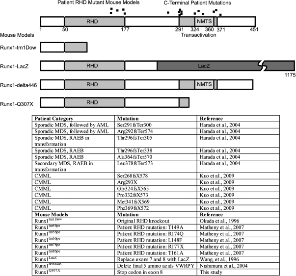
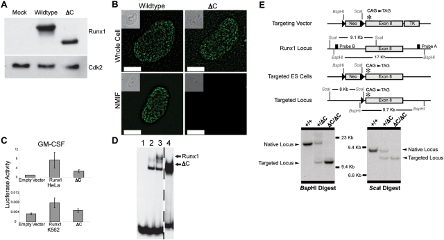
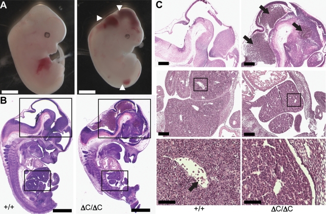
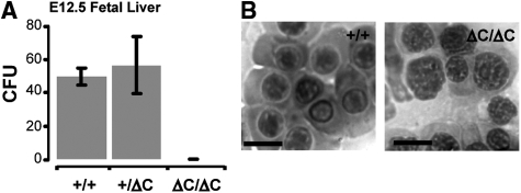
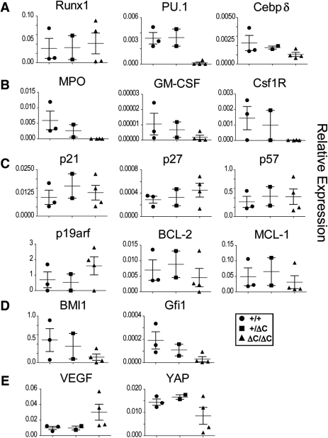
Runx1 is a key hematopoietic transcription factor required for definitive hematopoiesis and is a frequent target of leukemia-related chromosomal translocations. The resulting fusion proteins, while retaining DNA binding activity, display loss of subnuclear targeting and associated transactivation functions encoded by the C-terminus of the protein. To define the precise contribution of the Runx1 C-terminus in development and leukemia, we created a knock-in mouse with a C-terminal truncation by introducing a single nucleic acid substitution in the native Runx1 locus. This mutation (Runx1(Q307X)) models genetic lesions observed in patients with leukemia and myeloproliferative disorders. The Runx1(Q307X) homozygous mouse exhibits embryonic lethality at E12.5 due to central nervous system hemorrhages and a complete lack of hematopoietic stem cell function. While able to bind DNA, Runx1(Q307X) is unable to activate target genes, resulting in deregulation of various hematopoietic markers. Thus, we demonstrate that the subnuclear targeting and transcriptional regulatory activities of the Runx1 C-terminus are critical for hematopoietic development. We propose that compromising the C-terminal functions of Runx1 is a common mechanism for the pathological consequences of a variety of somatic mutations and Runx1-related leukemic fusion proteins observed in human patients.


Figures

Figure 1.
Figure 1.
Runx1 patient mutations and mouse models. Diagram of Runx1 with regions of interest highlighted. Runx1Q307X was designed to model several human mutations observed in MDS, AML, RAEB and CMML. Also illustrated are the first RHD knock out mouse, existing mouse models bearing patient mutations in the RHD and previous models with mutations in the Runx1 C-terminus. Runx1-LacZ is broken to show the full length of the chimeric fusion protein.
Figure 2.
Figure 2.
In vitro characterization of Runx1Q307X and generation of Runx1Q307X mice. (A) Western blot showing expression of wild-type Runx1 or Runx1Q307X in transfected HeLa cells. (B) Whole cell (upper panels) or NMIF preparations (lower panels) of HeLa cells transfected with wild-type Runx1 or Runx1Q307X. Anti-Runx1 immunofluorescence images are shown with phase contrast insets. Scale bars are 100 µm. The signal for Runx1Q307X cells in NMIF preparations decreased by 4.5-fold compared with wild-type Runx1, based on observation of at least 200 cells per sample with representative cells shown. (C) Luciferase expression from GM-CSF promoter construct in the presence of empty vector, wild-type or Runx1Q307X in HeLa and K562 cells. (D) EMSA showing DNA binding activity of Runx1 and Runx1Q307X from transduced HeLa cell nuclear extracts: lane 1, free probe; lane 2, empty vector; lane 3, wild-type Runx1; lane 4, Runx1Q307X. (E) Targeting strategy to replace a portion of exon 8 with exon 8 containing the point mutation to cause a stop codon after amino acid 307 (CAG to TAG). Probes A and B and restriction enzyme cut sites are shown. The Neomycin resistance gene (Neo) was removed by using embryonic stem cells containing the sperm-specific PC3Cre, excising Neo during generation of founder mice. Southern blot analysis of genomic DNA from tails of E12.5 embryos was performed using restriction enzymes and probes shown.
Figure 3.
Figure 3.
Histology of Runx1Q307X at E12.5. (A) Gross image of wild-type (left panel) and Runx1Q307X (right panel) E12.5 embryos. White arrowheads note sites of hemorrhage in the Runx1Q307X embryo. Scale bars are 1 mm. (B) Whole mount hematoxylin and eosin staining of embryos in A. Scale bars are 1 mm. (C) Magnification of heads of E12.5, noting hemorrhages with black arrows (upper panels, scale bars are 500 µm). Magnification of fetal livers of E12.5 embryos (middle panels, scale bars are 250 µm) and magnification showing liver sinusoids (lower panels, scale bars are 100 µm) with arrow noting the hematopoietic precursor cells in the wild-type sinusoids that are absent from Runx1Q307X.
Figure 4.
Figure 4.
Lack of hematopoietic progenitor cell function in Runx1Q307X. (A) Colony forming unit assays using fetal liver cells from wild-type, heterozygous or Runx1Q307X homozygous E12.5 embryos. (B) Nucleated erythroid progenitors are morphologically normal in peripheral blood smears of wild-type and Runx1Q307X E12.5 embryos. Scale bars are 10 µm.
Figure 5.
Figure 5.
Runx1Q307X causes deregulation of hematopoietic genes. Relative expression of hematopoietic transcription factors (A), phenotypic markers (B), proliferation and cell survival markers (C), genes required for HSC maintenance (D), and signaling molecules (E) by RT–PCR in wild-type, heterozygous and homozygous Runx1Q307X E12.5 mice (N= 3, 2 and 4, respectively). All RT–PCR results were normalized to GAPDH expression. See Materials and Methods for specific primers.

Similar articles

Cited by

References

    1. Okuda T., van Deursen J., Hiebert S.W., Grosveld G., Downing J.R. AML1, the target of multiple chromosomal translocations in human leukemia, is essential for normal fetal liver hematopoiesis. Cell. 1996;84:321–330. - PubMed
    1. Wang Q., Stacy T., Binder M., Marin-Padilla M., Sharpe A.H., Speck N.A. Disruption of the Cbfa2 gene causes necrosis and hemorrhaging in the central nervous system and blocks definitive hematopoiesis. Proc. Natl Acad. Sci. USA. 1996;93:3444–3449. - PMC - PubMed
    1. Motoda L., Osato M., Yamashita N., Jacob B., Chen L.Q., Yanagida M., Ida H., Wee H.J., Sun A.X., Taniuchi I., et al. Runx1 protects hematopoietic stem/progenitor cells from oncogenic insult. Stem Cells. 2007;25:2976–2986. - PubMed
    1. Kuo M.C., Liang D.C., Huang C.F., Shih Y.S., Wu J.H., Lin T.L., Shih L.Y. RUNX1 mutations are frequent in chronic myelomonocytic leukemia and mutations at the C-terminal region might predict acute myeloid leukemia transformation. Leukemia. 2009;23:1426–1431. - PubMed
    1. Look A.T. Oncogenic transcription factors in the human acute leukemias. Science. 1997;278:1059–1064. - PubMed

Publication types

MeSH terms

Substances