. 2010 Mar 15;19(6):1058-65.
doi: 10.1093/hmg/ddp569. Epub 2009 Dec 24.

Loss of nuclear poly(A)-binding protein 1 causes defects in myogenesis and mRNA biogenesis

Affiliations

Loss of nuclear poly(A)-binding protein 1 causes defects in myogenesis and mRNA biogenesis

Luciano H Apponi et al. Hum Mol Genet. .

Abstract

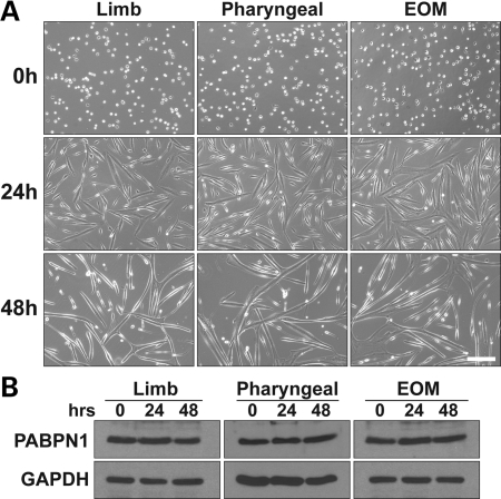
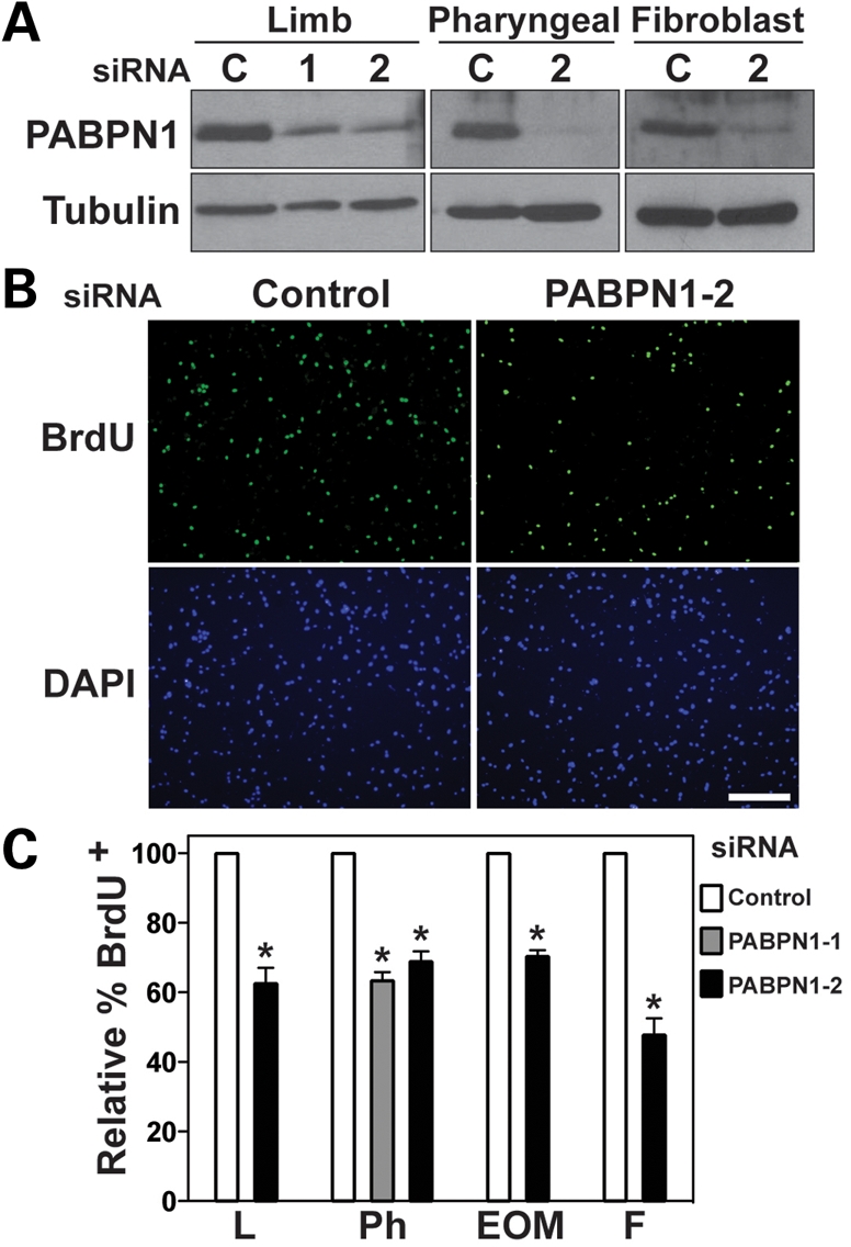
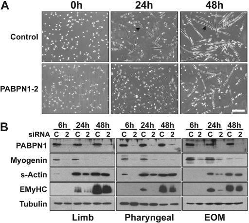
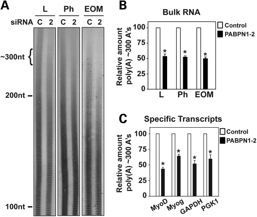
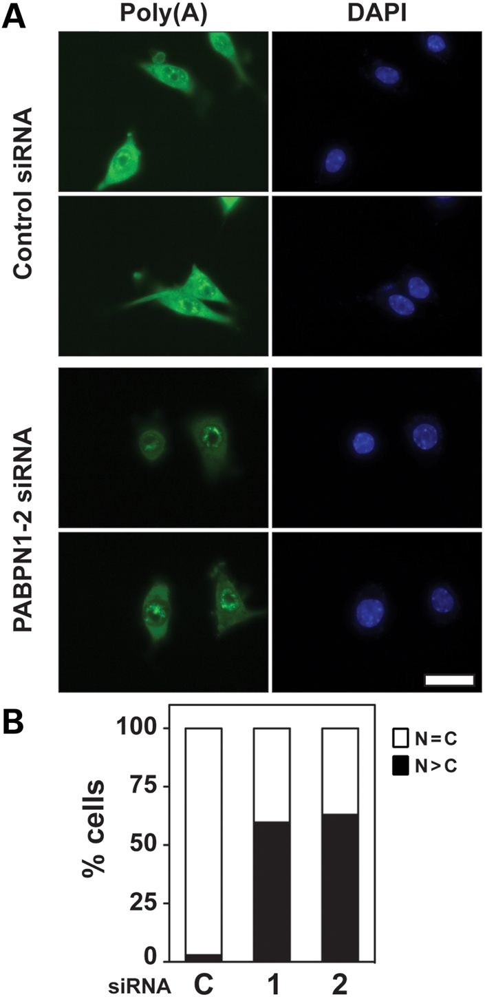
The nuclear poly(A)-binding protein 1 (PABPN1) is a ubiquitously expressed protein that plays a critical role in polyadenylation. Short expansions of the polyalanine tract in the N-terminus of PABPN1 lead to oculopharyngeal muscular dystrophy (OPMD), which is an adult onset disease characterized by eyelid drooping, difficulty in swallowing and weakness in the proximal limb muscles. Although significant data from in vitro biochemical assays define the function of PABPN1 in control of poly(A) tail length, little is known about the role of PABPN1 in mammalian cells. To assess the function of PABPN1 in mammalian cells and specifically in cells affected in OPMD, we examined the effects of PABPN1 depletion using siRNA in primary mouse myoblasts from extraocular, pharyngeal and limb muscles. PABPN1 knockdown significantly decreased cell proliferation and myoblast differentiation during myogenesis in vitro. At the molecular level, PABPN1 depletion in myoblasts led to a shortening of mRNA poly(A) tails, demonstrating the cellular function of PABPN1 in polyadenylation control in a mammalian cell. In addition, PABPN1 depletion caused nuclear accumulation of poly(A) RNA, revealing that PABPN1 is required for proper poly(A) RNA export from the nucleus. Together, these experiments demonstrate that PABPN1 plays an essential role in myoblast proliferation and differentiation, suggesting that it is required for muscle regeneration and maintenance in vivo.


Figures

Figure 1.
Figure 1.
PABPN1 expression is not altered during in vitro myogenesis. Pure cultures of primary mouse myoblasts from limb, pharyngeal and extraocular (EOM) muscles were differentiated for 0, 24 and 48 h. (A) Representative phase-contrast images of muscle cells after 0, 24 and 48 h of differentiation are shown (bar, 100 µm). (B) Protein extracts were analyzed by immunoblotting for PABPN1 expression. GAPDH was used as a loading control. Similar results were obtained from at least three independent assays.
Figure 2.
Figure 2.
PABPN1 is required for normal myoblast proliferation. Pure cultures of primary mouse myoblasts from limb, pharyngeal and extraocular (EOM) muscles were transfected with one of two siRNA oligonucleotides for PABPN1 [PABPN1-1 (1) or PABPN1-2 (2)] or a scrambled control (C). Cells were labeled with BrdU and immunostained with anti-BrdU antibody. (A) Protein extracts were analyzed by immunoblotting for PABPN1 to confirm knockdown. Alpha-tubulin was used as a loading control. (B) Representative BrdU immunostaining after siRNA transfection (bar, 100 µm). DAPI staining was used for nuclear visualization. (C) The relative percentage of BrdU+ nuclei was determined in limb (L), pharyngeal (Ph) and extraocular (EOM) myoblasts and fibroblasts (F) transfected with siRNA. PABPN1 knockdown results in ∼40% decrease in the percentage of BrdU+ cells. Data are mean ± SE, *P < 0.05, n = 3.
Figure 3.
Figure 3.
PABPN1 is required for proper myoblast differentiation. Pure cultures of primary mouse myoblasts from limb, pharyngeal and extraocular (EOM) muscles were transfected with PABPN1-2 (2) or control scrambled (C) siRNA. (A) Representative phase-contrast images of limb muscle cells after 0, 24 and 48 h of differentiation (bar, 100 µm). PABPN1 siRNA cells exhibit defects in myotube formation at 24 and 48 h. Arrows indicate myotubes in control siRNA cells. (B) Protein extracts were analyzed by immunoblotting after 6, 24 and 48 h of differentiation for the differentiation markers myogenin, sarcomeric actin and embryonic myosin heavy chain (eMyHC). Knockdown of PABPN1 was also determined and alpha-tubulin was used as a loading control. Biochemical differentiation is defective in all three types of muscle cells following PABPN1 knockdown. Similar effects were observed in three independent assays.
Figure 4.
Figure 4.
PABPN1 is required for proper poly(A) tail length in muscle cells. Pure cultures of primary mouse myoblasts from limb (L), pharyngeal (Ph) and extraocular (EOM) muscles were transfected with PABPN1-2 (2) or control scrambled (C) siRNA. Cells were collected and total RNA was isolated. (A) Representative distribution of bulk poly(A) tails from different types of myoblasts following PABPN1 knockdown. Total RNA was labeled with [32P] cytidine 3′,5′-bis(phosphate) using T4 RNA ligase and digested with RNase A/T1. Samples were resolved by electrophoresis in denaturing polyacrylamide gels and exposed to radiographic film. Cells treated with PABPN1 siRNA show a significant decrease in poly(A) tract length of ∼300 nt. (B) Densitometric quantification of poly(A) tracts of ∼300 nt as normalized to poly(A) tracts of ∼100 nt for control siRNA versus PABPN1-2 siRNA. A 50% decrease in the relative number of ∼300 nt poly(A) tails is observed following PABPN1 knockdown. Data are mean ± SD, *P < 0.05, n = 5. (C) Linker ligation-mediated poly(A) tail (LLM-PAT) assays were used to determine poly(A) tail length of specific transcripts following myoblast transfection with control or PABPN1-2 siRNA. mRNA transcripts of myogenin, MyoD, GAPDH and PGK1 showed a significant decrease in poly(A) tract length of ∼300 nt. Data are mean ± SD, *P < 0.05, n = 3.
Figure 5.
Figure 5.
PABPN1 is required for proper poly(A) RNA export from the nucleus. Pure cultures of primary mouse myoblasts from limb were transfected with PABPN1-1 or 2 or control scrambled (C) siRNA and poly(A) localization was determined by fluorescent in situ hybridization (FISH) using an oligo(dT) probe. (A) Representative image of poly(A) RNA localization after siRNA transfection (bar, 20 µm). DAPI staining was used for nuclear visualization. (B) Relative distribution of cells showing normal diffuse nuclear (N) and cytoplasmic (C) localization (n = C) or nuclear accumulation (n > C) of poly(A) RNA after siRNA transfection. Following transfection with either PABPN1-1 or -2 siRNA, more than 50% of myoblasts displayed nuclear poly(A) RNA accumulation. Data are the percentage of nuclear poly(A) RNA distribution for at least 70 cells. Similar results were obtained in three independent experiments.

References

    1. Kuhn U., Wahle E. Structure and function of poly(A) binding proteins. Biochim. Biophys. Acta. 2004;1678:67–84. - PubMed
    1. Kuhn U., Nemeth A., Meyer S., Wahle E. The RNA binding domains of the nuclear poly(A)-binding protein. J. Biol. Chem. 2003;278:16916–16925. - PubMed
    1. Fan X., Dion P., Laganiere J., Brais B., Rouleau G.A. Oligomerization of polyalanine expanded PABPN1 facilitates nuclear protein aggregation that is associated with cell death. Hum. Mol. Genet. 2001;10:2341–2351. - PubMed
    1. Calado A., Tome F.M., Brais B., Rouleau G.A., Kuhn U., Wahle E., Carmo-Fonseca M. Nuclear inclusions in oculopharyngeal muscular dystrophy consist of poly(A) binding protein 2 aggregates which sequester poly(A) RNA. Hum. Mol. Genet. 2000;9:2321–2328. - PubMed
    1. Calado A., Kutay U., Kuhn U., Wahle E., Carmo-Fonseca M. Deciphering the cellular pathway for transport of poly(A)-binding protein II. RNA. 2000;6:245–256. - PMC - PubMed

Publication types