. 2010 Feb;176(2):812-26.
doi: 10.2353/ajpath.2010.090450. Epub 2009 Dec 24.

Biglycan and fibromodulin have essential roles in regulating chondrogenesis and extracellular matrix turnover in temporomandibular joint osteoarthritis

Affiliations

Biglycan and fibromodulin have essential roles in regulating chondrogenesis and extracellular matrix turnover in temporomandibular joint osteoarthritis

Mildred C Embree et al. Am J Pathol. 2010 Feb.

Abstract

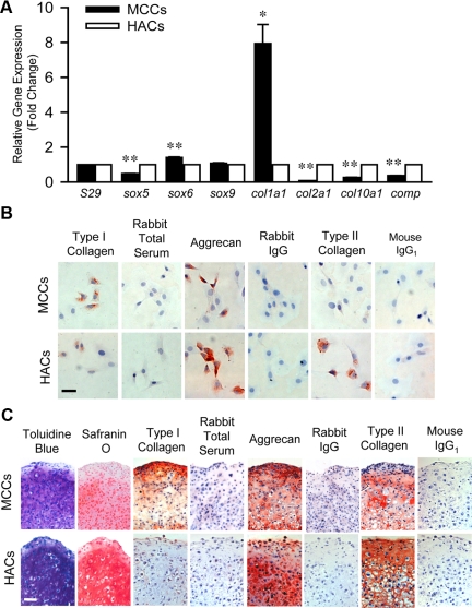
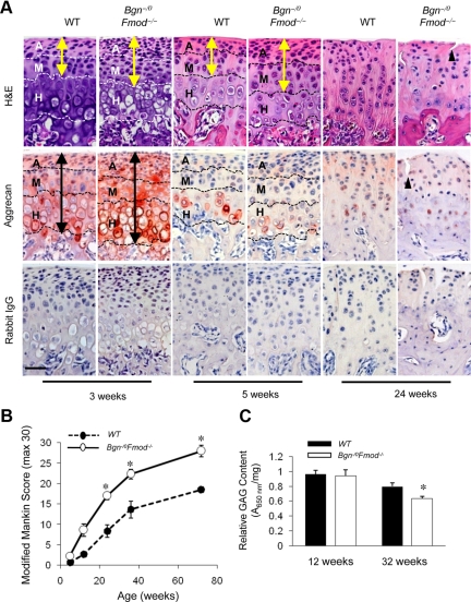
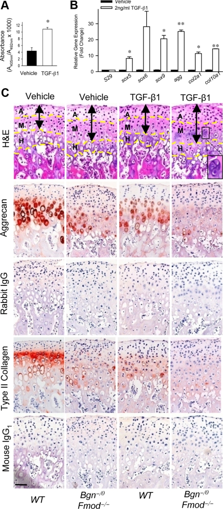
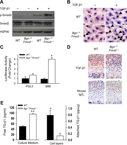
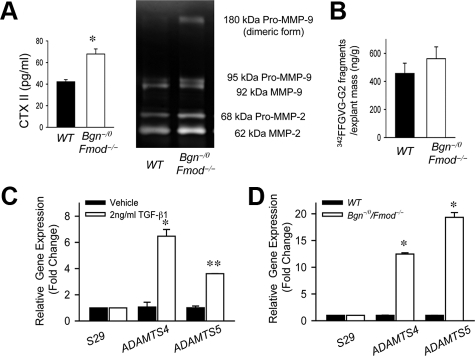
The temporomandibular joint is critical for jaw movements and allows for mastication, digestion of food, and speech. Temporomandibular joint osteoarthritis is a degenerative disease that is marked by permanent cartilage destruction and loss of extracellular matrix (ECM). To understand how the ECM regulates mandibular condylar chondrocyte (MCC) differentiation and function, we used a genetic mouse model of temporomandibular joint osteoarthritis that is deficient in two ECM proteins, biglycan and fibromodulin (Bgn(-/0)Fmod(-/-)). Given the unavailability of cell lines, we first isolated primary MCCs and found that they were phenotypically unique from hyaline articular chondrocytes isolated from the knee joint. Using Bgn(-/0) Fmod(-/-) MCCs, we discovered the early basis for temporomandibular joint osteoarthritis arises from abnormal and accelerated chondrogenesis. Transforming growth factor (TGF)-beta1 is a growth factor that is critical for chondrogenesis and binds to both biglycan and fibromodulin. Our studies revealed the sequestration of TGF-beta1 was decreased within the ECM of Bgn(-/0) Fmod(-/-) MCCs, leading to overactive TGF-beta1 signal transduction. Using an explant culture system, we found that overactive TGF-beta1 signals induced chondrogenesis and ECM turnover in this model. We demonstrated for the first time a comprehensive study revealing the importance of the ECM in maintaining the mandibular condylar cartilage integrity and identified biglycan and fibromodulin as novel key players in regulating chondrogenesis and ECM turnover during temoporomandibular joint osteoarthritis pathology.


Figures

Figure 1
Figure 1
Comparison of mandibular condylar chondrocytes (MCCs) and hyaline articular chondrocytes (HACs). A: Quantitative real-time RT-PCR analysis of cartilage-related genes sox5, sox6, sox 9, col1a1, col2a1, col10a1, and comp using total RNA isolated from HACs (open bars) and MCCs (black bars). Gene expression was normalized to the housekeeping gene S29. The expression of genes in MCCs was relative to that of HACs. Data are mean ± SEM of 2 experiments. *P < 0.02, **P < 0.006 MCCs versus HACs. B: Immunocytochemistry staining of cartilage-related proteins type I collagen, aggrecan, and type II collagen in MCCs and HACs. Rabbit total serum, rabbit IgG, and mouse IgG1 were used under the same conditions as negative controls, respectively. Scale bar = 50 μm. C: Chondrogenic differentiation of HACs and MCCs. Chondrogenic differentiation was induced by culturing pelleted HACs or MCCs in chondrogenic induction medium for 3 weeks and was assessed by toluidine blue and Safranin O staining. The expression of type I collagen, aggrecan, and type II collagen was assessed by immunhistochemistry. Rabbit total serum, rabbit IgG, and mouse IgG1 were used under the same conditions as negative controls, respectively. Scale bar = 50 μm.
Figure 2
Figure 2
Increased chondrogenesis in the absence of Bgn and Fmod. A: Quantitative real-time RT-PCR analysis compared the expression levels of chondrogenic-related genes using total RNA isolated from in wild-type (WT) and Bgn−/0Fmod−/− MCCs (left). Gene expression was normalized to the housekeeping gene S29. The expression of genes in wild-type MCCs was relative to that in Bgn−/0Fmod−/− MCCs. Data are mean ± SEM of 3 experiments. *P < 0.02, **P < 0.006 Bgn−/0Fmod−/− versus wild-type. Immunocytochemistry staining of aggrecan and type II collagen in wild-type and Bgn−/0Fmod−/− MCCs (right). Rabbit IgG1 and mouse IgG1 were used as negative controls, respectively. Scale bar = 20 μm. B: Chondrogenesis of MCCs was induced by culturing pelleted wild-type and Bgn−/0Fmod−/− MCCs in chondrogenic induction medium for 3 weeks. Real-time RT-PCR analysis of chondrogenic-related genes using total RNA isolated from wild-type and Bgn−/0Fmod−/− pellets (left). Gene expression was normalized to the housekeeping gene S29. The expression of genes in wild-type pellets was relative to that in Bgn−/0/Fmod−/− pellets. Data are mean ± SEM of 2 experiments. *P < 0.09 Bgn−/0Fmod−/− versus wild-type. Pellets were assessed by toluidine blue and Safranin O staining. The expression of type I collagen, aggrecan, type II collagen, and type X collagen were examined by immunohistochemistry (right). Scale bar = 50 μm. Rabbit total serum, rabbit IgG, and mouse IgG1 were used under the same conditions as negative controls. C: Type II collagen expression was assessed by immunohistochemistry in the mandibular condylar cartilage of 3-week-old wild-type and Bgn−/0Fmod−/− mice (left). Arrows indicate the position and extent of type II collagen expression. Mouse IgG1 was used under the same conditions as a negative control. Scale bar = 50 μm. Real-time RT-PCR analysis was used to compare the expression chondrogenenic-related genes Using total RNA isolated from the mandibular condylar cartilages of 3-week-old wild-type and Bgn−/0Fmod−/− mice (right). Gene expression was normalized to the housekeeping gene S29. The expression of individual genes in wild-type mandibular condylar cartilage was relative to that in Bgn−/0Fmod−/− mandibular condylar cartilage. Data are mean ± SEM of 3 experiments. *P < 0.003, **P < 0.0001 Bgn−/0Fmod−/− versus wild-type.
Figure 3
Figure 3
Accelerated degeneration of mandibular condylar cartilage and loss of aggrecan expression with aging in the absence of Bgn and Fmod. A: Comparable sections from wild-type (WT) and Bgn−/0Fmod−/− condylar cartilages at 3, 5, and 24 weeks of age were stained with H&E and aggrecan expression was assessed by immunohistochemistry. Rabbit IgG was used under the same conditions as negative controls. Dashed lines divide the condyle into articular zone (A), mature zone (M), and hypertrophic zone (H). Yellow arrows indicate extent of articular/mature zones and black arrows indicate extent of aggrecan immunostaining. Black triangle indicates tissue clefting. Scale bar = 50 μm. B: Semiquantitative modified Mankin score was used to assess histological changes in the wild-type and Bgn−/0Fmod−/− condylar cartilages on aging. Values are mean score ± SD of four mice. *P < 0.003 Bgn−/0Fmod−/− versus wild-type. C: The Blyscan glycosaminoglycan assay was used to determine the GAG content in wild-type and Bgn−/0Fmod−/− condylar cartilages at 12 and 32 weeks. The GAG content was normalized to the mass of the sample. Values are mean score ± SEM of 6 mice. *P < 0.008 Bgn−/0 Fmod−/− versus wild-type.
Figure 4
Figure 4
TGF-β1 accelerates both formation and degeneration of mandibular condylar cartilages in the absence of Bgn and Fmod. A: TGF-β1 induces proliferation of MCCs. BrdU ELISA was used to measure proliferation of MCCs treated with vehicle or TGF-β1. Data are mean ± SEM of 6 wells. *P < 0.005 vehicle versus TGF-β1. B: TGF-β1 induces the expression of chondrogenic-related genes in the MCCs. Quantitative real-time RT-PCR analysis compared the expression levels of chondrogenic-related genes using total RNA isolated from MCCs treated with vehicle or TGF-β1. Gene expression was normalized to the housekeeping gene S29. The expression of genes in MCCs treated with vehicle was relative to that in MCCs treated with TGF-β1. Data are mean ± SEM of 2 experiments. *P < 0.02, **P < 0.002 vehicle versus TGF-β1. C: TGF-β1 accelerates loss of aggrecan content in mandibular condyle explants. Whole mandibles were isolated from wild-type and Bgn−/0 Fmod−/− mice and explants were cultured for 48 hours with vehicle or 2 ng/ml TGF-β1. Comparable histological sections from explants were stained with H&E. Aggrecan and type II collagen expression was examined by immunohistochemistry. Rabbit IgG and mouse IgG1 were used under the same conditions as negative controls, respectively. Dashed yellow lines divide the condyle into articular zone (A), mature zone (M) and hypertrophic zone (H). Black double-ended arrows indicate extent of articular and mature zones. Black square indicates chondrocyte cluster. Scale bar = 50 μm.
Figure 5
Figure 5
Decreased TGF-β1 sequestration leads to overactivation of TGF-β1 signaling in the absence of Bgn and Fmod. A: Increased phosphorylation of Smad2 in MCCs in the absence of Bgn and Fmod. Western blot analysis was used to determine Smad2 and phosphorylated Smad2 (p-Smad2) levels in wild-type (WT) and Bgn−/0Fmod−/− MCCs treated with vehicle or 2 ng/ml TGF-β1. HSP90 was used as protein loading control. B: Increased Smad4 nuclear translocation in the absence of Bgn and Fmod. Immunocytochemistry was used to examine the nuclear localization of Smad4 (arrows) in wild-type and Bgn−/0Fmod−/− MCCs treated with vehicle or TGF-β1. Scale bar = 20 μm. C: Increased TGF-β1 responsive transcriptional activity in the absence of Bgn and Fmod. TGF-β1–induced transcriptional activity was determined by transfecting wild-type and Bgn−/0Fmod−/− MCCs with a reporter plasmid expressing a TGF-β responsive luciferase construct (SBE). pGL3 was used as control. Data are mean ± SEM of two to three transfections. *P < 0.006 Bgn−/0Fmod−/− versus wild-type. D: Decreased sequestration of active TGF-β1 in ECM of mandibular condylar cartilage in the absence of Bgn and Fmod. The expression of active TGF-β1 was examined by immunohistochemistry using comparable histological sections from 3-week-old wild-type and Bgn−/0Fmod−/− mandibular condylar cartilages. Mouse IgG1 was used under the same conditions as a negative control. Scale bar = 50 μm. E: Decreased TGF-β1 binding to ECM and cells in the absence of Bgn and Fmod. Confluent wild-type and Bgn−/0Fmod−/− MCCs were cultured with vehicle or 1 ng/ml TGF-β1. The concentration of unbound TGF-β1 in the culture media (left) and the levels of active TGF-β1 bound to the cell layer (cell surface and ECM; right) were measured by ELISA. Data are mean ± SEM of three to four wells. *P < 0.01, **P < 0.0001 Bgn−/0Fmod−/− versus wild-type.
Figure 6
Figure 6
MMP and aggrecanase expression and activity are increased in the absence of Bgn and Fmod. A: Increased release of CTX II and MMP activity in the absence of Bgn and Fmod. Type II collagen degradation was determined using CTX-II ELISA, which measured the release of the MMP-derived type II collagen fragment CTX-II in culture medium of wild-type (WT) and Bgn−/0Fmod−/− explants (left). Gelatin zymography was used to determine the MMP activity in the culture medium of wild-type and Bgn−/0Fmod−/− explants (right). Data are mean ± SEM of 12 explants. *P < 0.008 Bgn−/0 Fmod−/− versus wild-type. B: MMP mediated aggrecan degradation was unaffected in the absence of Bgn and Fmod. The release of the MMP mediated aggrecan fragment 342FFGVG-G2 in the culture media of wild-type and Bgn−/0Fmod−/− explants was measured by ELISA. Data are mean ± SEM of nine to twelve explants. C: TGF-β1 up-regulates the expression of ADAMTS4 and ADAMTS5 in MCCs. Quantitative real-time RT-PCR analysis was used to examine the expression of ADAMTS4 and ADAMTS5 using total RNA isolated from MCCs treated with vehicle or TGF-β1. Gene expression was normalized to the housekeeping gene S29. The gene expression levels in cultures with TGF-β1 were relative to the cultures with vehicle. Data are mean ± SEM of 2 experiments. *P < 0.02, **P < 0.003 TGF-β1 versus vehicle. D: Increased expression of ADAMTS4 and ADAMTS5 in the absence of Bgn and Fmod. Quantitative real-time RT-PCR analysis was used to examine the expression of ADAMTS4 and ADAMTS5 using total RNA isolated from wild-type and Bgn−/0Fmod−/− cultured MCCs. Gene expression was normalized to the housekeeping gene S29. The gene expression levels in Bgn−/0 Fmod−/− MCCs were relative to wild-type MCCs. Data are mean ± SEM of 3 experiments. *P < 0.0001 Bgn−/0Fmod−/− versus wild-type MCCs.

Similar articles

Cited by

References

    1. Scrivani SJ, Keith DA, Kaban LB. Temporomandibular disorders. N Engl J Med. 2008;359:2693–2705. - PubMed
    1. Zarb GA, Carlsson GE. Temporomandibular disorders: osteoarthritis. J Orofac Pain. 1999;13:295–306. - PubMed
    1. Bosshardt-Luehrs CP, Luder HU. Cartilage matrix production and chondrocyte enlargement as contributors to mandibular condylar growth in monkeys (Macaca fascicularis). Am J Orthod Dentofacial Orthop. 1991;100:362–369. - PubMed
    1. Meikle MC. Remodeling the dentofacial skeleton: the biological basis of orthodontics and dentofacial orthopedics. J Dent Res. 2007;86:12–24. - PubMed
    1. Almarza AJ, Athanasiou KA. Design characteristics for the tissue engineering of cartilaginous tissues. Ann Biomed Eng. 2004;32:2–17. - PubMed

Publication types

MeSH terms