. 2010 Feb;176(2):850-60.
doi: 10.2353/ajpath.2010.090551. Epub 2009 Dec 24.

Inhibition of CD36-dependent phagocytosis by prostaglandin E2 contributes to the development of endometriosis

Affiliations

Inhibition of CD36-dependent phagocytosis by prostaglandin E2 contributes to the development of endometriosis

Pei-Chin Chuang et al. Am J Pathol. 2010 Feb.

Abstract

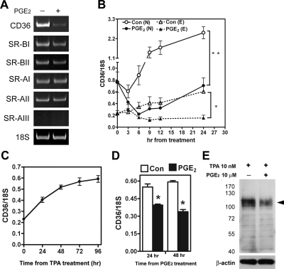
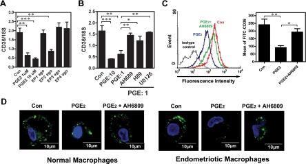
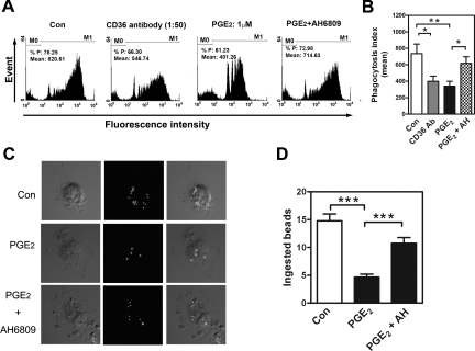
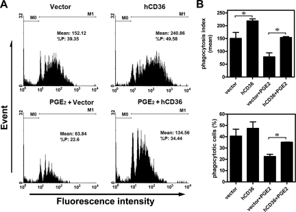
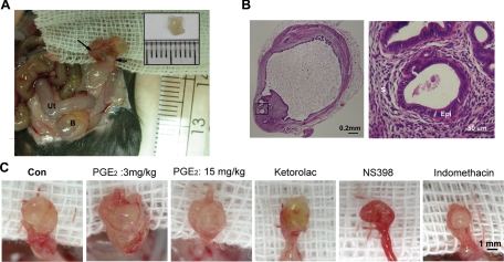
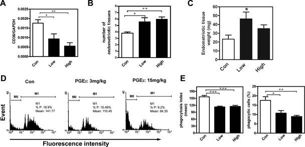
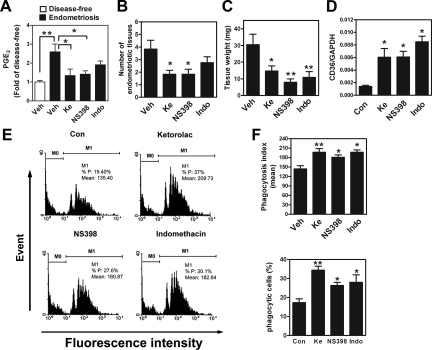
Dysfunction in macrophage-mediated phagocytosis of aberrant cells that undergo retrograde transport to the peritoneal cavity is considered an important factor in the development of endometriosis. However, the mechanisms responsible for the loss of function of macrophages remain largely unknown. Herein, we report that prostaglandin (PG) E(2), via the EP2 receptor-dependent signaling pathway, inhibits the expression of CD36 in peritoneal macrophages, resulting in reduced phagocytic ability. PGE(2)-mediated inhibition of macrophage phagocytic capability was restored by ectopic expression of CD36. Treatment with PGE(2) inhibited CD36-dependent phagocytosis of peritoneal macrophages and increased the number and size of endometriotic lesions in mice. In contrast, blockade of PGE(2) production by cyclooxygenase inhibitors enhanced the phagocytic ability of peritoneal macrophages and reduced endometriotic lesion formation. Taken together, our findings reveal a potential mechanism of immune dysfunction during endometriosis development and may contribute to the design of an effective prevention/treatment regimen.


Figures

Figure 1
Figure 1
PGE2 inhibits CD36 expression in normal and endometriotic macrophages. A: Macrophages from normal women were treated with 10 μM PGE2 or vehicle for 24 hr, and expression of scavenger receptors was determined by RT-PCR. This experiment was done four times using different batches of macrophages, and the results were similar. B: Normal (N) and endometriotic (E) macrophages were cultured for different periods as indicated in the absence (control) or presence of PGE2, and levels of CD36 were quantified by real-time quantitative RT-PCR. Data represent the mean and standard deviation (SD) of four independent experiments using different batches of cells and were analyzed by ANOVA with repeated measurement. C: U937 monocytic cells were treated with 10 nM TPA for various periods as indicated, and CD36 mRNA expression was quantified by real-time quantitative RT-PCR. Data represent the mean and SD of three independent experiments. D, E: U937 cells were incubated with TPA for 48 hr and then treated with PGE2 (10 μM) for 24 hr or 48 hr. Expression of CD36 mRNA (D) and protein (E) was determined (β-actin served as a loading control). The arrowhead indicates the glycosylated membrane form of CD36. Molecular size markers (kDa) are shown to the left. *P < 0.05, **P < 0.01.
Figure 2
Figure 2
Inhibition of CD36 by PGE2 is mediated by the EP2 receptor signaling pathway. A: Macrophages isolated from normal women were treated with vehicle, PGE2 (1 μM), or selective agonists for EP1 (ONO-D1-004, 10 μM), EP2 (ONO-AE1-259-01, 10 μM), EP3 (ONO-AE-248, 10 μM), or EP4 (ONO-AE1-329, 10 μM) for 12 hr. CD36 mRNA expression was detected by real-time quantitative RT-PCR. B: Macrophages isolated from normal women were treated with PGE2 (1 or 10 μM) or PGE2 plus inhibitors as indicated for 12 hr, and CD36 mRNA expression was determined by real-time quantitative RT-PCR. C: Macrophages isolated from normal women were treated with vehicle (Con), PGE2, or PGE2+AH6809 (10 μM) for 96 hr, and CD36 expression was assessed by staining with anti-CD36 antibody or isotype-matched FITC-IgA (isotype control) followed by quantification via flow cytometry. Representative flow cytometric data are shown in the left panel. The right panel shows the mean and SD of four independent experiments using different batches of peritoneal macrophages. D: Representative confocal images show CD36 staining in macrophages isolated from normal women treated with vehicle (Con), PGE2, or PGE2+AH6809 as described in (C). This experiment was done four times using different batches of cells. *P < 0.05, **P < 0.01, ***P < 0.001.
Figure 3
Figure 3
PGE2 inhibits phagocytosis via the EP2 receptor–dependent signaling pathway. A: Macrophages isolated from normal women were treated with vehicle (Con), anti-CD36, PGE2 (1 μM), or PGE2 plus EP2 antagonist (AH6809, 10 μM) for 24 hr, and fluorescence intensity was determined by flow cytometry. M0: no bead was ingested; M1: with at least one bead ingested. B: Mean and SD of M1 obtained from four experiments using different batches of cells. C: Representative confocal microscopic images of phagocytic macrophages isolated from normal patients. Macrophages were treated with vehicle (Con), PGE2, or PGE2+AH6809 and then incubated with FITC-conjugated beads for 1 h. D: Mean and SD of the number of beads phagocytosed by macrophages isolated from normal women after treatment with vehicle (Con), PGE2, or PGE2+AH6809. Four independent experiments using different batches of macrophages were conducted, and at least 20 cells per experiment were counted. *P < 0.05, **P < 0.01, ***P < 0.001.
Figure 4
Figure 4
Ectopic expression of CD36 rescues PGE2-inhibited phagocytosis. A: Macrophages isolated from women with endometriosis were transfected with vector (as a control) or vector containing human CD36 cDNA (hCD36) for 24 hr. Cells were then treated without or with PGE2, and phagocytic ability was determined by flow cytometry. M0: no bead was ingested; M1: with at least one bead ingested. B: Mean and SD of phagocytic ability (upper panel) and percent of phagocytic cells (lower panel) from four independent experiments using different batches of macrophages. *P < 0.05.
Figure 5
Figure 5
Macroscopic and microscopic images of endometriotic-like lesions in female C57BL/6NCrj recipients after syngeneic uterine tissue transfer. A: Four weeks after injection, a recipient displayed significant gross vascularity and hyperemia within the lesion and marked vascular recruitment along the periphery of the peritoneal attachment site (arrows). Ut: uterus; B: bladder. The inset shows the endometrial tissue peeled from a donor mouse. Eight pieces of such endometrial fragments were injected into the peritoneal cavity of each recipient. B: Representative images showing H&E–stained adhesive endometriotic tissues in a surgery-induced endometriotic mouse. The right panel is a high-magnification image from the square indicated in the left panel. S: stroma, Epi: epithelium. C: Macroscopic images of endometriotic-like lesions in mice treated with vehicle (Con), PGE2 (3 mg/kg-body weight), PGE2 (15 mg/kg-body weight), COX-1 inhibitor (ketorolac, 10 mg/kg body weight), COX-2 inhibitor (NS398, 10 mg/kg body weight), or nonselective COX-1/COX-2 inhibitor (indomethacin, 10 mg/kg body weight).
Figure 6
Figure 6
PGE2 inhibits the phagocytic ability of peritoneal macrophages and enhances endometriosis formation in vivo (n = 10 animals per group). A: Mean and SD of levels of CD36 mRNA on peritoneal macrophages freshly isolated from mice treated with vehicle (Con), 3 mg/kg body weight PGE2 (Low), or 15 mg/kg body weight PGE2 (High). B: Mean and SD of the number of endometriotic-like lesions in mice treated without or with PGE2 for 4 weeks. C: Mean and SD of total wet-weight of endometriotic-like lesions in mice treated without or with PGE2 for 4 weeks. D: Representative flow cytometric results of the phagocytic ability of peritoneal macrophages freshly isolated from mice subjected to surgery-induced endometriosis. M0: no bead was ingested; M1: with at least one bead ingested. E: Percent of phagocytic macrophages and mean phagocytosis index of peritoneal macrophages freshly isolated from mice subjected to surgery-induced endometriosis. *P < 0.05, **P < 0.01, ***P < 0.001, as compared with control.
Figure 7
Figure 7
Inhibition of PGE2 production increases the phagocytic ability of macrophages and reduces endometriosis formation in vivo (n=10 animals per group). A: Concentrations of PGE2 in the peritoneal fluid of mice without (disease-free) or with surgery-induced endometriosis. Mice with endometriosis were further divided into four groups and treated with vehicle (Veh), COX-1 inhibitor (ketorolac, Ke, 10 mg/kg body weight), COX-2 inhibitor (NS398, 10 mg/kg body weight), or COX-1/-2 inhibitor (indomethacin, Indo, 10 mg/kg body weight) for 4 weeks. B: Mean and SD of the number of endometriotic-like lesions in mice treated with COX inhibitors. C: Mean and SD of total wet-weight of endometriotic lesions in mice treated as described above. D: CD36 expression in mouse peritoneal macrophages after surgery-induced endometriosis and drug treatment. E: Representative flow cytometric results of phagocytic ability of freshly isolated peritoneal macrophages after surgery-induced endometriosis and drug treatment. F: Mean and SD of the percent of phagocytic macrophages (lower panel) and mean phagocytosis index (upper panel) in peritoneal macrophages freshly isolated from surgery-induced endometriotic mice. M0: no bead was ingested; M1: with at least one bead ingested *P < 0.05, **P < 0.01, ***P < 0.001, as compared with control.

Similar articles

Cited by

References

    1. Simoens S, Hummelshoj L, D'Hooghe T. Endometriosis: cost estimates and methodological perspective. Hum Reprod Update. 2007;13:395–404. - PubMed
    1. Sampson JA. Peritoneal endometriosis due to the menstrual dissemination of endometrial tissue into the peritoneal cavity. Am J Obstet Gynecol. 1927;14:422–425. - PubMed
    1. Wu MH, Shoji Y, Chuang PC, Tsai SJ. Endometriosis: disease pathophysiology and the role of prostaglandins. Expert Rev Mol Med. 2007;9:1–20. - PubMed
    1. Ozkan S, Murk W, Arici A. Endometriosis and infertility: epidemiology and evidence-based treatments. Ann NY Acad Sci. 2008;1127:92–100. - PubMed
    1. Dunselman GA, Hendrix MG, Bouckaert PX, Evers JL. Functional aspects of peritoneal macrophages in endometriosis of women. J Reprod Fertil. 1988;82:707–710. - PubMed

Publication types

MeSH terms