Review
. 2010 May;67(9):1383-96.
doi: 10.1007/s00018-009-0227-8. Epub 2009 Dec 25.

The role of synapsins in neuronal development

Affiliations
Review

The role of synapsins in neuronal development

Eugenio F Fornasiero et al. Cell Mol Life Sci. 2010 May.

Abstract

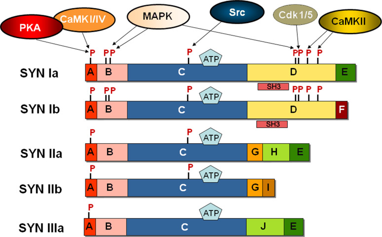
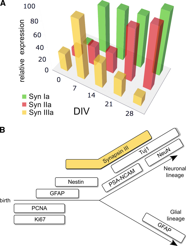
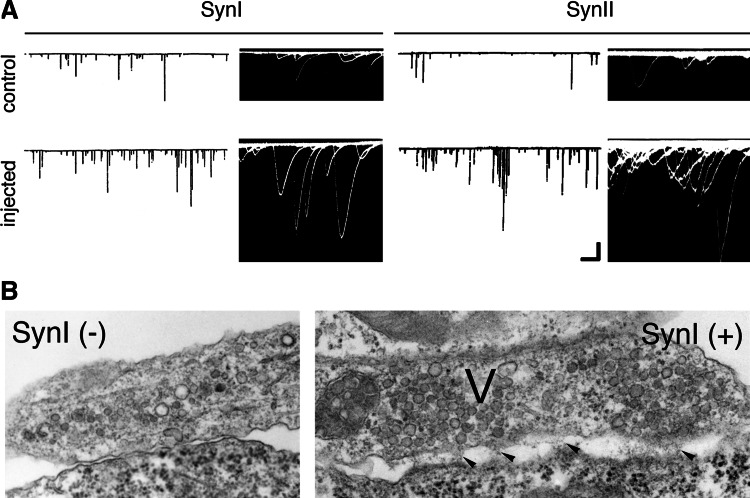
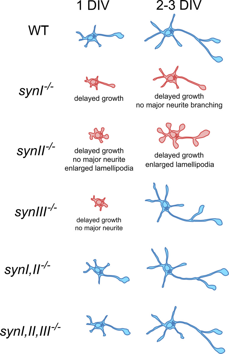
The synapsins, the first identified synaptic vesicle-specific proteins, are phosphorylated on multiple sites by a number of protein kinases and are involved in neurite outgrowth and synapse formation as well as in synaptic transmission. In mammals, the synapsin family consists of at least 10 isoforms encoded by 3 distinct genes and composed by a mosaic of conserved and variable domains. The synapsins are highly conserved evolutionarily, and orthologues have been found in invertebrates and lower vertebrates. Within nerve terminals, synapsins are implicated in multiple interactions with presynaptic proteins and the actin cytoskeleton. Via these interactions, synapsins control several mechanisms important for neuronal homeostasis. In this review, we describe the main functional features of the synapsins, in relation to the complex role played by these phosphoproteins in neuronal development.


Figures

Fig. 1
Fig. 1
Schematic protein domain model of the mammalian synapsin family with functional properties and protein kinase phosphorylation sites. Within domain A, site 1 (Ser9 in synapsin I) is phosphorylated by PKA and CaMKI/IV [–35]. Within domain B, sites 4 and 5 (Ser62 and Ser67, respectively) are phosphorylated by MAPKs [36]. All synapsin isoforms have a conserved tyrosine in domain C (site 8) which has been shown to be phosphorylated by the tyrosine kinase Src in synapsins I and II [29]. Domain C also contains putative ATP binding residues [26]. The D domain includes the proline-rich strand binding SH3 domains [29, 89] and the phosphorylation sites 2 and 3 (Ser566 and Ser603, respectively) that are phosphorylated by CaMKII [35]. Within domain D, site 6 (Ser549) is phosphorylated by MAPKs, Cdk1/5 [36, 37]. The latter kinase also phosphorylates an adjacent site (site 7, Ser551 in synapsin I) [37, 38]. Only one isoform is represented for Syn III, although multiple synIII products have been described in the adult brain [90]
Fig. 2
Fig. 2
Temporal expression pattern of synapsin isoforms. a Expression level during in vitro development of cultured hippocampal neurons of synapsins I, II, and III. Neuronal extracts were prepared from E16 mouse dissociated hippocampal neurons kept in culture for up to 30 days in vitro (DIV). The proteins were separated by electrophoresis and immunoblots were reacted with specific antibodies and densitometrically analyzed. Results are plotted as percentage of the highest level detected. While synapsin III has the highest peak of expression at 7 DIV, when synaptogenesis is still ongoing, synapsins I and II levels increase with the onset of synaptic activity. Modified with permission from [91]; based on data from [9]. b Expression of synapsin III and lineage markers during development of neuronal precursor cells. The mitotic markers PCNA and Ki67 are expressed during proliferation; nestin is expressed before the cells become committed to either the neuronal or the glial lineage; GFAP is expressed in undifferentiated precursors and mature glial cells; PSA-NCAM is expressed in immature neurons, whereas NeuN is expressed in mature neurons. Tuj1 begins its expression in immature neurons and persists in mature neurons. The temporal expression profile for synapsin III is based on its colocalization with the other established markers [10]. Note the precocious expression of synapsin III, which is already present in nestin-positive cells prior to neuronal commitment. Reprinted with permission of John Wiley and Sons from [10]
Fig. 3
Fig. 3
Effects of synapsin I or II overexpression on the maturation of Xenopus neuromuscular synapses in culture. a Spontaneous acetylcholine release from Xenopus neurons after 1 DIV. Shown are spontaneous synaptic currents recorded from whole-cell voltage clamped myocytes that were innervated by either a control neuron or by a neuron injected with either exogenous synapsin I [64] or recombinant synapsin IIa [65]. Inward currents are shown as downward deflections at low (white background) and high (black background) time resolution. Note the higher frequency and amplitude of the spontaneous currents recorded in synapsin-injected neurons (scale bars 250 pA, 40 s, and 100 pA, 10 ms for the slow and fast traces, respectively). Reprinted with permission from [64] (Copyright 1992, Cell Press, USA.) and from [65] (Copyright 1994, National Academy of Sciences, USA.). b Fine structure of two neuromuscular synapses formed by either an uninjected neuron (SynI (−), left) or a neuron injected with purified rat synapsin I (SynI (+), right). Magnification: ×40,000 SynI (−) and ×45,000 SynI (+). Note the cluster of SVs (V) in the injected axoplasm in correspondence with basal lamina deposition (arrowheads). Modified with permission from [78]
Fig. 4
Fig. 4
Effects of the absence of various synapsin isoforms on early stages of rodent neuronal development in vitro. Blue color designates normal development, while red color indicates developmental defects. Wild-type (WT) rodent hippocampal neurons in culture undergo a series of well-characterized developmental stages [92]. Between 1 and 3 days DIV, stage III neurons show a longer neuritic process that has the highest probability to become the axon. Mouse synI −/− hippocampal neurons at 1 and 3 DIV show an overall growth delay accompanied by decreased branching of the primary neurite [53, 72]. Mouse synII −/− neurons have a stronger phenotype, being devoid of a major neurite at 1 DIV and showing a slower growth phenotype [70]. In addition synII −/− neurons have enlarged lamellipodia veils. Mouse hippocampal synIII −/− neurons display a milder phenotype at 1 DIV and are completely normal at 2 DIV [57]. Neurons knocked out for multiple synapsin genes are normal [19, 70]. However, a comprehensive comparative work with all these genotypes is still missing
Fig. 5
Fig. 5
Effects of synapsin phosphorylation on neuronal development. a Images of spinal nerves from the thoracic and abdominal areas derived from Xenopus embryos injected unilaterally at the two-cell stage with WT or mutated Xenopus synapsin IIa (SynII) mRNA. The serine to alanine mutation (SynII S9A, upper panel) mimics unphosphorylated synapsin, while the serine to glutamic acid mutation (SynII S9E, middle panel) mimics phosphorylated synapsin. Each image is the summation of a Z-stack of optical slices spaced 4 μm apart from each side of the embryo. Green, spinal nerves from injected side; red, nerves from uninjected side. WT synapsin II injection does not change the growth rate of Xenopus spinal nerves. Following injection of the non-phosphorylatable S9A mutant nerves are significantly shorter than the contralateral controls. Conversely, the pseudophosphorylated S9E mutant increases the outgrowth of injected nerves. These data indicate that the pattern of synapsin activation is more important than the increase of its total level in the determination of cytoskeletal rearrangements. Scale bar 100 μm. Reproduced with permission from [70]. b Images of growth cones of hippocampal neurons derived from synI −/− mice coexpressing synaptophysin I-EYFP (SypI-EYFP) and either WT synapsin I (SynI, upper two rows) or its non-phosphorylatable S9A mutant (SynI S9A, bottom two rows). The white traces in the left row outline the distal edges of the peripheral domain, as determined based on differential interference contrast images. Growth cones were imaged before and after a 5-min incubation with forskolin, an adenylate cyclase activator that increases intracellular cAMP levels and activates PKA. In the right column, SypI–EYFP image gray scales were transposed into a pseudocolor spectrum surface plot, with warmer hues corresponding to pixels of higher fluorescence intensity. Forskolin induces dispersion of SypI–EYFP-positive SVs in growth cones expressing WT, but not S9A, SynI, confirming that phosphorylation of synapsin I at site 1 by PKA is required for mobilization of SVs from the central domain of the growth cone. Scale bar 10 μm. Modified with permission from [87]
Fig. 6
Fig. 6
Differential role of synapsin isoforms in SV clustering in growth cones. Hippocampal neurons from either WT, synI −/−, synII −/−, or synI,II,III −/− were fixed and stained with an anti-synaptotagmin I antibody (green in the merged images) and with fluorescently-tagged phalloidin that specifically labels F-actin (red in the merged images). In culture, neuronal growth cones show two main cytoskeleton defined regions: a central domain that is enriched in organelles and microtubules, and a peripheral domain that is characterized by the absence of organelles an the deposition of filamentous actin [86]. Synaptotagmin I-positive SV precursors are confined to the central domain in WT and synII −/− neurons, but they disperse throughout the growth cone in synI −/− and synI,II,III −/− neurons. The lack of phenotype in synII −/− neurons reinforces the idea that synapsin I is the major determinant for the localization of SV precursors in neuronal growth cones [87]. The white traces in the left row outline the distal edges of the peripheral domain, as determined based on differential interference contrast images. Scale bar 5 μm. Original from Bonanomi and Valtorta, unpublished

Similar articles

Cited by

References

    1. De Camilli P, Benfenati F, Valtorta F, Greengard P. The synapsins. Annu Rev Cell Biol. 1990;6:433–460. doi: 10.1146/annurev.cb.06.110190.002245. - DOI - PubMed
    1. Greengard P, Valtorta F, Czernik AJ, Benfenati F. Synaptic vesicle phosphoproteins and regulation of synaptic function. Science. 1993;259:780–785. doi: 10.1126/science.8430330. - DOI - PubMed
    1. Fdez E, Hilfiker S. Vesicle pools and synapsins: new insights into old enigmas. Brain Cell Biol. 2006;35:107–115. doi: 10.1007/s11068-007-9013-4. - DOI - PubMed
    1. De Camilli P, Cameron R, Greengard P. Synapsin I (protein I), a nerve terminal-specific phosphoprotein. I. Its general distribution in synapses of the central and peripheral nervous system demonstrated by immunofluorescence in frozen and plastic sections. J Cell Biol. 1983;96:1337–1354. doi: 10.1083/jcb.96.5.1337. - DOI - PMC - PubMed
    1. De Camilli P, Harris SM, Jr, Huttner WB, Greengard P. Synapsin I (protein I), a nerve terminal-specific phosphoprotein. II. Its specific association with synaptic vesicles demonstrated by immunocytochemistry in agarose-embedded synaptosomes. J Cell Biol. 1983;96:1355–1373. doi: 10.1083/jcb.96.5.1355. - DOI - PMC - PubMed

Publication types

LinkOut - more resources