. 2010 Feb;8(1):27-36.
doi: 10.1089/adt.2009.0208.

Microbead arrays for the analysis of ErbB receptor tyrosine kinase activation and dimerization in breast cancer cells

Affiliations

Microbead arrays for the analysis of ErbB receptor tyrosine kinase activation and dimerization in breast cancer cells

Imran H Khan et al. Assay Drug Dev Technol. 2010 Feb.

Abstract

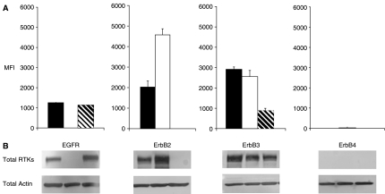
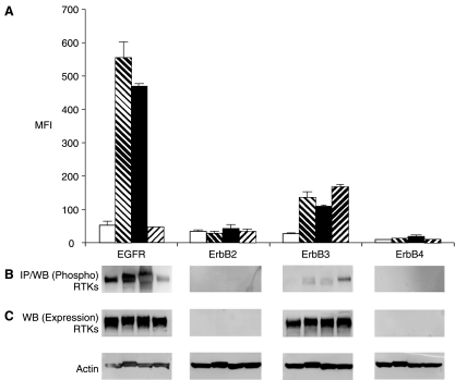
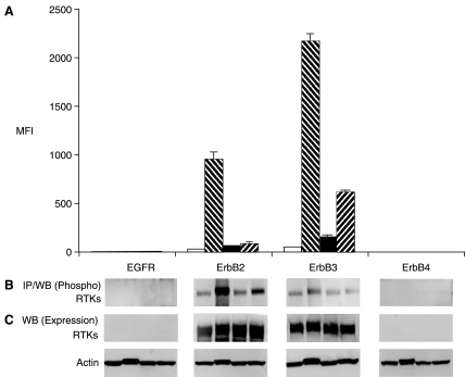
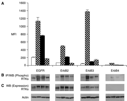
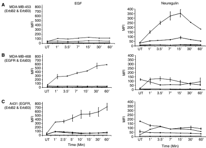
Receptor tyrosine kinases (RTKs) in the ErbB family (EGFR, ErbB2, ErbB3, and ErbB4) are implicated in a variety of human malignancies. Accordingly, determination of both expression and activation (dimerization/heterodimerization and phosphorylation) of ErbB proteins is critical in defining their functional role in cancer. Efficient and comprehensive methods to study molecular functions of ErbB family of RTKs are needed not only for improvements in diagnostics but also for early screening of targeted drugs (eg, small molecule inhibitors and therapeutic antibodies). We report development of 3 multiplex microbead immunoassays for simultaneous detection of expression, protein-protein interactions, and phosphorylation of these RTKs. These novel multiplex immunoassays were used to study ErbB RTKs under different cell activation conditions in 2 breast cancer cell lines (MDA-MB-453 and MDA-MB-468) and an epidermoid cancer cell line (A431). The results were confirmed by immunoprecipitation/western blot. Importantly, the multiplex immunoassay facilitated time-course studies in these cell lines after cell activation with EGF and neuregulin, revealing the kinetics of phosphorylation of the ErbB family RTKs. This study demonstrates the utility of the Luminex(R) multiplex system as an efficient and comprehensive approach to study different aspects of molecular roles of these RTKs. Importantly, the study provides proof-of-concept for the utility of the multiplex microbead immunoassay approach for potential use in efficient, robust, and rapid screening of drugs, particularly those targeting functional aspects of these potent signaling molecules. In addition, the assays described here may be useful for cancer diagnostics and monitoring efficacy of therapy targeting the ErbB family of RTKs.


Figures

Fig. 1.
Fig. 1.
(A) Expression levels of ErbB family of receptors analyzed by multiplex microbead suspension immunoassay in MDA-MB-453 (hashed bars), MDA-MB-468 (open bars), and A431 (closed bars) cells. A mixture of microbeads coated with antibodies to EGFR, ErbB2, ErbB3, and ErbB4 were incubated with lysates from nonactivated cells. Detection of protein expression was achieved by a second antibody against each individual receptor protein. Error bars represent standard error of n = 4 values. (B) Western blot analysis of EGFR, ErbB2, ErbB3, and ErbB4 expression. Actin expression shows equal loading. Abbreviations: MFI, median fluorescence intensity; RTK, receptor tyrosine kinase.
Fig. 2.
Fig. 2.
(A) Phosphoproteomic analysis of RTKs by multiplex microbead suspension array immunoassay in MDA-MB-468 cells. Cells were used as untreated (empty bars), treated with 6.6 mM sodium pervanadate for 5 min (hashed bars), 16.5 nM EGF (closed bars), or 1:500 neuregulin for 7 min (reverse hashed bars). A mixture of microbeads coated with antibodies to EGFR, ErbB2, ErbB3, and ErbB4 were incubated with the cell lysates. Error bars represent standard error of n = 4 values. (B) Immunoprecipitation and western blot analysis of phosphorylation and total ErbB receptors in MDA-MB-468 cells. Antibodies used for immunoprecipitation were the same as those coated on the microbeads for multiplex analysis. (C) Western blot analyses were performed for the detection of total RTK proteins (actin shows equal loading). Abbreviations: MFI, median fluorescence intensity; RTK, receptor tyrosine kinase.
Fig. 3.
Fig. 3.
Phosphoproteomic analysis of RTKs by multiplex microbead immunoassay in MDA-MB-453 cells. Details are the same as described for Figure 2. Abbreviations: MFI, median fluorescence intensity; RTK, receptor tyrosine kinase.
Fig. 4.
Fig. 4.
Phosphoproteomic analysis of RTKs by multiplex microbead immunoassay in A431 cells. Details are the same as described for Figure 2. Abbreviations: MFI, median fluorescence intensity; RTK, receptor tyrosine kinase.
Fig. 5.
Fig. 5.
Kinetics of phosphorylation of RTKs in cells treated with EGF or neuregulin. (A) MDA-MB-453, (B) MDA-MB-468, and (C) A431 cells were activated with 16.5 nM EGF or 1:500 neuregulin for various times ranging from 1 to 60 min. Microbeads used in the multiplex assay were coated with antibodies against EGFR (diamond), ErbB2 (square), ErbB3 (triangle), and ErbB4 (cross). Multiplex microbead immunoassay was performed as described for Figure 2. Detection of phosphorylation was performed by 4G10. Error bars represent standard error of n = 4 values. Abbreviations: MFI, median fluorescence intensity; RTK, receptor tyrosine kinase.
Fig. 6.
Fig. 6.
Detection of heterodimerization of ErbB family of receptors. Cells were used as untreated (open bars), treated with 6.6 mM sodium pervanadate for 5 min (hashed bars), 16.5 nM EGF (closed bars), or 1:500 neuregulin for 7 min (reverse hashed bars). A second antibody (biotinylated), either specific to ErbB2 (A) or ErbB3 (B), was used for detection of the respective proteins. Treatment of cells by pervanadate, EGF, or neuregulin is indicated. Error bars represent standard deviation error of n = 4 values. Significant difference (P value < 0.01) in detection signal for ErbB2 and ErbB3 (between untreated control and activated cell lysates) is indicated by an encircled asterisk.

References

    1. Stern DF. ERBB3/HER3 and ERBB2/HER2 duet in mammary development and breast cancer. J Mammary Gland Biol Neoplasia. 2008;13:215–223. - PMC - PubMed
    1. Lo HW. Hsu SC. Hung MC. EGFR signaling pathway in breast cancers: from traditional signal transduction to direct nuclear translocalization. Breast Cancer Res Treat. 2006;95:211–218. - PubMed
    1. Ono M. Kuwano M. Molecular mechanisms of epidermal growth factor receptor (EGFR) activation and response to gefitinib and other EGFR-targeting drugs. Clin Cancer Res. 2006;12:7242–7251. - PubMed
    1. Sweeney C. Miller JK. Shattuck DL. Carraway KL. ErbB receptor negative regulatory mechanisms: implications in cancer. J Mammary Gland Biol Neoplasia. 2006;11:89–99. - PubMed
    1. Agrawal A. Gutteridge E. Gee JM. Nicholson RI. Robertson JF. Overview of tyrosine kinase inhibitors in clinical breast cancer. Endocr Relat Cancer. 2005;12(Suppl 1):S135–S144. - PubMed

Publication types

MeSH terms