. 2010 Feb;8(1):96-105.
doi: 10.1089/adt.2009.0217.

A time-resolved fluorescence-resonance energy transfer assay for identifying inhibitors of hepatitis C virus core dimerization

Affiliations

A time-resolved fluorescence-resonance energy transfer assay for identifying inhibitors of hepatitis C virus core dimerization

Smitha Kota et al. Assay Drug Dev Technol. 2010 Feb.

Abstract

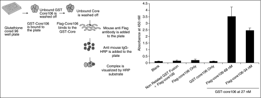
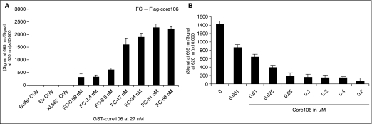
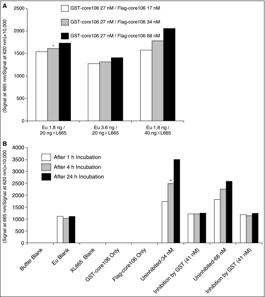
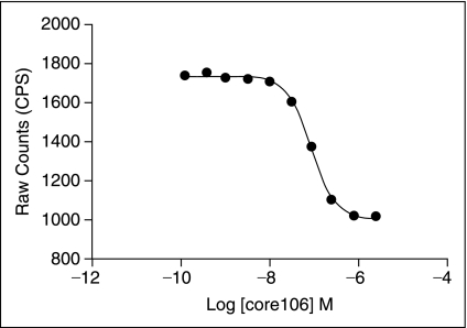
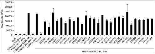
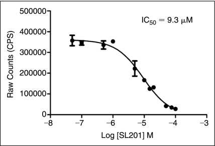
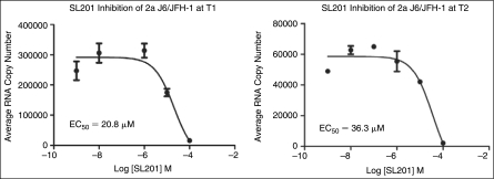
Binding of hepatitis C virus (HCV) RNA to core, the capsid protein, results in the formation of the nucleocapsid, the first step in the assembly of the viral particle. A novel assay was developed to discover small molecule inhibitors of core dimerization. This assay is based on time-resolved fluorescence resonance energy transfer (TR-FRET) between anti-tag antibodies labeled with either europium cryptate (Eu) or allophycocyanin (XL-665). The N-terminal 106-residue portion of core protein (core106) was tagged with either glutathione-S-transferase (GST) or a Flag peptide. Tag-free core106 was selected as the reference inhibitor. The assay was used to screen the library of pharmacologically active compounds (LOPAC) consisting of 1,280 compounds and a 2,240-compound library from the Center for Chemical Methodology and Library Development at Boston University (CMLD-BU). Ten of the 28 hits from the primary TR-FRET run were confirmed in a secondary amplified luminescent proximity homogeneous assay (ALPHA screen). One hit was further characterized by dose-response analysis yielding an IC(50) of 9.3 microM. This 513 Da compound was shown to inhibit HCV production in cultured hepatoma cells.


Figures

Fig. 1.
Fig. 1.
ELISA scheme: glutathione-coated 96-well plates were coated with GST-core106 and incubated overnight at 4°C. The unbound protein–antibody was washed off at every coating step. Different concentrations of Flag-core106 were added to the plate and incubated for 2 h at 37°C. Then, mouse anti-Flag antibody was added and incubated for 2 h at room temperature. Anti-mouse IgG was added and incubated for 1 h at room temperature. The complex was visualized by the addition of Ultra TMB-HRP substrate. Figure 1 data: confirming GST-core106/Flag-core106 dimerization: GST-core106 was kept constant at 27 nM/well and Flag-core106 was dosed at 68 and 34 nM/well. Buffer only, GST-core106 alone, Flag-core106 alone were included in the assay as background controls. An unrelated GST-tagged protein (1 μg) was adsorbed on a glutathione-coated plate and when mixed with Flag-core106 gave a signal similar to the background in the assay. Error bars represent standard deviations (SDs) of n = 3 values in 3 assays.
Fig. 2.
Fig. 2.
(A) Time-resolved fluorescence–resonance energy transfer (TR-FRET) assays confirming core dimerization. Concentration of GST-core106 was kept constant at 27 nM. Flag-core106 concentrations ranged from 0.068 to 680 nM. The transfer of fluorescence was measured as ratio of (signal at 665 nm/signal at 620 nm) × 10,000. (B) TR-FRET assay showing the reference inhibition/competition of core dimerization by N-terminal 106-residue portion of core protein (core106). Core106 concentrations ranged from 0.001 to 0.6 μM. The graph was plotted after europium cryptate (Eu) background correction. This TR-FRET assay is representative of 5 independent experiments. Error bars represent standard deviation (SD) of n = 7 values in 1 assay.
Fig. 3.
Fig. 3.
(A) Optimization of europium cryptate-tagged anti-GST-and XL 665-tagged anti-Flag- in 384-well format. Eu-anti-GST antibody was analyzed at 2 different concentrations: 1.8 ng/well and 3.6 ng/well. Allophycocyanin (XL-665)-anti-Flag antibody was analyzed at 20 ng/well and 40 ng/well. The asterisk indicates the condition used for the medium-throughput Center for Chemical Methodology and Library Development at Boston University (CMLD-BU) run. The reaction conditions are discussed in the results section. (B) Optimization of incubation times for N-terminal 106-residue portion of core protein (core106) time-resolved fluorescence–resonance energy transfer (TR-FRET) assay in 384-well format. GST-core106 and Flag-core106 were kept constant at 27 and 34 nM, respectively. The assay was analyzed at 1, 4, and 24 h. Free GST at 41 nM was included as a control inhibitor. The asterisk indicates the incubation time selected for the CMLD-BU run.
Fig. 4.
Fig. 4.
Dose–response analysis of N-terminal 106-residue portion of core protein (core106) inhibition on GST-core106 and Flag-core106 dimerization. The x-axis shows the molar concentration of core106. The y-axis shows the time-resolved fluorescence–resonance energy transfer (TR-FRET) ratio. The inhibitory concentration at 50% (IC50) for the free core inhibition of the core dimerization was calculated to be 89 nM. The standard deviation (SD) was not significant hence not represented by error bars.
Fig. 5.
Fig. 5.
Amplified luminescent proximity homogeneous assay (ALPHA screen) confirming hits from of primary time-resolved fluorescence–resonance energy transfer (TR-FRET)-based Center for Chemical Methodology and Library Development at Boston University (CMLD-BU) run. N-terminal 106-residue portion of core protein (Core106) ALPHA screen assay was used as a secondary confirmation to validate the 28 hits from the primary TR-FRET screen. GST-core106 (GC) and Flag-core106 (FC) were kept constant at 150 nM each. Core106 was added as a 100% inhibition control. DMSO was included as a control because the compounds were dissolved in DMSO. Ten of the 28 hits were confirmed as potential inhibitors of core106 dimerization, indicated with asterisks. Error bars represent standard deviation (SD) of n = 2 in 2 assays.
Fig. 6.
Fig. 6.
Dose–response analysis of compound SL201, using the N-terminal 106-residue portion of core protein (core106) amplified luminescent proximity homogeneous assay (ALPHA screen) assay. The compounds were dosed from 0.05 to 100 μM. The IC50 was calculated to be 9.3 μM. Error bars represent standard deviation (SD) of n = 2 in 2 assays.
Fig. 7.
Fig. 7.
Effect of SL201 on HCV 2a J6/JFH-1 virus. Inhibition assay of SL201 was performed on HCV 2a J6/JFH-1 virus by adding serially diluted SL201 and virus onto naïve Huh-7.5 cells and incubated for 3 days (early stage—T1). Culture supernatant (T1) was transferred onto naïve Huh-7.5 cells and incubated for an additional 3 days (late stage—T2). RNA was purified from lysed cells from both T1 and T2. Real-time RT-PCR was performed on purified RNA. EC50 for SL201 were calculated to be 20.8 and 36.3 μM, respectively, at T1 and T2.

Similar articles

  • Peptide inhibitors of hepatitis C virus core oligomerization and virus production.
    Kota S, Coito C, Mousseau G, Lavergne JP, Strosberg AD. Kota S, et al. J Gen Virol. 2009 Jun;90(Pt 6):1319-1328. doi: 10.1099/vir.0.008565-0. Epub 2009 Mar 4. J Gen Virol. 2009. PMID: 19264632
  • Cell-based hepatitis C virus infection fluorescence resonance energy transfer (FRET) assay for antiviral compound screening.
    Yu X, Uprichard SL. Yu X, et al. Curr Protoc Microbiol. 2010 Aug;Chapter 17(1):Unit 17.5.. doi: 10.1002/9780471729259.mc1705s18. Curr Protoc Microbiol. 2010. PMID: 20812217 Free PMC article.
  • Identification of Compounds Targeting Hepatitis B Virus Core Protein Dimerization through a Split Luciferase Complementation Assay.
    Wei XF, Gan CY, Cui J, Luo YY, Cai XF, Yuan Y, Shen J, Li ZY, Zhang WL, Long QX, Hu Y, Chen J, Tang N, Guo H, Huang AL, Hu JL. Wei XF, et al. Antimicrob Agents Chemother. 2018 Nov 26;62(12):e01302-18. doi: 10.1128/AAC.01302-18. Print 2018 Dec. Antimicrob Agents Chemother. 2018. PMID: 30224531 Free PMC article.
  • Ebselen, a Small-Molecule Capsid Inhibitor of HIV-1 Replication.
    Thenin-Houssier S, de Vera IM, Pedro-Rosa L, Brady A, Richard A, Konnick B, Opp S, Buffone C, Fuhrmann J, Kota S, Billack B, Pietka-Ottlik M, Tellinghuisen T, Choe H, Spicer T, Scampavia L, Diaz-Griffero F, Kojetin DJ, Valente ST. Thenin-Houssier S, et al. Antimicrob Agents Chemother. 2016 Mar 25;60(4):2195-208. doi: 10.1128/AAC.02574-15. Print 2016 Apr. Antimicrob Agents Chemother. 2016. PMID: 26810656 Free PMC article.
  • Compound-Mediated Assay Interferences in Homogeneous Proximity Assays.
    Coussens NP, Auld D, Roby P, Walsh J, Baell JB, Kales S, Hadian K, Dahlin JL. Coussens NP, et al. 2020 Feb 1. In: Markossian S, Grossman A, Baskir H, Arkin M, Auld D, Austin C, Baell J, Brimacombe K, Chung TDY, Coussens NP, Dahlin JL, Devanarayan V, Foley TL, Glicksman M, Gorshkov K, Grotegut S, Hall MD, Hoare S, Inglese J, Iversen PW, Lal-Nag M, Li Z, Manro JR, McGee J, Norvil A, Pearson M, Riss T, Saradjian P, Sittampalam GS, Tarselli MA, Trask OJ Jr, Weidner JR, Wildey MJ, Wilson K, Xia M, Xu X, editors. Assay Guidance Manual [Internet]. Bethesda (MD): Eli Lilly & Company and the National Center for Advancing Translational Sciences; 2004–. 2020 Feb 1. In: Markossian S, Grossman A, Baskir H, Arkin M, Auld D, Austin C, Baell J, Brimacombe K, Chung TDY, Coussens NP, Dahlin JL, Devanarayan V, Foley TL, Glicksman M, Gorshkov K, Grotegut S, Hall MD, Hoare S, Inglese J, Iversen PW, Lal-Nag M, Li Z, Manro JR, McGee J, Norvil A, Pearson M, Riss T, Saradjian P, Sittampalam GS, Tarselli MA, Trask OJ Jr, Weidner JR, Wildey MJ, Wilson K, Xia M, Xu X, editors. Assay Guidance Manual [Internet]. Bethesda (MD): Eli Lilly & Company and the National Center for Advancing Translational Sciences; 2004–. PMID: 32045180 Free Books & Documents. Review.

Cited by

References

    1. WHO. Hepatitis C: global prevalence. Wkly Epidemiol Rec. 1997;72:341–344. - PubMed
    1. Giannini C. Brechot C. Hepatitis C virus biology. Cell Death Differ. 2003;10(Suppl. 1):S27–S38. - PubMed
    1. Choo Q. Kuo G. Weiner A. Overby L. Bradley D. Houghton M. Isolation of a cDNA clone derived from a blood-borne non-A, non-B viral hepatitis genome. Science. 1989;244(4902):359. - PubMed
    1. McGovern BH. Hepatitis C in the HIV-infected patient. J Acquir Immune Defic Syndr. 2007;45(Suppl 2):S47–S56. - PubMed
    1. Cristina J. Moreno-del Pilar M. Moratorio G. Hepatitis C virus genetic variability in patients undergoing antiviral therapy. Virus Res. 2007;127(2):185–194. - PubMed

Publication types

MeSH terms