. 2010 Apr 2;208(2):415-24.
doi: 10.1016/j.bbr.2009.12.015. Epub 2009 Dec 24.

Cognitive recovery in the aged rat after stroke and anti-Nogo-A immunotherapy

Affiliations

Cognitive recovery in the aged rat after stroke and anti-Nogo-A immunotherapy

Rebecca L Gillani et al. Behav Brain Res. .

Abstract

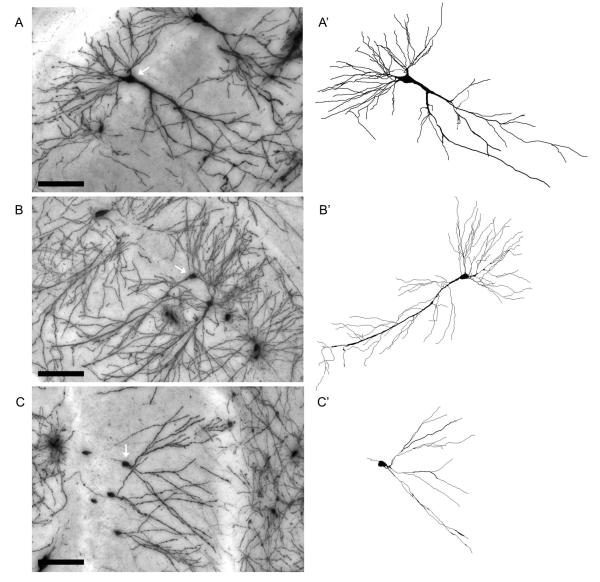
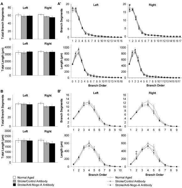
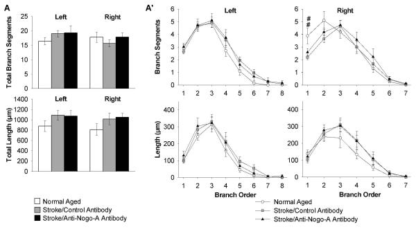
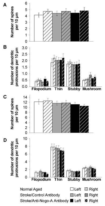
We have previously shown that immunotherapy directed against the protein Nogo-A leads to recovery on a skilled forelimb reaching task in rats after sensorimotor cortex stroke, which correlated with axonal and dendritic plasticity. Here we investigated anti-Nogo-A immunotherapy as an intervention to improve performance on a spatial memory task in aged rats after stroke, and whether cognitive recovery was correlated with structural plasticity. Aged rats underwent a unilateral distal permanent middle cerebral artery occlusion and one week later were treated with an anti-Nogo-A or control antibody. Nine weeks post-stroke, treated rats and normal aged rats were tested on the Morris water maze task. Following testing rats were sacrificed and brains processed for the Golgi-Cox method. Hippocampal CA3 and CA1 pyramidal and dentate gyrus granule cells were examined for dendritic length and number of branch segments, and CA3 and CA1 pyramidal cells were examined for spine density and morphology. Anti-Nogo-A immunotherapy given one week following stroke in aged rats improved performance on the reference memory portion of the Morris water maze task. However, this improved performance was not correlated with structural changes in the hippocampal neurons examined. Our finding of improved performance on the Morris water maze in aged rats after stroke and treatment with anti-Nogo-A immunotherapy demonstrates the promising therapeutic potential for anti-Nogo-A immunotherapy to treat cognitive deficits after stroke. The identification of sites of axonal and dendritic plasticity in the aged brain after stroke and treatment with anti-Nogo-A immunotherapy is still under investigation.


Figures

Figure 1
Figure 1
Improved performance on a spatial reference memory task after stroke and treatment with anti-Nogo-A immunotherapy. (A) Timeline of experiments. (B) Representative right sided stroke lesion one day post-stroke in an aged rat (scale bar=1 cm). In the TTC (2,3,5-triphenyl-2H-tetrazolium chloride)-reacted coronal brain sections viable tissue appears red (shown here in gray) and the ischemic infarction appears white demonstrating sensorimotor cortex involvement and subcortical sparing. (C) The stroke lesion size quantified in Golgi-Cox stained tissue, represented as percent of the intact hemisphere, did not differ between the two stroke groups (p=0.706, t-test). (D) Time to locate the hidden platform during the Morris water maze place task. Average curves of the fitted functions for each group. (D’) Averages of the individual parameters of the functions. Time at the start of testing, or the y intercept, was the same for all groups. As testing continued normal aged and stroke/anti-Nogo-A antibody treated rats acquired the location of the platform faster than stroke/control antibody treated rats, as shown by the slope parameter (p < 0.001, likelihood based χ2 test of random-effects simple exponential two-parameter model, η(x) = θ1eθ2x, where x = day – 1 [28]). (E) Distance to locate the hidden platform during the Morris water maze place task. Average curves of the fitted functions for each group. (E’) Averages of the individual parameters of the functions. Distance at the start of testing, or the y intercept, was the same for all groups. As testing continued all three groups acquired the location of the platform at significantly different rates, with the stroke/anti-Nogo-A antibody treated rats having faster rates to acquire the platform location than stroke/control antibody treated rats (p<0.001 for stroke/anti-Nogo-A antibody and stroke/control antibody, p<0.01 for normal aged and stroke/anti-Nogo-A antibody, p<0.001 for normal aged and stroke/control antibody, likelihood based χ2 test of random-effects simple exponential two-parameter model, η(x) = θ1eθ2x , where x = day –1). (F) During the probe trial all groups equally preferred the quadrant that contained the platform during the place task (p=0.876, one-way ANOVA). (G) During the matching-to-place (MTP) task all groups found the platform faster on Trial 2 than on Trial 1 (p<0.001), and there were no significant differences between groups (p=0.4387 for trial 1, p=0.0550 for trial 2, one-way ANOVA). Error bars denote ± standard error of the mean. **p<0.01, ***p<0.001.
Figure 2
Figure 2
Behaviors in the Morris water maze that were unique to rats with stroke regardless of antibody treatment. (A) Thigmotaxis, time spent in the periphery, during the place task. Average curves of the fitted functions for each group. (A’) Averages of the individual parameters of the functions. Thigmotaxis at the start of testing, or the y intercept, was the same for all groups. As testing continued both stroke groups showed more thigmotaxis behavior than normal aged rats (p<0.001, likelihood based χ2 test of random-effects simple exponential three-parameter model, η(x) = θ1eθ2x + θ3 , where x = day – 1 [28]). Thigmotaxis at the end of testing, or the lower asymptote, was the same for all groups. (B) The path circuity or direct distance to the platform/ the actual distance swam to the platform during a trial during the place task. Both stroke groups had significantly more circuitous paths than the normal aged group (p<0.05, repeated measures ANOVA, Bonferroni test for post-hoc comparison). (C) Swim velocity during the Morris water maze place task did not significantly differ across groups (p=0.2476, repeated measure ANOVA). Error bars denote ± standard error of the mean. *p<0.05, ***p<0.001.
Figure 3
Figure 3
Representative Golgi-Cox stained hippocampal CA3 (A) and CA1 pyramidal cells (B) and dentate gyrus granule cell (C, white arrows), and the corresponding Neurolucida tracings (A’, B’, C’). Images were acquired from the hippocampus of a normal aged rat. Scale bar=100 μm.
Figure 4
Figure 4
Decreased dendritic arbor complexity of CA3 pyramidal cells in both stroke groups ipsilateral to the stroke (right). (A) Quantification of apical total number of branch segments and total dendritic length, and (A’) apical branch segments and dendritic length in each branch order. (B) Quantification of basilar total number of branch segments and total dendritic length, and (B’) basilar branch segments and dendritic length in each branch order. Error bars denote ± standard error of the mean. *p<0.05, ** p<0.01, ***p<0.001 for normal aged vs. both stroke groups, and #p<0.05 for normal aged vs. stroke/control antibody (one-way ANOVA p values reported, Student-Newman-Keuls test for post-hoc comparison).
Figure 5
Figure 5
Decreased dendritic arbor complexity of CA1 pyramidal cells in both stroke groups ipsilateral to the stroke (right). (A) Quantification of apical total number of branch segments and total dendritic length, and (A’) apical branch segments and dendritic length in each branch order. (B) Quantification of basilar total number of branch segments and total dendritic length, and (B’) basilar branch segments and dendritic length in each branch order. Error bars denote ± standard error of the mean. #p<0.05 for normal aged vs. stroke/control antibody (one-way ANOVA p value reported, Student-Newman-Keuls test for post-hoc comparison). ‡p<0.05 for normal aged vs. stroke/anti-Nogo-A antibody (Kruskal Wallis one-way ANOVA on ranks p value reported, Dunn’s method for post-hoc comparison).
Figure 6
Figure 6
Decreased dendritic arbor complexity of dentate gyrus granule cells in the stroke/control antibody group ipsilateral to the stroke (right). (A) Quantification of total number of branch segments and total dendritic length, and (A’) branch segments and dendritic length in each branch order. Error bars denote ± standard error of the mean. ##p<0.01 for normal aged vs. stroke/control antibody (Kruskal Wallis one-way ANOVA on ranks p value reported, Dunn’s method for post-hoc comparison).
Figure 7
Figure 7
Apical dendritic spine density and morphology in CA3 and CA1 pyramidal cells did not significantly differ across groups. (A) Quantification of CA3 pyramidal cell dendritic spine density, and (B) CA3 pyramidal cell dendritic protrusion morphology. (C) Quantification of CA1 pyramidal cell dendritic spine density, and (D) CA1 pyramidal cell dendritic protrusion morphology. Error bars denote ± standard error of the mean.

Similar articles

Cited by

References

    1. Recommendations for Standards Regarding Preclinical Neuroprotective and Restorative Drug Development. Stroke. 1999;30:2752–2758. - PubMed
    1. Andersen MB, Zimmer J, Sams-Dodd F. Specific behavioral effects related to age and cerebral ischemia in rats. Pharmacol Biochem Behav. 1999;62:673–682. - PubMed
    1. Badan I, Buchhold B, Hamm A, Gratz M, Walker LC, Platt D, Kessler C, Popa-Wagner A. Accelerated glial reactivity to stroke in aged rats correlates with reduced functional recovery. J Cereb Blood Flow Metab. 2003;23:845–854. - PubMed
    1. Bourne JN, Harris KM. Balancing Structure and Function at Hippocampal Dendritic Spines. Annu Rev Neurosci. 2008;31:47–67. - PMC - PubMed
    1. Brenneman MM, Wagner SJ, Cheatwood JL, Heldt SA, Corwin JV, Reep RL, Kartje GL, Mir AK, Schwab ME. Nogo-A inhibition induces recovery from neglect in rats. Behav Brain Res. 2008;187:262–272. - PMC - PubMed

Publication types

MeSH terms