

Metabolic regulation of ghrelin O-acyl transferase (GOAT) expression in the mouse hypothalamus, pituitary, and stomach
- PMID: 20035826
- PMCID: PMC2819060
- DOI: 10.1016/j.mce.2009.12.023
Metabolic regulation of ghrelin O-acyl transferase (GOAT) expression in the mouse hypothalamus, pituitary, and stomach
Abstract
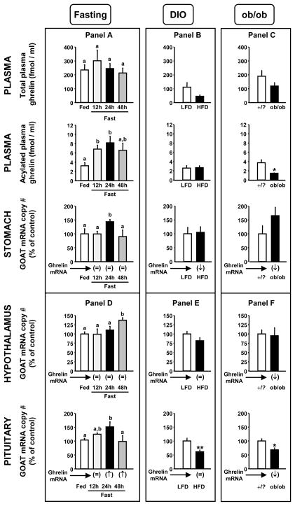
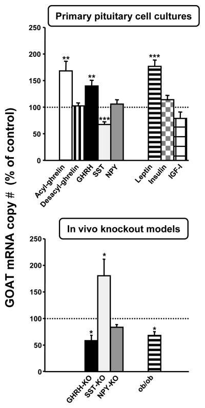
Ghrelin acts as an endocrine link connecting physiological processes regulating food intake, body composition, growth, and energy balance. Ghrelin is the only peptide known to undergo octanoylation. The enzyme mediating this process, ghrelin O-acyltransferase (GOAT), is expressed in the gastrointestinal tract (GI; primary source of circulating ghrelin) as well as other tissues. The present study demonstrates that stomach GOAT mRNA levels correlate with circulating acylated-ghrelin levels in fasted and diet-induced obese mice. In addition, GOAT was found to be expressed in both the pituitary and hypothalamus (two target tissues of ghrelin's actions), and regulated in response to metabolic status. Using primary pituitary cell cultures as a model system to study the regulation of GOAT expression, we found that acylated-ghrelin, but not desacyl-ghrelin, increased GOAT expression. In addition, growth-hormone-releasing hormone (GHRH) and leptin increased, while somatostatin (SST) decreased GOAT expression. The physiologic relevance of these later results is supported by the observation that pituitary GOAT expression in mice lacking GHRH, SST and leptin showed opposite changes to those observed after in vitro treatment with the corresponding peptides. Therefore, it seems plausible that these hormones directly contribute to the regulation of pituitary GOAT. Interestingly, in all the models studied, pituitary GOAT expression paralleled changes in the expression of a dominant spliced-variant of ghrelin (In2-ghrelin) and therefore this transcript may be a primary substrate for pituitary GOAT. Collectively, these observations support the notion that the GI tract is not the only source of acylated-ghrelin, but in fact locally produced des-acylated-ghrelin could be converted to acylated-ghrelin within target tissues by locally active GOAT, to mediate its tissue-specific effects.
Figures
Similar articles
-
The expression of ghrelin O-acyltransferase (GOAT) in human tissues.Endocr J. 2011;58(8):707-10. doi: 10.1507/endocrj.k11e-117. Epub 2011 Jun 4. Endocr J. 2011. PMID: 21646729
-
Ghrelin octanoylation mediated by an orphan lipid transferase.Proc Natl Acad Sci U S A. 2008 Apr 29;105(17):6320-5. doi: 10.1073/pnas.0800708105. Epub 2008 Apr 28. Proc Natl Acad Sci U S A. 2008. PMID: 18443287 Free PMC article.
-
Effect of Deletion of Ghrelin-O-Acyltransferase on the Pulsatile Release of Growth Hormone in Mice.J Neuroendocrinol. 2015 Dec;27(12):872-86. doi: 10.1111/jne.12327. J Neuroendocrinol. 2015. PMID: 26442444
-
Ghrelin O-acyltransferase (GOAT) and energy metabolism.Sci China Life Sci. 2016 Mar;59(3):281-91. doi: 10.1007/s11427-015-4973-6. Epub 2016 Jan 6. Sci China Life Sci. 2016. PMID: 26732975 Review.
-
Ghrelin acylation and metabolic control.Peptides. 2011 Nov;32(11):2301-8. doi: 10.1016/j.peptides.2011.08.020. Epub 2011 Aug 27. Peptides. 2011. PMID: 21893140 Review.
Cited by
-
Analysis of brain nuclei accessible to ghrelin present in the cerebrospinal fluid.Neuroscience. 2013 Dec 3;253:406-15. doi: 10.1016/j.neuroscience.2013.09.008. Epub 2013 Sep 13. Neuroscience. 2013. PMID: 24042041 Free PMC article.
-
Abdominal surgery inhibits circulating acyl ghrelin and ghrelin-O-acyltransferase levels in rats: role of the somatostatin receptor subtype 2.Am J Physiol Gastrointest Liver Physiol. 2011 Aug;301(2):G239-48. doi: 10.1152/ajpgi.00018.2011. Epub 2011 Jun 2. Am J Physiol Gastrointest Liver Physiol. 2011. PMID: 21636529 Free PMC article.
-
Therapeutic Potential of Targeting the Ghrelin Pathway.Int J Mol Sci. 2017 Apr 11;18(4):798. doi: 10.3390/ijms18040798. Int J Mol Sci. 2017. PMID: 28398233 Free PMC article. Review.
-
Ghrelin system is involved in improvements in glucose metabolism mediated by hyperbaric oxygen treatment in a streptozotocin‑induced type 1 diabetes mouse model.Mol Med Rep. 2020 Nov;22(5):3767-3776. doi: 10.3892/mmr.2020.11481. Epub 2020 Sep 2. Mol Med Rep. 2020. PMID: 32901885 Free PMC article.
-
Sensing of fatty acids for octanoylation of ghrelin involves a gustatory G-protein.PLoS One. 2012;7(6):e40168. doi: 10.1371/journal.pone.0040168. Epub 2012 Jun 29. PLoS One. 2012. PMID: 22768248 Free PMC article.
References
-
- Ahmad I, Finkelstein JA, Downs TR, Frohman LA. Obesity-associated decrease in growth hormone-releasing hormone gene expression: a mechanism for reduced growth hormone mRNA levels in genetically obese Zucker rats. Neuroendocrinology. 1993;58(3):332–7. - PubMed
-
- Alba M, Salvatori R. A mouse with targeted ablation of the growth hormone-releasing hormone gene: a new model of isolated growth hormone deficiency. Endocrinology. 2004;145(9):4134–43. - PubMed
-
- Ariyasu H, Takaya K, Tagami T, Ogawa Y, Hosoda K, Akamizu T, Suda M, Koh T, Natsui K, Toyooka S, Shirakami G, Usui T, Shimatsu A, Doi K, Hosoda H, Kojima M, Kangawa K, Nakao K. Stomach is a major source of circulating ghrelin, and feeding state determines plasma ghrelin-like immunoreactivity levels in humans. J Clin Endocrinol Metab. 2001;86(10):4753–8. - PubMed
-
- Casanueva FF, Dieguez C. Ghrelin: the link connecting growth with metabolism and energy homeostasis. Rev Endocr Metab Disord. 2002;3(4):325–38. - PubMed
-
- Cummings DE. Ghrelin and the short- and long-term regulation of appetite and body weight. Physiol Behav. 2006;89(1):71–84. - PubMed
Publication types
MeSH terms
Substances
Grants and funding
LinkOut - more resources
Full Text Sources
