Multicenter Study
. 2010 Mar;149(3):405-15.
doi: 10.1016/j.ajo.2009.09.013. Epub 2009 Dec 29.

A simplified quantitative method for assessing keratoconjunctivitis sicca from the Sjögren's Syndrome International Registry

Collaborators, Affiliations
Multicenter Study

A simplified quantitative method for assessing keratoconjunctivitis sicca from the Sjögren's Syndrome International Registry

John P Whitcher et al. Am J Ophthalmol. 2010 Mar.

Abstract

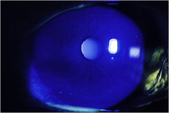
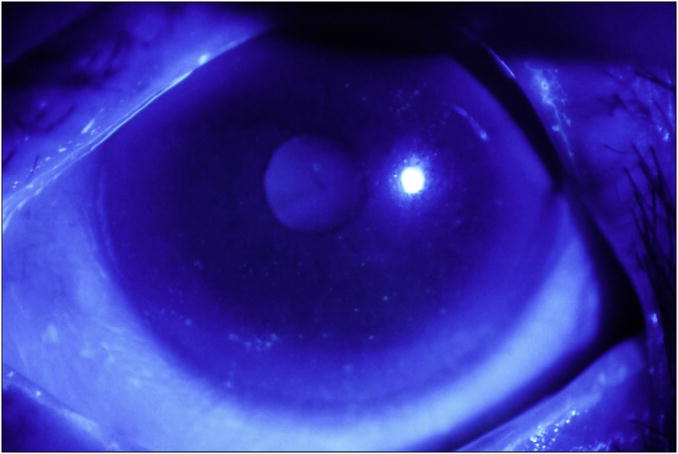
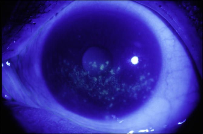
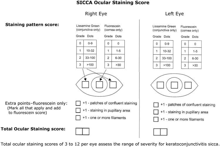
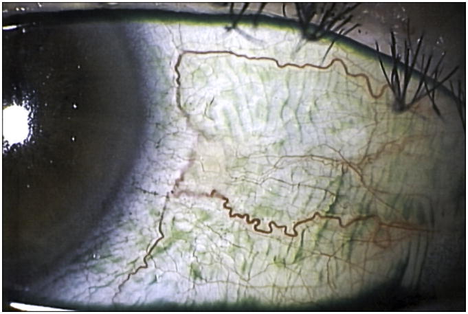
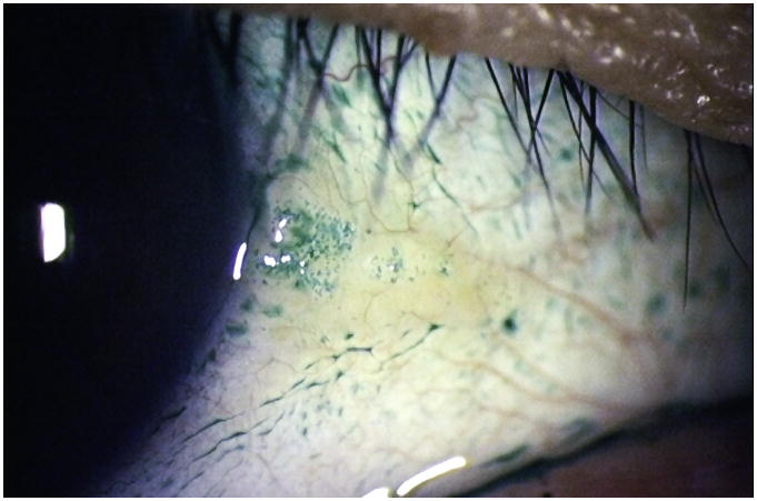
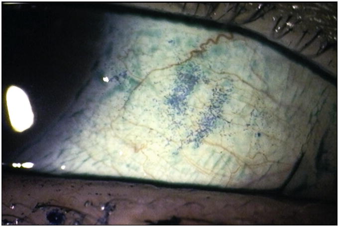
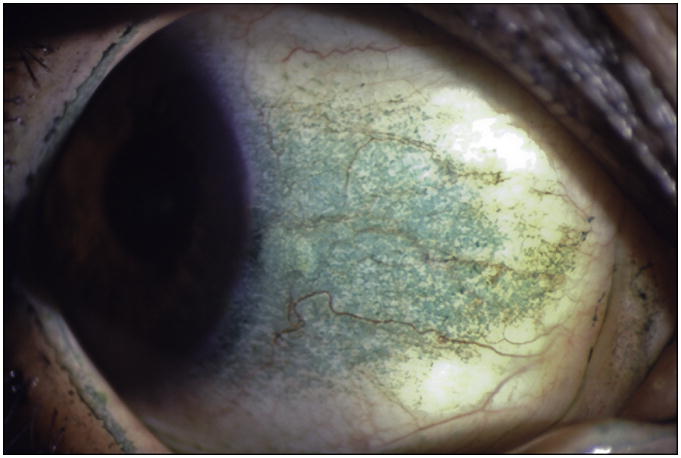
Purpose: To describe, apply, and test a new ocular grading system for assessing keratoconjunctivitis sicca (KCS) using lissamine green and fluorescein.

Design: Prospective, observational, multicenter cohort study.

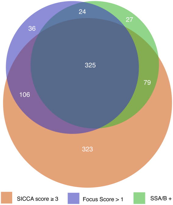
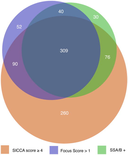
Methods: The National Institutes of Health-funded Sjögren's Syndrome International Registry (called Sjögren's International Collaborative Clinical Alliance [SICCA]) is developing standardized classification criteria for Sjögren syndrome (SS) and is creating a biospecimen bank for future research. Eight SICCA ophthalmologists developed a new quantitative ocular grading system (SICCA ocular staining score [OSS]), and we analyzed OSS distribution among the SICCA cohort and its association with other phenotypic characteristics of SS. The SICCA cohort includes participants ranging from possibly early SS to advanced disease. Procedures include sequenced unanesthetized Schirmer test, tear break-up time, ocular surface staining, and external eye examination at the slit lamp. Using statistical analyses and proportional Venn diagrams, we examined interrelationships between abnormal OSS (>or=3) and other characteristics of SS (labial salivary gland [LSG] biopsy with focal lymphocytic sialadenitis and focus score >1 positive anti-SS A antibodies, anti-SS B antibodies, or both).

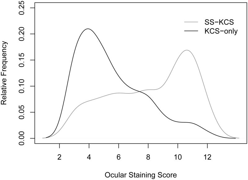
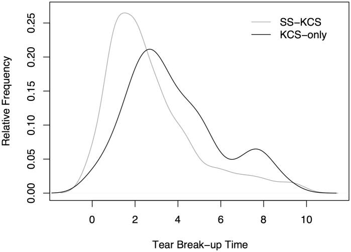
Results: Among 1208 participants, we found strong associations between abnormal OSS, positive serologic results, and positive LSG focus scores (P < .0001). Analysis of the overlapping relationships of these 3 measures defined a large group of participants who had KCS without other components of SS, representing a clinical entity distinct from the KCS associated with SS.

Conclusions: This new method for assessing KCS will become the means for diagnosing the ocular component of SS in future classification criteria. We find 2 forms of KCS whose causes may differ.


Figures

Figure 1
Figure 1
Fluorescein staining of the cornea in keratoconjunctivitis sicca: Figure 1a. Fluorescein staining of the cornea in keratoconjunctivitis sicca, Score 1 (1-5 very fine discrete punctate epithelial erosions seen inferiorly). Photograph courtesy of Dr. Kitagawa. Figure 1b. Fluorescein staining of the cornea in keratoconjunctivitis sicca, Score 3 (6-30 punctate epithelial erosions seen inferiorly, score 2, plus one punctate epithelial erosion present in the papillary area, for a total of 3). Photograph courtesy of Dr. Kitagawa. Figure 1c. Fluorescein staining of the cornea in keratoconjunctivitis sicca, Score 5 (>30 punctate epithelial erosions, many patches of confluent staining, and staining in the papillary area; no filaments). Photograph courtesy of Dr. Kitagawa.
Figure 1
Figure 1
Fluorescein staining of the cornea in keratoconjunctivitis sicca: Figure 1a. Fluorescein staining of the cornea in keratoconjunctivitis sicca, Score 1 (1-5 very fine discrete punctate epithelial erosions seen inferiorly). Photograph courtesy of Dr. Kitagawa. Figure 1b. Fluorescein staining of the cornea in keratoconjunctivitis sicca, Score 3 (6-30 punctate epithelial erosions seen inferiorly, score 2, plus one punctate epithelial erosion present in the papillary area, for a total of 3). Photograph courtesy of Dr. Kitagawa. Figure 1c. Fluorescein staining of the cornea in keratoconjunctivitis sicca, Score 5 (>30 punctate epithelial erosions, many patches of confluent staining, and staining in the papillary area; no filaments). Photograph courtesy of Dr. Kitagawa.
Figure 2
Figure 2. Sjögren's International Collaborative Clinical Alliance (SICCA) ocular staining score form
Figure 3
Figure 3
Lissamine green staining of the bulbar conjunctiva in keratoconjunctivitis sicca Figure 3a. Lissamine green background staining of the bulbar conjunctiva, Score 0. (Some pooling of dye is present; but <10 discrete individual green dots are seen in the interpalpebral area). Photograph courtesy of Dr. Kitagawa. Figure 3b. Lissamine green staining of the bulbar conjunctiva in keratoconjunctivitis sicca, Score 1 with >10 and <33 green dots in the interpalpebral area. (Staining on the surface of the adjacent pingueculum is considered an artifact and is therefore ignored). Photograph courtesy of Dr. Kitagawa. Figure 3c.. Lissamine green staining of the bulbar conjunctiva in keratoconjunctivitis sicca, Score 2 with 33 to100 green dots present in the interpalpebral area. (The two areas of partial confluent staining are each < 4 mm2 and are therefore scored as 33 to 100 dots – see text). Photograph courtesy of Dr. Kitagawa. Figure 3d. Lissamine green staining of the bulbar conjunctiva in keratoconjunctivitis sicca, Score 3 with >100 individual green dots in the interpalpebral area associated with many areas of confluent staining. Photograph courtesy of Dr. Kitagawa.
Figure 3
Figure 3
Lissamine green staining of the bulbar conjunctiva in keratoconjunctivitis sicca Figure 3a. Lissamine green background staining of the bulbar conjunctiva, Score 0. (Some pooling of dye is present; but <10 discrete individual green dots are seen in the interpalpebral area). Photograph courtesy of Dr. Kitagawa. Figure 3b. Lissamine green staining of the bulbar conjunctiva in keratoconjunctivitis sicca, Score 1 with >10 and <33 green dots in the interpalpebral area. (Staining on the surface of the adjacent pingueculum is considered an artifact and is therefore ignored). Photograph courtesy of Dr. Kitagawa. Figure 3c.. Lissamine green staining of the bulbar conjunctiva in keratoconjunctivitis sicca, Score 2 with 33 to100 green dots present in the interpalpebral area. (The two areas of partial confluent staining are each < 4 mm2 and are therefore scored as 33 to 100 dots – see text). Photograph courtesy of Dr. Kitagawa. Figure 3d. Lissamine green staining of the bulbar conjunctiva in keratoconjunctivitis sicca, Score 3 with >100 individual green dots in the interpalpebral area associated with many areas of confluent staining. Photograph courtesy of Dr. Kitagawa.
Figure 4
Figure 4. Area-proportional Venn diagrams visualizing the interrelationships between abnormal Ocular Staining Score, labial salivary gland focus scores > 1, and positive anti-SSA and/or anti-SSB antibodies
Figure 4a. Compares the three measures using an Ocular Staining Score cut-off value of ≥ 3 Figure 4b. Compares the three measures using an Ocular Staining Score cut-off value of ≥ 4
Figure 5
Figure 5
Distribution of the Ocular Staining Score (OSS) among participants with Keratoconjunctivitis Sicca-Only and those with Sjögren's Syndrome-Keratoconjunctivitis Sicca. Keratoconjunctivitis Sicca-Only cases have abnormal OSS ≥ 3, but no focal lymphocytic sialadenitis or have focus scores <1 and have negative serology to anti-SS-A or B antibodies. Sjögren's Syndrome-Keratoconjunctivitis Sicca cases have OSS ≥ 3, focal lymphocytic sialadenitis with focus score >1 and/or positive anti-SS-A or B antibodies. The estimated relative frequency among the two groups extends slightly beyond the ranges of the OSS as a result of “smoothing” used in the estimation process.
Figure 6
Figure 6
Distribution of the tear break-up time among participants with Keratoconjunctivitis Sicca-Only and those with Sjögren's Syndrome-Keratoconjunctivitis Sicca. Keratoconjunctivitis Sicca-Only cases have abnormal ocular staining score (OSS ≥ 3), but no focal lymphocytic sialadenitis or have focus scores <1 and have negative serology to anti-SS-A or B antibodies. Sjögren's Syndrome-Keratoconjunctivitis Sicca cases have OSS ≥ 3, focal lymphocytic sialadenitis with focus score >1 and/or positive anti-SS-A or B antibodies.

References

    1. Sjögren H. Zur Kenntis der Keratoconjunctivitis Sicca (Keratitis filiformis bei Hypofunktion der Tranendrusen) Acta Ophthalmol. 1933;(2):1–151.
    1. fluger P. Zur Ernahrung der cornea. Klin Monatsbl Augenheilkd. 1882;20:69–81.
    1. Holm S. Keratoconjunctivitis sicca and the sicca syndrome. Acta Ophthamologica. 1949;33(Suppl):1–230.
    1. van Bijsterveld OP. Diagnostic tests in the sicca syndrome. Arch Ophthal. 1969;82:10–14. - PubMed
    1. Norn MS. Lissamine green: Vital staining of cornea and conjunctiva. Acta Ophthalmologica. 1973;51:483–91. - PubMed

Publication types