. 2010;261(2):81-92.
doi: 10.1016/j.cellimm.2009.11.004. Epub 2009 Dec 24.

Thymic nurse cells exhibit epithelial progenitor phenotype and create unique extra-cytoplasmic membrane space for thymocyte selection

Affiliations

Thymic nurse cells exhibit epithelial progenitor phenotype and create unique extra-cytoplasmic membrane space for thymocyte selection

Tonya M Hendrix et al. Cell Immunol. 2010.

Abstract

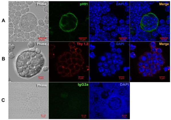
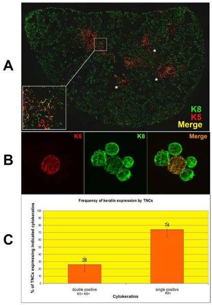
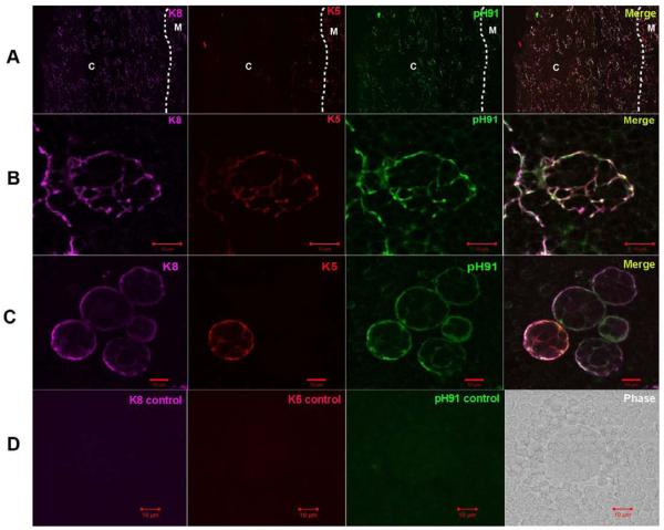
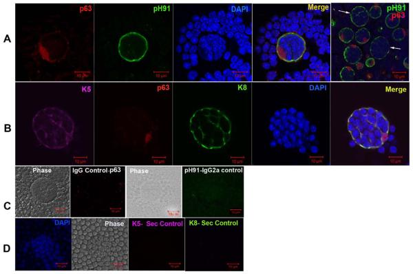
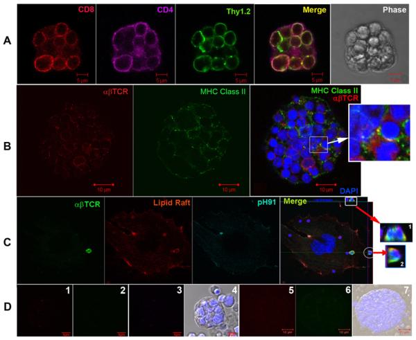
Thymic nurse cells (TNCs) are epithelial cells in the thymic cortex that contain as many as 50 thymocytes within specialized cytoplasmic vacuoles. The function of this cell-in-cell interaction has created controversy since their discovery in 1980. Further, some skepticism exists about the idea that apoptotic thymocytes within the TNC complex result from negative selection, a process believed to occur exclusively within the medulla. In this report, we have microscopic evidence that defines a unique membranous environment wherein lipid raft aggregates around the alphabetaTCR expressed on captured thymocytes and class II MHC molecules expressed on TNCs. Further, immunohistological examination of thymic sections show TNCs located within the cortico-medullary junction to express cytokeratins five and eight (K5 and K8), and the transcription factor Trp-63, the phenotype defined elsewhere as the thymic epithelial progenitor subset. Our results suggest that the microenvironment provided by TNCs plays an important role in thymocyte selection as well as the potential for TNCs to be involved in the maintenance of thymic epithelia.


Figures

Figure 1
Figure 1
PH91 identifies the TNC lymphostromal complex. (A) Phase image, pH91 staining (green), DAPI staining (blue), and overlay of a freshly isolated TNC. Scale bar represents 10 μm. (B) Phase image, Thy 1.2 staining (red), DAPI staining (blue), and overlay of a freshly isolated TNC. Scale bar represents 5 μm. (C) Panel shows phase image and isotype control for pH91 (IgG2a). Data is representative of three independent experiments.
Figure 2
Figure 2
K8+K5+ cells exist at the cortico-medullary junction and are a subset of TNCs. Confocal analysis of keratin and pH91 staining of thymic sections and freshly isolated TNCs. (A) Thymic section stained with K5 (red) and K8 (green). Yellow regions (inset and asterisks) show double positive (K8+K5+) epithelial cells. Original magnification: 40X. (B) Freshly isolated TNCs stained with K5 (red) and K8 (green). The double positive TNC appears as yellow in the merge. Original magnification: 40X. (C) Frequency of K8+ and K8+K5+ populations in freshly isolated TNCs determined by manual counting of 1000 cytospun TNCs after immunostaining. No K5+ single positive cells with the TNC complex morphology were detected. Data for (A) and (B) is representative of three independent experiments.
Figure 3
Figure 3
Expression of K8 and K5 by pH91-labeled TNCs. (A) Thymic sections stained with anti-K8 (magenta), anti-K5 (red), and pH91 (green) Abs. The cortex and medulla are indicated by “C” and “M”, respectively, and the cortico-medullary junction by a dotted line. The merge shows areas of co-localization. Original magnification: 40X. (B) Magnified area of section in panel A. The triple stained TNC exhibits a fenestrated structure. (C) Freshly isolated TNCs stained with anti-K8 (magenta), anti-K5 (red), and pH91 (green) Abs. Both K8+K5+ and K8+ only TNCs are visible. (D) Shows secondary Ab controls for K8, K5, and pH91 as well as a phage image. Data is representative of three independent experiments.
Figure 4
Figure 4
Trp63 expression of freshly isolated TNCs. (A) Confocal micrograph of a TNC labeled with Ab against p63 (red) and pH91 (green). Merge shows cell surface staining with pH91, nuclear localization of p63, and DAPI stained nuclei. Last panel shows that not all TNCs (white arrows) express p63. (B) Confocal micrograph of a TNC labeled with Ab against p63 (red), K5 (magenta), and K8 (green) with DAPI stained nuclei (blue). Merge shows cell surface staining with pH91, nuclear localization of p63, and DAPI stained nuclei. (C) TNCs stained with secondary controls for p63 (IgG) and pH91 (IgG2a) with phase image. (D) TNCs stained with secondary controls for K5 and K8 showing phase image and DAPI stained nuclei. Data is representative of three independent experiments.
Figure 5
Figure 5
Thymic nurse cells facilitate the inward and outward migration of thymocytes. tsTNC-1 cells were co-incubated with freshly isolated thymocytes for 5 hours. Panels A-C show the stepwise internalization of a thymocyte (arrow). Panels D-F show the outward movement of a different thymocyte (arrow). The movement of a macrophage within a TNC complex is shown in panels G-I. The “N” indicates the location of the nucleus of the thymic nurse cell.
Figure 6
Figure 6
SEM of tsTNC-1 cells co-incubated with thymocytes for up to 10 hours. (A) An individual TNC displaying membrane extensions polarized to one side of the cell. The body of the cell is indicated by “*b”. (Time of incubation = 0 hour) (B) TNCs co-incubated with thymocytes for 1 hours display larger areas of membrane extensions. (C) Thymocytes are trapped within the folds of the membrane extensions after 2 hours of incubation (arrow). (D) Membrane extensions of TNCs wrapped around the surface bound thymocytes (arrow). Cells were incubated for 2 hours. (E) After 4 hours of incubation, TNC no longer displays prolific membrane extensions but does contain internalized thymocytes. An arrow points to a partially internalized thymocyte. (F) Large TNC complex contains internalized thymocytes. (G) Confocal micrograph of tsTNC co-incubated with thymocytes for 10 hours is shown in inset. Prior to co-incubation the TNCs were labeled with CFDA (green) and the thymocytes were labeled with CFSE (red). The TNC nucleus (N) is visible as are internalized thymocytes (T).
Figure 7
Figure 7
SEM of TNCs isolated from murine thymus and incubated over a 3 hours time period. (A) Thirty minute after isolation, the cell exhibits a circular morphology and layers of membrane folds (see inset). (B) Membrane begins to unfold and wraps around released thymocytes (see inset) (60 minutes post isolation). (C) Membrane layer is completely unfolds exposing thymocytes located within the fenestra (inset) (90 minutes post isolation). (D) TNC membrane is completely unfolded (120 minutes post isolation). (E) (150 min) Thymocytes are then gradually released from the TNC. (F) No thymocytes are associated with the TNC. (180 min) (G) TEM image of a freshly isolated TNC corresponds to cell shown in panel A.
Figure 8
Figure 8
Transmission electron micrographs of TNCs containing internalized thymocytes. (A) Freshly isolated TNC fixed and prepared for TEM analysis immediately following isolation. Membrane partially encloses a thymocyte (asterisk). (B) Thymocytes and tsTNC-1 cells were incubated for 10 hours and then analyzed by TEM. Thymocytes at different stages of apoptosis (insets 1 and 2) are visible within the cytoplasmic vacuoles.
Figure 9
Figure 9
Microscopic identification of membrane extensions and cage structure of TNCs during thymocyte binding and internalization. (A) Membrane extensions of TNCs visualized with SEM (panel 1) and TEM (panel 2). The circle in panel 2 corresponds to the membrane extensions seen in panel 1. TNC membrane extensions, equivalent to those in panels 1 and 2, are wrapped around a thymocyte (asterisk, panel 3) with a large cytoplasmic vesicle directly beneath (arrow). (B) The cage structure of TNCs visualized with SEM (panel 1), TEM (panel 2), and confocal microscopy of thymic section stained with pH91 (panel 3). The inset in panel 1 shows thymocytes residing within the cage like structure. The circle in panel 2 surrounds the cage as it appears in TEM. The arrow points to its SEM equivalent in panel 1. (C) Phase contrast video microscopy of TNC - thymocyte interaction. Thymocytes bound to the TNC surface are phase bright. Thymocytes trapped within the cage-like structure (circle, B and C) are phase dark. Thymocyte movement within cage was also detected. The arrow shows initial contact of thymocytes by membrane extensions from TNC. Membrane extensions pull thymocytes to the TNC complex.
Figure 10
Figure 10
TNCs participate in the MHC restriction of thymocytes. (A) Freshly isolated TNCs were stained with mAbs against CD8 (red), CD4 (magenta), and Thy 1.2 (green). Merge panel shows captured thymocytes to be CD4+CD8+. (B) Freshly isolated TNC stained with mAbs to αβTCR (red) and MHC Class II (green). Thymocyte nuclei are visualized using DAPI staining (blue). Insets show co-localization of MHC and αβTCR (yellow). (C) Co-localization of thymocyte lipid raft with αβTCR. tsTNC-1 cells were co-incubated with freshly isolated thymocytes for 4 hours. Cells were stained with cholera toxin subunit B (red) for presence of lipid rafts and mAbs to αβTCR (green) and pH91 (magenta). Merge images (insets 1 and 2) are orthogonal projections that show the contact areas between αβTCR and lipid raft (yellow). (D) Panels 1, 2, and 3 show controls for CD8, CD4, and Thy 1, respectively. Panels 5 and 6 show mAb controls for αβTCR and MHC class II. Data is representative of three independent experiments.

Similar articles

Cited by

References

    1. Overholtzer M, Brugge JS. The cell biology of cell-in-cell structures. Nat Rev Mol Cell Biol. 2008;9:796–809. - PubMed
    1. Wekerle H, Ketelsen UP. Thymic nurse cells--Ia-bearing epithelium involved in T-lymphocyte differentiation? Nature. 1980;283:402–4. - PubMed
    1. Wekerle H, Ketelsen UP, Ernst M. Thymic nurse cells. Lymphoepithelial cell complexes in murine thymuses: morphological and serological characterization. J Exp Med. 1980;151:925–44. - PMC - PubMed
    1. Ritter MA, Sauvage CA, Cotmore SF. The human thymus microenvironment: in vivo identification of thymic nurse cells and other antigenically-distinct subpopulations of epithelial cells. Immunology. 1981;44:439–46. - PMC - PubMed
    1. Boyd RL, Oberhuber G, Hala K, Wick G. Obese strain (OS) chickens with spontaneous autoimmune thyroiditis have a deficiency in thymic nurse cells. J Immunol. 1984;132:718–24. - PubMed

Publication types